Advertisement

Main menu

  • Home
  • Content
    • Current Issue
    • Accepted Manuscripts
    • Article Preview
    • Past Issue Archive
    • Video Articles
    • AJNR Case Collection
    • Case of the Week Archive
    • Case of the Month Archive
    • Classic Case Archive
  • Special Collections
    • AJNR Awards
    • Low-Field MRI
    • Alzheimer Disease
    • ASNR Foundation Special Collection
    • Photon-Counting CT
    • View All
  • Multimedia
    • AJNR Podcasts
    • AJNR SCANtastic
    • Trainee Corner
    • MRI Safety Corner
    • Imaging Protocols
  • For Authors
    • Submit a Manuscript
    • Submit a Video Article
    • Submit an eLetter to the Editor/Response
    • Manuscript Submission Guidelines
    • Statistical Tips
    • Fast Publishing of Accepted Manuscripts
    • Graphical Abstract Preparation
    • Imaging Protocol Submission
    • Author Policies
  • About Us
    • About AJNR
    • Editorial Board
    • Editorial Board Alumni
  • More
    • Become a Reviewer/Academy of Reviewers
    • Subscribers
    • Permissions
    • Alerts
    • Feedback
    • Advertisers
    • ASNR Home

User menu

  • Alerts
  • Log in

Search

  • Advanced search
American Journal of Neuroradiology

American Journal of Neuroradiology

ASHNR American Society of Functional Neuroradiology ASHNR American Society of Pediatric Neuroradiology ASSR
  • Alerts
  • Log in

Advanced Search

  • Home
  • Content
    • Current Issue
    • Accepted Manuscripts
    • Article Preview
    • Past Issue Archive
    • Video Articles
    • AJNR Case Collection
    • Case of the Week Archive
    • Case of the Month Archive
    • Classic Case Archive
  • Special Collections
    • AJNR Awards
    • Low-Field MRI
    • Alzheimer Disease
    • ASNR Foundation Special Collection
    • Photon-Counting CT
    • View All
  • Multimedia
    • AJNR Podcasts
    • AJNR SCANtastic
    • Trainee Corner
    • MRI Safety Corner
    • Imaging Protocols
  • For Authors
    • Submit a Manuscript
    • Submit a Video Article
    • Submit an eLetter to the Editor/Response
    • Manuscript Submission Guidelines
    • Statistical Tips
    • Fast Publishing of Accepted Manuscripts
    • Graphical Abstract Preparation
    • Imaging Protocol Submission
    • Author Policies
  • About Us
    • About AJNR
    • Editorial Board
    • Editorial Board Alumni
  • More
    • Become a Reviewer/Academy of Reviewers
    • Subscribers
    • Permissions
    • Alerts
    • Feedback
    • Advertisers
    • ASNR Home
  • Follow AJNR on Twitter
  • Visit AJNR on Facebook
  • Follow AJNR on Instagram
  • Join AJNR on LinkedIn
  • RSS Feeds

AJNR Awards, New Junior Editors, and more. Read the latest AJNR updates

Getting new auth cookie, if you see this message a lot, tell someone!
Research ArticleExtracranial Vascular
Open Access

Characterization of Craniocervical Artery Dissection by Simultaneous MR Noncontrast Angiography and Intraplaque Hemorrhage Imaging at 3T

Q. Li, J. Wang, H. Chen, X. Gong, N. Ma, K. Gao, L. He, M. Guan, Z. Chen, R. Li, D. Mi, C. Yuan, X. Zhao and X.H. Zhao
American Journal of Neuroradiology September 2015, 36 (9) 1769-1775; DOI: https://doi.org/10.3174/ajnr.A4348
Q. Li
aFrom the Departments of Neurology (Q.L., X.G., D.M., X.Z.)
cDepartment of Neurology (Q.L.), People's Hospital of Xinjiang Uygur Autonomous Region, Urumqi, China
  • Find this author on Google Scholar
  • Find this author on PubMed
  • Search for this author on this site
J. Wang
dClinical Sites Research Program (J.W.), Philips Research North America, Briarcliff Manor, New York
  • Find this author on Google Scholar
  • Find this author on PubMed
  • Search for this author on this site
H. Chen
eDepartment of Biomedical Engineering (H.C., L.H., Z.C., R.L., C.Y., X.H.Z.), Center for Biomedical Imaging Research, Tsinghua University School of Medicine, Beijing, China
  • Find this author on Google Scholar
  • Find this author on PubMed
  • Search for this author on this site
X. Gong
aFrom the Departments of Neurology (Q.L., X.G., D.M., X.Z.)
  • Find this author on Google Scholar
  • Find this author on PubMed
  • Search for this author on this site
N. Ma
bInterventional Neuroradiology (N.M., K.G.), Beijing Tiantan Hospital, Capital Medical University, Beijing, China
  • Find this author on Google Scholar
  • Find this author on PubMed
  • Search for this author on this site
K. Gao
bInterventional Neuroradiology (N.M., K.G.), Beijing Tiantan Hospital, Capital Medical University, Beijing, China
  • Find this author on Google Scholar
  • Find this author on PubMed
  • Search for this author on this site
L. He
eDepartment of Biomedical Engineering (H.C., L.H., Z.C., R.L., C.Y., X.H.Z.), Center for Biomedical Imaging Research, Tsinghua University School of Medicine, Beijing, China
  • Find this author on Google Scholar
  • Find this author on PubMed
  • Search for this author on this site
M. Guan
fDepartment of Radiology (M.G.), Yangzhou First People's Hospital, Yangzhou, China
  • Find this author on Google Scholar
  • Find this author on PubMed
  • Search for this author on this site
Z. Chen
eDepartment of Biomedical Engineering (H.C., L.H., Z.C., R.L., C.Y., X.H.Z.), Center for Biomedical Imaging Research, Tsinghua University School of Medicine, Beijing, China
  • Find this author on Google Scholar
  • Find this author on PubMed
  • Search for this author on this site
R. Li
eDepartment of Biomedical Engineering (H.C., L.H., Z.C., R.L., C.Y., X.H.Z.), Center for Biomedical Imaging Research, Tsinghua University School of Medicine, Beijing, China
  • Find this author on Google Scholar
  • Find this author on PubMed
  • Search for this author on this site
D. Mi
aFrom the Departments of Neurology (Q.L., X.G., D.M., X.Z.)
  • Find this author on Google Scholar
  • Find this author on PubMed
  • Search for this author on this site
C. Yuan
eDepartment of Biomedical Engineering (H.C., L.H., Z.C., R.L., C.Y., X.H.Z.), Center for Biomedical Imaging Research, Tsinghua University School of Medicine, Beijing, China
gDepartment of Radiology (C.Y.), University of Washington, Seattle, Washington.
  • Find this author on Google Scholar
  • Find this author on PubMed
  • Search for this author on this site
X. Zhao
aFrom the Departments of Neurology (Q.L., X.G., D.M., X.Z.)
  • Find this author on Google Scholar
  • Find this author on PubMed
  • Search for this author on this site
X.H. Zhao
eDepartment of Biomedical Engineering (H.C., L.H., Z.C., R.L., C.Y., X.H.Z.), Center for Biomedical Imaging Research, Tsinghua University School of Medicine, Beijing, China
  • Find this author on Google Scholar
  • Find this author on PubMed
  • Search for this author on this site
  • Article
  • Figures & Data
  • Info & Metrics
  • Responses
  • References
  • PDF
Loading

Article Figures & Data

Figures

  • Tables
  • Fig 1.
    • Download figure
    • Open in new tab
    • Download powerpoint
    Fig 1.

    Excellent agreement between SNAP imaging and TOF MRA in measuring stenosis. Mild stenosis in the basilar artery is determined by MRA images derived from SNAP imaging (A), which corresponds to the TOF MRA image (B). The hyperintense signal on the SNAP vessel wall image (C) indicates intramural hematoma.

  • Fig 2.
    • Download figure
    • Open in new tab
    • Download powerpoint
    Fig 2.

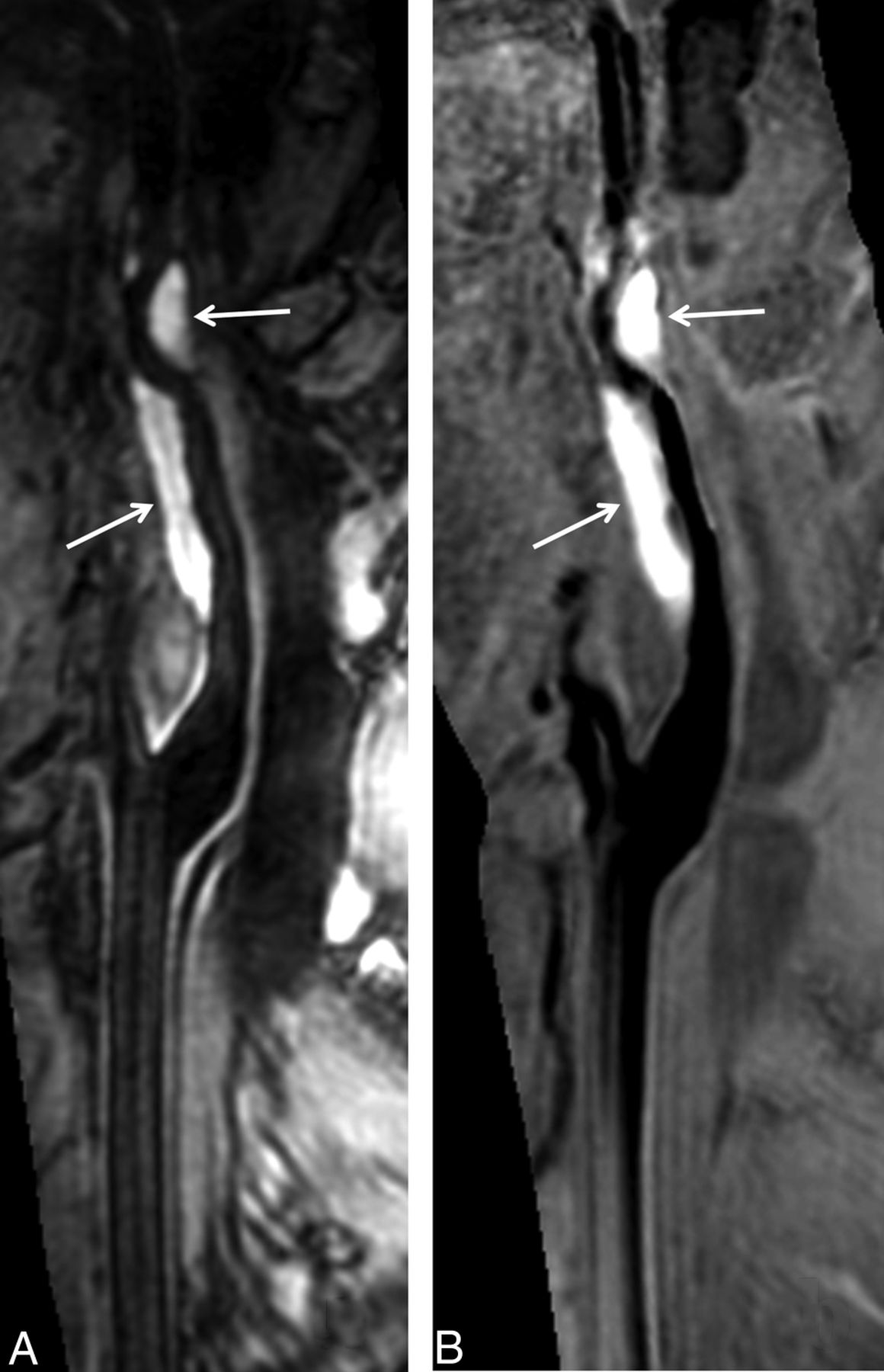
    SNAP imaging depicts intramural hematoma. The intramural hematoma in the internal carotid artery appears hyperintense on both 3D MERGE (A, white arrows) and SNAP images (B, white arrows).

  • Fig 3.
    • Download figure
    • Open in new tab
    • Download powerpoint
    Fig 3.

    SNAP imaging delineates the intimal flaps. The SNAP MRA image (A) shows a dilated lumen (pseudoaneurysm) in the internal carotid artery. The intimal flaps (hollow arrow) and double lumen (white arrow) are noted on vessel wall images derived from SNAP imaging in the coronal (B) and axial (C) views after MPR reconstruction. The white line on the coronal view indicates the location of the axial view acquisition.

  • Fig 4.
    • Download figure
    • Open in new tab
    • Download powerpoint
    Fig 4.

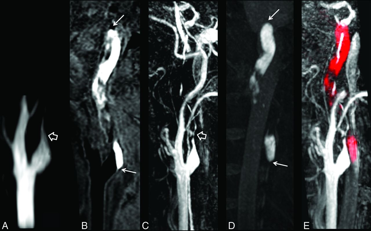
    The luminal narrowing and intramural hematoma are jointly visualized in a single SNAP image. Total occlusion in the internal carotid artery is demonstrated by TOF MRA (A, hollow arrow) and SNAP MRA (C, hollow arrow). A vessel wall image (B) derived from SNAP imaging shows a hyperintense lesion in the vessel wall (white arrows), indicating intramural hematoma. The intramural hematoma is well-depicted by maximum intensity projection of SNAP images (D). The MRA and vessel wall images derived from SNAP imaging are naturally registered to jointly visualize luminal narrowing and intramural hematoma in red (E) after color-coded processing.

Tables

  • Figures
    • View popup
    Table 1:

    Clinical characteristics of study population

    CharacteristicsMean or No. (%)Range (if Applicable)
    Age (yr)45.0 ± 16.114–70
    Male sex21 (87.5)
    Weight (kg)75.1 ± 8.560–90
    Height (cm)171.2 ± 5.5160–183
    Smoking10 (41.7)
    Hypertension13 (54.2)
    Diabetes6 (25)
    High-density lipoprotein (mmol/L)1.0 ± 0.20.5–1.8
    Low-density lipoprotein (mmol/L)2.3 ± 0.81.0–5.7
    Total cholesterol (mmol/L)4.0 ± 1.12.2–7.8
    Triglycerides (mmol/L)1.5 ± 0.70.6–3.0
    Headache4 (16.7)
    Neck pain3 (12.5)
    Ischemic stroke20 (83.3)
    Transient ischemic attack4 (16.7)
    • View popup
    Table 2:

    Characteristics of 24 patients with CCAD on multisequence MR imaging

    Patient No.Sex/Age (yr)LocationLength (mm)Category of StenosisaPresence or Absence
    IMHPseudo-aneurysmIntimal Flaps and Double Lumen
    1M/65BA16.7Moderate+––
    2M/24MCA8.1Occlusion+––
    3M/14MCA6.0Mild+––
    4M/53ICA (C1)35.8No stenosis–++
    5M/20BA12.7No stenosis––+
    6M/40ICA (C1)70.2Occlusion+––
    7M/62BA and VA44.1Occlusion+––
    8M/59BA22.2No stenosis–+–
    9M/36MCA10.7No stenosis––+
    10F/18CCA23.3Occlusion+––
    11M/34VA10.1Moderate–––
    12F/38ICA (C1)42.4Severe+––
    13F/66CCA44.8No stenosis+––
    14M/43VA7.5Occlusion+––
    15M/62ICA (C1)40Severe+––
    16M/68CCA37.1Occlusion+––
    17M/42VA24.8Occlusion+––
    18M/35ICA (C1)63.4Occlusion+––
    19M/51BA and VA50.8Mild+––
    20M/69ICA (C1)57Occlusion+––
    21M/56ICA (C1)66.4Severe+––
    22M/40ICA (C1)13.6Occlusion+––
    23M/41VA42.7Occlusion+––
    24M/43BA11.6Moderate+–+
    • Note:—BA indicates basilar artery; VA, vertebral artery; CCA, common carotid artery; +, positive; –, negative.

    • ↵a Mild stenosis, 1%–49%; moderate stenosis, 50%–69%; severe stenosis, 70%–99%; occlusion, 100%.

PreviousNext
Back to top

In this issue

American Journal of Neuroradiology: 36 (9)
American Journal of Neuroradiology
Vol. 36, Issue 9
1 Sep 2015
  • Table of Contents
  • Index by author
  • Complete Issue (PDF)
Advertisement
Print
Download PDF
Email Article

Thank you for your interest in spreading the word on American Journal of Neuroradiology.

NOTE: We only request your email address so that the person you are recommending the page to knows that you wanted them to see it, and that it is not junk mail. We do not capture any email address.

Enter multiple addresses on separate lines or separate them with commas.
Characterization of Craniocervical Artery Dissection by Simultaneous MR Noncontrast Angiography and Intraplaque Hemorrhage Imaging at 3T
(Your Name) has sent you a message from American Journal of Neuroradiology
(Your Name) thought you would like to see the American Journal of Neuroradiology web site.
CAPTCHA
This question is for testing whether or not you are a human visitor and to prevent automated spam submissions.
Cite this article
Q. Li, J. Wang, H. Chen, X. Gong, N. Ma, K. Gao, L. He, M. Guan, Z. Chen, R. Li, D. Mi, C. Yuan, X. Zhao, X.H. Zhao
Characterization of Craniocervical Artery Dissection by Simultaneous MR Noncontrast Angiography and Intraplaque Hemorrhage Imaging at 3T
American Journal of Neuroradiology Sep 2015, 36 (9) 1769-1775; DOI: 10.3174/ajnr.A4348

Citation Manager Formats

  • BibTeX
  • Bookends
  • EasyBib
  • EndNote (tagged)
  • EndNote 8 (xml)
  • Medlars
  • Mendeley
  • Papers
  • RefWorks Tagged
  • Ref Manager
  • RIS
  • Zotero
0 Responses
Respond to this article
Share
Bookmark this article
Characterization of Craniocervical Artery Dissection by Simultaneous MR Noncontrast Angiography and Intraplaque Hemorrhage Imaging at 3T
Q. Li, J. Wang, H. Chen, X. Gong, N. Ma, K. Gao, L. He, M. Guan, Z. Chen, R. Li, D. Mi, C. Yuan, X. Zhao, X.H. Zhao
American Journal of Neuroradiology Sep 2015, 36 (9) 1769-1775; DOI: 10.3174/ajnr.A4348
del.icio.us logo Twitter logo Facebook logo Mendeley logo
  • Tweet Widget
  • Facebook Like
  • Google Plus One
Purchase

Jump to section

  • Article
    • Abstract
    • ABBREVIATIONS:
    • Materials and Methods
    • Results
    • Discussion
    • Conclusions
    • Footnotes
    • References
  • Figures & Data
  • Info & Metrics
  • Responses
  • References
  • PDF

Related Articles

  • No related articles found.
  • PubMed
  • Google Scholar

Cited By...

  • 3D T1-weighted black blood sequence at 3.0 Tesla for the diagnosis of cervical artery dissection
  • Crossref
  • Google Scholar

This article has not yet been cited by articles in journals that are participating in Crossref Cited-by Linking.

More in this TOC Section

Extracranial Vascular

  • Proximal Vertebral Artery Variants and Embryology
  • High-Risk Plaque Features in Carotid MRI
  • Nonstenotic Carotid Plaques and Stroke Review
Show more Extracranial Vascular

Head and Neck Imaging

  • MRI of the Parasellar Ligaments
  • ASL Sensitivity for Head and Neck Paraganglioma
  • Post SRS Peritumoral Hyperintense Signal of VSs
Show more Head and Neck Imaging

Neurointervention

  • Rescue Reentry in Carotid Near-Occlusion
  • Contour Neurovascular System: Five Year Follow Up
  • Effect of SARS-CoV2 on Endovascular Thrombectomy
Show more Neurointervention

Similar Articles

Advertisement

Indexed Content

  • Current Issue
  • Accepted Manuscripts
  • Article Preview
  • Past Issues
  • Editorials
  • Editor's Choice
  • Fellows' Journal Club
  • Letters to the Editor
  • Video Articles

Cases

  • Case Collection
  • Archive - Case of the Week
  • Archive - Case of the Month
  • Archive - Classic Case

More from AJNR

  • Trainee Corner
  • Imaging Protocols
  • MRI Safety Corner
  • Book Reviews

Multimedia

  • AJNR Podcasts
  • AJNR Scantastics

Resources

  • Turnaround Time
  • Submit a Manuscript
  • Submit a Video Article
  • Submit an eLetter to the Editor/Response
  • Manuscript Submission Guidelines
  • Statistical Tips
  • Fast Publishing of Accepted Manuscripts
  • Graphical Abstract Preparation
  • Imaging Protocol Submission
  • Evidence-Based Medicine Level Guide
  • Publishing Checklists
  • Author Policies
  • Become a Reviewer/Academy of Reviewers
  • News and Updates

About Us

  • About AJNR
  • Editorial Board
  • Editorial Board Alumni
  • Alerts
  • Permissions
  • Not an AJNR Subscriber? Join Now
  • Advertise with Us
  • Librarian Resources
  • Feedback
  • Terms and Conditions
  • AJNR Editorial Board Alumni

American Society of Neuroradiology

  • Not an ASNR Member? Join Now

© 2025 by the American Society of Neuroradiology All rights, including for text and data mining, AI training, and similar technologies, are reserved.
Print ISSN: 0195-6108 Online ISSN: 1936-959X

Powered by HighWire