Advertisement

Main menu

  • Home
  • Content
    • Current Issue
    • Accepted Manuscripts
    • Article Preview
    • Past Issue Archive
    • Video Articles
    • AJNR Case Collection
    • Case of the Week Archive
    • Case of the Month Archive
    • Classic Case Archive
  • Special Collections
    • AJNR Awards
    • Low-Field MRI
    • Alzheimer Disease
    • ASNR Foundation Special Collection
    • Photon-Counting CT
    • View All
  • Multimedia
    • AJNR Podcasts
    • AJNR SCANtastic
    • Trainee Corner
    • MRI Safety Corner
    • Imaging Protocols
  • For Authors
    • Submit a Manuscript
    • Submit a Video Article
    • Submit an eLetter to the Editor/Response
    • Manuscript Submission Guidelines
    • Statistical Tips
    • Fast Publishing of Accepted Manuscripts
    • Graphical Abstract Preparation
    • Imaging Protocol Submission
    • Author Policies
  • About Us
    • About AJNR
    • Editorial Board
    • Editorial Board Alumni
  • More
    • Become a Reviewer/Academy of Reviewers
    • Subscribers
    • Permissions
    • Alerts
    • Feedback
    • Advertisers
    • ASNR Home

User menu

  • Alerts
  • Log in

Search

  • Advanced search
American Journal of Neuroradiology

American Journal of Neuroradiology

ASHNR American Society of Functional Neuroradiology ASHNR American Society of Pediatric Neuroradiology ASSR
  • Alerts
  • Log in

Advanced Search

  • Home
  • Content
    • Current Issue
    • Accepted Manuscripts
    • Article Preview
    • Past Issue Archive
    • Video Articles
    • AJNR Case Collection
    • Case of the Week Archive
    • Case of the Month Archive
    • Classic Case Archive
  • Special Collections
    • AJNR Awards
    • Low-Field MRI
    • Alzheimer Disease
    • ASNR Foundation Special Collection
    • Photon-Counting CT
    • View All
  • Multimedia
    • AJNR Podcasts
    • AJNR SCANtastic
    • Trainee Corner
    • MRI Safety Corner
    • Imaging Protocols
  • For Authors
    • Submit a Manuscript
    • Submit a Video Article
    • Submit an eLetter to the Editor/Response
    • Manuscript Submission Guidelines
    • Statistical Tips
    • Fast Publishing of Accepted Manuscripts
    • Graphical Abstract Preparation
    • Imaging Protocol Submission
    • Author Policies
  • About Us
    • About AJNR
    • Editorial Board
    • Editorial Board Alumni
  • More
    • Become a Reviewer/Academy of Reviewers
    • Subscribers
    • Permissions
    • Alerts
    • Feedback
    • Advertisers
    • ASNR Home
  • Follow AJNR on Twitter
  • Visit AJNR on Facebook
  • Follow AJNR on Instagram
  • Join AJNR on LinkedIn
  • RSS Feeds

AJNR Awards, New Junior Editors, and more. Read the latest AJNR updates

Research ArticleNeurointervention

The Preventive Effect of Endovascular Treatment for Recurrent Hemorrhage in Patients with Spinal Cord Arteriovenous Malformations

Y. Niimi, H. Matsukawa, N. Uchiyama and A. Berenstein
American Journal of Neuroradiology September 2015, 36 (9) 1763-1768; DOI: https://doi.org/10.3174/ajnr.A4396
Y. Niimi
aFrom the Departments of Neuroendovascular Therapy (Y.N.)
  • Find this author on Google Scholar
  • Find this author on PubMed
  • Search for this author on this site
H. Matsukawa
bNeurosurgery (H.M.), St. Luke's International Hospital, Tokyo, Japan
  • Find this author on Google Scholar
  • Find this author on PubMed
  • Search for this author on this site
N. Uchiyama
cDepartment of Neurosurgery (N.U.), Kanazawa University, Kanazawa, Japan
  • Find this author on Google Scholar
  • Find this author on PubMed
  • Search for this author on this site
  • ORCID record for N. Uchiyama
A. Berenstein
dCenter for Endovascular Surgery (A.B.), Roosevelt Hospital, New York, New York.
  • Find this author on Google Scholar
  • Find this author on PubMed
  • Search for this author on this site
  • Article
  • Figures & Data
  • Info & Metrics
  • Responses
  • References
  • PDF
Loading

Article Figures & Data

Figures

  • Tables
  • Fig 1.
    • Download figure
    • Open in new tab
    • Download powerpoint
    Fig 1.

    Exclusion criteria of the present study. SVM indicates spinal vascular malformation.

  • Fig 2.
    • Download figure
    • Open in new tab
    • Download powerpoint
    Fig 2.

    Kaplan-Meier plot of cumulative freedom from the progression by endovascular embolization.
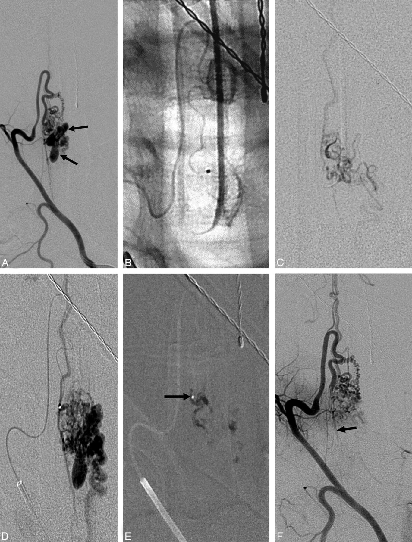
  • Fig 3.
    • Download figure
    • Open in new tab
    • Download powerpoint
    Fig 3.

    A, Posteroanterior view of the right T2 intercostal artery angiogram showing a nidus-type SCAVM supplied by the anterior spinal artery with multiple intranidal aneurysms (arrows). B, Nonsubtracted image of the microcatheter inserted into a feeder to the SCAVM. C, Superselective angiogram from the microcatheter inserted into the same feeder as B. No aneurysms are visualized. D, Superselective angiogram from a microcatheter placed in the ASA at the origin of the feeder to the SCAVM. Intranidal aneurysms and the distal anterior spinal artery are visualized. Embolization was performed from this catheter position using NBCA. E, Cast of NBCA. A small amount of NBCA is in the ASA axis near the catheter tip (arrow). F, Posteroanterior view of the right T2 intercostal artery after embolization. There is decreased visualization of the nidus without opacification of the intranidal aneurysms. The distal ASA is seen through the collateral (arrow).

Tables

  • Figures
    • View popup
    Table 1:

    Duration between initial and recurrent hemorrhage in patients with SCAVMs

    DurationNo. (n = 35)Percentage
    <7 Days618
    8–31 Days514
    1–6 Months25.7
    6 Months to 1 year411
    1–2 Years25.7
    2–3 Years38.6
    >3 Years1337
    • View popup
    Table 2:

    Univariate freedom from recurrent hemorrhage of SCAVMsa

    VariablesTotal (n = 80)Recurrent HemorrhageUnadjusted P Value
    (+) (n = 35)(−) (n = 45)
    Baseline characteristics
        Median age (yr) (IQR)20 (10–32)18 (8–25)21 (15–38)
            Pediatric30 (38)16 (46)14 (31).87
        Male31 (39)12 (34)19 (42).73
        Endovascular embolizationb41 (51)1 (2.9)40 (89)<.0001
    Radiologic characteristics
        Subclassification.51
            SCAVM63 (79)31 (89)32 (71)
            SCAVF, single hole7 (8.8)1 (2.9)6 (13)
            SCAVF, multiple holes10 (13)4 (11)6 (13)
        SCAVM level.92
            Cranial-cervical2 (2.5)1 (2.9)1 (2.2)
            Cervical33 (41)16 (46)17 (39)
            Cervical-thoracic1 (1.3)01 (2.2)
            Thoracic31 (39)13 (37)18 (40)
            Thoracic-lumbar9 (11)4 (11)5 (11)
            Lumbar4 (5.0)1 (2.9)3 (6.7)
        Associated aneurysmb56 (70)31 (89)25 (56).049
        Venous thrombosisb7 (8.8)1 (2.9)6 (13).19
        Venous stricture2 (2.5)02 (4.4).21
        Venous ectasia16 (20)7 (20)9 (20).24
        Venous hypertension16 (20)7 (20)9 (20).66
    • Note:—SCAVF indicates spinal cord AVF; IQR, interquartile range 25th–75th percentile.

    • ↵a Data are expressed as number of lesions (%), unless otherwise indicated.

    • ↵b Variables related to the recurrent hemorrhage by log-rank test (P < .20).

    • View popup
    Table 3:

    Multivariate analyses using the Cox proportional hazards model for recurrent hemorrhage from SCAVMs

    VariableAdjusted HR95% CIMultivariable Adjusted P Value
    Endovascular treatmenta0.0270.0040–0.19<.0001
    Associated aneurysma3.41.2–11.044
    Venous thrombosis0.610.082–4.5.63
    • Note:—HR indicates hazard ratio.

    • ↵a Variables related to recurrent hemorrhage.

PreviousNext
Back to top

In this issue

American Journal of Neuroradiology: 36 (9)
American Journal of Neuroradiology
Vol. 36, Issue 9
1 Sep 2015
  • Table of Contents
  • Index by author
  • Complete Issue (PDF)
Advertisement
Print
Download PDF
Email Article

Thank you for your interest in spreading the word on American Journal of Neuroradiology.

NOTE: We only request your email address so that the person you are recommending the page to knows that you wanted them to see it, and that it is not junk mail. We do not capture any email address.

Enter multiple addresses on separate lines or separate them with commas.
The Preventive Effect of Endovascular Treatment for Recurrent Hemorrhage in Patients with Spinal Cord Arteriovenous Malformations
(Your Name) has sent you a message from American Journal of Neuroradiology
(Your Name) thought you would like to see the American Journal of Neuroradiology web site.
CAPTCHA
This question is for testing whether or not you are a human visitor and to prevent automated spam submissions.
Cite this article
Y. Niimi, H. Matsukawa, N. Uchiyama, A. Berenstein
The Preventive Effect of Endovascular Treatment for Recurrent Hemorrhage in Patients with Spinal Cord Arteriovenous Malformations
American Journal of Neuroradiology Sep 2015, 36 (9) 1763-1768; DOI: 10.3174/ajnr.A4396

Citation Manager Formats

  • BibTeX
  • Bookends
  • EasyBib
  • EndNote (tagged)
  • EndNote 8 (xml)
  • Medlars
  • Mendeley
  • Papers
  • RefWorks Tagged
  • Ref Manager
  • RIS
  • Zotero
0 Responses
Respond to this article
Share
Bookmark this article
The Preventive Effect of Endovascular Treatment for Recurrent Hemorrhage in Patients with Spinal Cord Arteriovenous Malformations
Y. Niimi, H. Matsukawa, N. Uchiyama, A. Berenstein
American Journal of Neuroradiology Sep 2015, 36 (9) 1763-1768; DOI: 10.3174/ajnr.A4396
del.icio.us logo Twitter logo Facebook logo Mendeley logo
  • Tweet Widget
  • Facebook Like
  • Google Plus One
Purchase

Jump to section

  • Article
    • Abstract
    • ABBREVIATIONS:
    • Materials and Methods
    • Results
    • Discussion
    • Conclusions
    • References
  • Figures & Data
  • Info & Metrics
  • Responses
  • References
  • PDF

Related Articles

  • No related articles found.
  • PubMed
  • Google Scholar

Cited By...

  • Long term follow-up of endovascular management of spinal cord arteriovenous malformations with emphasis on particle embolization
  • Crossref
  • Google Scholar

This article has not yet been cited by articles in journals that are participating in Crossref Cited-by Linking.

More in this TOC Section

Neurointervention

  • Rescue Reentry in Carotid Near-Occlusion
  • Contour Neurovascular System: Five Year Follow Up
  • Effect of SARS-CoV2 on Endovascular Thrombectomy
Show more Neurointervention

Spine Imaging and Spine Image-Guided Interventions

  • Optimization of Photon Counting CT Myelography
  • Characteristics of SIH Type I Culprit Lesions
  • Management Outcomes For VO Spine Biopsy
Show more Spine Imaging and Spine Image-Guided Interventions

Similar Articles

Advertisement

Indexed Content

  • Current Issue
  • Accepted Manuscripts
  • Article Preview
  • Past Issues
  • Editorials
  • Editor's Choice
  • Fellows' Journal Club
  • Letters to the Editor
  • Video Articles

Cases

  • Case Collection
  • Archive - Case of the Week
  • Archive - Case of the Month
  • Archive - Classic Case

More from AJNR

  • Trainee Corner
  • Imaging Protocols
  • MRI Safety Corner
  • Book Reviews

Multimedia

  • AJNR Podcasts
  • AJNR Scantastics

Resources

  • Turnaround Time
  • Submit a Manuscript
  • Submit a Video Article
  • Submit an eLetter to the Editor/Response
  • Manuscript Submission Guidelines
  • Statistical Tips
  • Fast Publishing of Accepted Manuscripts
  • Graphical Abstract Preparation
  • Imaging Protocol Submission
  • Evidence-Based Medicine Level Guide
  • Publishing Checklists
  • Author Policies
  • Become a Reviewer/Academy of Reviewers
  • News and Updates

About Us

  • About AJNR
  • Editorial Board
  • Editorial Board Alumni
  • Alerts
  • Permissions
  • Not an AJNR Subscriber? Join Now
  • Advertise with Us
  • Librarian Resources
  • Feedback
  • Terms and Conditions
  • AJNR Editorial Board Alumni

American Society of Neuroradiology

  • Not an ASNR Member? Join Now

© 2025 by the American Society of Neuroradiology All rights, including for text and data mining, AI training, and similar technologies, are reserved.
Print ISSN: 0195-6108 Online ISSN: 1936-959X

Powered by HighWire