Advertisement

Main menu

  • Home
  • Content
    • Current Issue
    • Accepted Manuscripts
    • Article Preview
    • Past Issue Archive
    • Video Articles
    • AJNR Case Collection
    • Case of the Week Archive
    • Case of the Month Archive
    • Classic Case Archive
  • Special Collections
    • AJNR Awards
    • Low-Field MRI
    • Alzheimer Disease
    • ASNR Foundation Special Collection
    • Photon-Counting CT
    • View All
  • Multimedia
    • AJNR Podcasts
    • AJNR SCANtastic
    • Trainee Corner
    • MRI Safety Corner
    • Imaging Protocols
  • For Authors
    • Submit a Manuscript
    • Submit a Video Article
    • Submit an eLetter to the Editor/Response
    • Manuscript Submission Guidelines
    • Statistical Tips
    • Fast Publishing of Accepted Manuscripts
    • Graphical Abstract Preparation
    • Imaging Protocol Submission
    • Author Policies
  • About Us
    • About AJNR
    • Editorial Board
    • Editorial Board Alumni
  • More
    • Become a Reviewer/Academy of Reviewers
    • Subscribers
    • Permissions
    • Alerts
    • Feedback
    • Advertisers
    • ASNR Home

User menu

  • Alerts
  • Log in

Search

  • Advanced search
American Journal of Neuroradiology

American Journal of Neuroradiology

ASHNR American Society of Functional Neuroradiology ASHNR American Society of Pediatric Neuroradiology ASSR
  • Alerts
  • Log in

Advanced Search

  • Home
  • Content
    • Current Issue
    • Accepted Manuscripts
    • Article Preview
    • Past Issue Archive
    • Video Articles
    • AJNR Case Collection
    • Case of the Week Archive
    • Case of the Month Archive
    • Classic Case Archive
  • Special Collections
    • AJNR Awards
    • Low-Field MRI
    • Alzheimer Disease
    • ASNR Foundation Special Collection
    • Photon-Counting CT
    • View All
  • Multimedia
    • AJNR Podcasts
    • AJNR SCANtastic
    • Trainee Corner
    • MRI Safety Corner
    • Imaging Protocols
  • For Authors
    • Submit a Manuscript
    • Submit a Video Article
    • Submit an eLetter to the Editor/Response
    • Manuscript Submission Guidelines
    • Statistical Tips
    • Fast Publishing of Accepted Manuscripts
    • Graphical Abstract Preparation
    • Imaging Protocol Submission
    • Author Policies
  • About Us
    • About AJNR
    • Editorial Board
    • Editorial Board Alumni
  • More
    • Become a Reviewer/Academy of Reviewers
    • Subscribers
    • Permissions
    • Alerts
    • Feedback
    • Advertisers
    • ASNR Home
  • Follow AJNR on Twitter
  • Visit AJNR on Facebook
  • Follow AJNR on Instagram
  • Join AJNR on LinkedIn
  • RSS Feeds

AJNR Awards, New Junior Editors, and more. Read the latest AJNR updates

LetterLetter

Injury of the Contralateral Lower Ascending Reticular Activating System by an Intracerebral Hemorrhage

S.H. Jang and Y.S. Seo
American Journal of Neuroradiology August 2015, 36 (8) E58-E59; DOI: https://doi.org/10.3174/ajnr.A4446
S.H. Jang
aDepartment of Physical Medicine and Rehabilitation College of Medicine, Yeungnam University Daegu, Republic of Korea
  • Find this author on Google Scholar
  • Find this author on PubMed
  • Search for this author on this site
Y.S. Seo
aDepartment of Physical Medicine and Rehabilitation College of Medicine, Yeungnam University Daegu, Republic of Korea
  • Find this author on Google Scholar
  • Find this author on PubMed
  • Search for this author on this site
  • Article
  • Figures & Data
  • Info & Metrics
  • Responses
  • References
  • PDF
Loading

A 56-year-old female patient underwent decompressive craniectomy for removal of an intracerebral hematoma in the right temporoparietal lobe and spontaneous subarachnoid hemorrhage due to a ruptured right middle cerebral artery bifurcation aneurysm at the department of neurosurgery of a university hospital (Fig 1A). At 3 weeks after onset, she was transferred to the rehabilitation department. The patient exhibited impaired consciousness, with a Glasgow Coma Scale score of 8 (eye opening, 3; best verbal response, 1; and best motor response, 4). Seven age-matched control subjects (3 men; mean age, 55.28 years) were recruited. All subjects provided signed, informed consent, and the study protocol was approved by our institutional review board.

Fig 1.
  • Download figure
  • Open in new tab
  • Download powerpoint
Fig 1.

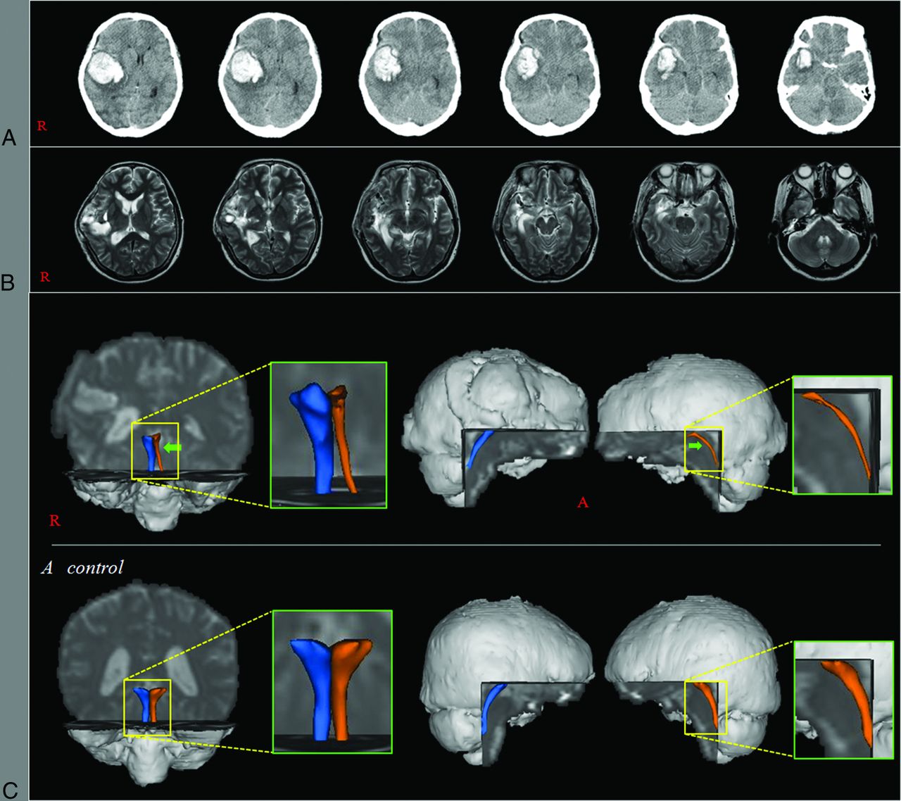
A, Brain CT images at onset show an intracerebral hemorrhage in the right frontotemporal lobes and subarachnoid hemorrhage. B, Brain MR images obtained at 3 weeks after onset show a malactic lesion in the right frontotemporal lobes. C, Results of diffusion tensor tractography. The left lower ascending reticular activating system is thinner than the right one of the patient and those of a control subject (a 54-year-old woman).

Diffusion tensor imaging data were acquired at 3 weeks after onset by using a 6-channel head coil on a 1.5T Gyroscan Intera scanner (Philips Healthcare, Best, the Netherlands). For each of the 32 noncollinear diffusion-sensitizing gradients, we acquired 67 contiguous sections. Imaging parameters were as follows: TR = 10,726 ms, TE = 76 ms, b=1000 s/mm2, NEX = 1, section thickness = 2.3 mm. Fiber tracking was performed by using a probabilistic tractography method based on a multifiber model (FSL, http://www.fmrib.ox.ac.uk/fsl; 5000 streamline samples; 0.5-mm step lengths; curvature thresholds = 0.2). The seed ROI was placed at the reticular formation of the pons, and the target ROI was placed at the intralaminar nuclei of the thalamus.1⇓–3 Fractional anisotropy (FA) and tract volume (TV) of the lower portion of the ascending reticular activation system (ARAS) were measured. In the patient, TV of the left ARAS was decreased >2 SDs compared with that of control subjects. The left lower ARAS was thinner than the right one of the patient and those of control subjects.

In this study, by using diffusion tensor tractography, we evaluated the lower portion of the ARAS between the pontine reticular formation and the thalamus in a patient with brain injury. Only the TV of the left lower ARAS was decreased compared with that in the control group. The TV is determined by the number of voxels contained within a neural tract; therefore, the decrement of the TV without a change of FA value appears to indicate injury of the left lower ARAS. On the configuration of diffusion tensor tractography, thinning of the left lower ARAS appears to coincide with the decrement of the TV of the left lower ARAS. It appears that the left lower ARAS might have been injured by the Kernohan notch phenomenon similar to an injury mechanism following transtentorial herniation by the right intracerebral hematoma.4 Our results suggest that analysis of the ARAS by using DTT would be helpful in elucidating the cause of impaired consciousness in patients with brain injury.

Acknowledgments

This work was supported by the National Research Foundation of Korea Grant funded by the Korean Government (NRF-2015R1A2A2A01004073).

References

  1. 1.↵
    1. Jang SH,
    2. Kim HS
    . Aneurysmal subarachnoid hemorrhage causes injury of the ascending reticular activating system: relation to consciousness. AJNR Am J Neuroradiol 2015;36:667–71
    Abstract/FREE Full Text
  2. 2.↵
    1. Jang SH,
    2. Kim SH,
    3. Lim HW, et al
    . Injury of the lower ascending reticular activating system in patients with hypoxic-ischemic brain injury: diffusion tensor imaging study. Neuroradiology 2014;56:965–70
    CrossRefPubMed
  3. 3.↵
    1. Jang SH,
    2. Kim SH,
    3. Lim HW, et al
    . Recovery of injured lower portion of the ascending reticular activating system in a patient with traumatic brain injury. Am J Phys Med Rehabil 2015;94:250–53
    CrossRefPubMed
  4. 4.↵
    1. Jang SH,
    2. Lim HW,
    3. Yeo SS
    . Injury of the ascending reticular activating system by transtentorial herniation in a patient with intracerebral haemorrhage: a diffusion tensor tractography study. J Neurol Neurosurg Psychiatry 2015 Jan 30. [Epub ahead of print]
  • © 2015 by American Journal of Neuroradiology
PreviousNext
Back to top

In this issue

American Journal of Neuroradiology: 36 (8)
American Journal of Neuroradiology
Vol. 36, Issue 8
1 Aug 2015
  • Table of Contents
  • Index by author
  • Complete Issue (PDF)
Advertisement
Print
Download PDF
Email Article

Thank you for your interest in spreading the word on American Journal of Neuroradiology.

NOTE: We only request your email address so that the person you are recommending the page to knows that you wanted them to see it, and that it is not junk mail. We do not capture any email address.

Enter multiple addresses on separate lines or separate them with commas.
Injury of the Contralateral Lower Ascending Reticular Activating System by an Intracerebral Hemorrhage
(Your Name) has sent you a message from American Journal of Neuroradiology
(Your Name) thought you would like to see the American Journal of Neuroradiology web site.
CAPTCHA
This question is for testing whether or not you are a human visitor and to prevent automated spam submissions.
Cite this article
S.H. Jang, Y.S. Seo
Injury of the Contralateral Lower Ascending Reticular Activating System by an Intracerebral Hemorrhage
American Journal of Neuroradiology Aug 2015, 36 (8) E58-E59; DOI: 10.3174/ajnr.A4446

Citation Manager Formats

  • BibTeX
  • Bookends
  • EasyBib
  • EndNote (tagged)
  • EndNote 8 (xml)
  • Medlars
  • Mendeley
  • Papers
  • RefWorks Tagged
  • Ref Manager
  • RIS
  • Zotero
0 Responses
Respond to this article
Share
Bookmark this article
Injury of the Contralateral Lower Ascending Reticular Activating System by an Intracerebral Hemorrhage
S.H. Jang, Y.S. Seo
American Journal of Neuroradiology Aug 2015, 36 (8) E58-E59; DOI: 10.3174/ajnr.A4446
del.icio.us logo Twitter logo Facebook logo Mendeley logo
  • Tweet Widget
  • Facebook Like
  • Google Plus One
Purchase

Jump to section

  • Article
    • Acknowledgments
    • References
  • Figures & Data
  • Info & Metrics
  • Responses
  • References
  • PDF

Related Articles

  • No related articles found.
  • PubMed
  • Google Scholar

Cited By...

  • No citing articles found.
  • Crossref
  • Google Scholar

This article has not yet been cited by articles in journals that are participating in Crossref Cited-by Linking.

More in this TOC Section

  • Letter to the Editor regarding “Automated Volumetric Software in Dementia: Help or Hindrance to the Neuroradiologist?”
  • Reply:
  • Brain AVM’s Nidus: What if We Hadn’t Understood Anything?
Show more LETTERS

Similar Articles

Advertisement

Indexed Content

  • Current Issue
  • Accepted Manuscripts
  • Article Preview
  • Past Issues
  • Editorials
  • Editor's Choice
  • Fellows' Journal Club
  • Letters to the Editor
  • Video Articles

Cases

  • Case Collection
  • Archive - Case of the Week
  • Archive - Case of the Month
  • Archive - Classic Case

More from AJNR

  • Trainee Corner
  • Imaging Protocols
  • MRI Safety Corner
  • Book Reviews

Multimedia

  • AJNR Podcasts
  • AJNR Scantastics

Resources

  • Turnaround Time
  • Submit a Manuscript
  • Submit a Video Article
  • Submit an eLetter to the Editor/Response
  • Manuscript Submission Guidelines
  • Statistical Tips
  • Fast Publishing of Accepted Manuscripts
  • Graphical Abstract Preparation
  • Imaging Protocol Submission
  • Evidence-Based Medicine Level Guide
  • Publishing Checklists
  • Author Policies
  • Become a Reviewer/Academy of Reviewers
  • News and Updates

About Us

  • About AJNR
  • Editorial Board
  • Editorial Board Alumni
  • Alerts
  • Permissions
  • Not an AJNR Subscriber? Join Now
  • Advertise with Us
  • Librarian Resources
  • Feedback
  • Terms and Conditions
  • AJNR Editorial Board Alumni

American Society of Neuroradiology

  • Not an ASNR Member? Join Now

© 2025 by the American Society of Neuroradiology All rights, including for text and data mining, AI training, and similar technologies, are reserved.
Print ISSN: 0195-6108 Online ISSN: 1936-959X

Powered by HighWire