Advertisement

Main menu

  • Home
  • Content
    • Current Issue
    • Accepted Manuscripts
    • Article Preview
    • Past Issue Archive
    • Video Articles
    • AJNR Case Collection
    • Case of the Week Archive
    • Case of the Month Archive
    • Classic Case Archive
  • Special Collections
    • AJNR Awards
    • Low-Field MRI
    • Alzheimer Disease
    • ASNR Foundation Special Collection
    • Photon-Counting CT
    • View All
  • Multimedia
    • AJNR Podcasts
    • AJNR SCANtastic
    • Trainee Corner
    • MRI Safety Corner
    • Imaging Protocols
  • For Authors
    • Submit a Manuscript
    • Submit a Video Article
    • Submit an eLetter to the Editor/Response
    • Manuscript Submission Guidelines
    • Statistical Tips
    • Fast Publishing of Accepted Manuscripts
    • Graphical Abstract Preparation
    • Imaging Protocol Submission
    • Author Policies
  • About Us
    • About AJNR
    • Editorial Board
    • Editorial Board Alumni
  • More
    • Become a Reviewer/Academy of Reviewers
    • Subscribers
    • Permissions
    • Alerts
    • Feedback
    • Advertisers
    • ASNR Home

User menu

  • Alerts
  • Log in

Search

  • Advanced search
American Journal of Neuroradiology

American Journal of Neuroradiology

ASHNR American Society of Functional Neuroradiology ASHNR American Society of Pediatric Neuroradiology ASSR
  • Alerts
  • Log in

Advanced Search

  • Home
  • Content
    • Current Issue
    • Accepted Manuscripts
    • Article Preview
    • Past Issue Archive
    • Video Articles
    • AJNR Case Collection
    • Case of the Week Archive
    • Case of the Month Archive
    • Classic Case Archive
  • Special Collections
    • AJNR Awards
    • Low-Field MRI
    • Alzheimer Disease
    • ASNR Foundation Special Collection
    • Photon-Counting CT
    • View All
  • Multimedia
    • AJNR Podcasts
    • AJNR SCANtastic
    • Trainee Corner
    • MRI Safety Corner
    • Imaging Protocols
  • For Authors
    • Submit a Manuscript
    • Submit a Video Article
    • Submit an eLetter to the Editor/Response
    • Manuscript Submission Guidelines
    • Statistical Tips
    • Fast Publishing of Accepted Manuscripts
    • Graphical Abstract Preparation
    • Imaging Protocol Submission
    • Author Policies
  • About Us
    • About AJNR
    • Editorial Board
    • Editorial Board Alumni
  • More
    • Become a Reviewer/Academy of Reviewers
    • Subscribers
    • Permissions
    • Alerts
    • Feedback
    • Advertisers
    • ASNR Home
  • Follow AJNR on Twitter
  • Visit AJNR on Facebook
  • Follow AJNR on Instagram
  • Join AJNR on LinkedIn
  • RSS Feeds

AJNR Awards, New Junior Editors, and more. Read the latest AJNR updates

Research ArticleAdult Brain
Open Access

High-Resolution DCE-MRI of the Pituitary Gland Using Radial k-Space Acquisition with Compressed Sensing Reconstruction

M.C. Rossi Espagnet, L. Bangiyev, M. Haber, K.T. Block, J. Babb, V. Ruggiero, F. Boada, O. Gonen and G.M. Fatterpekar
American Journal of Neuroradiology August 2015, 36 (8) 1444-1449; DOI: https://doi.org/10.3174/ajnr.A4324
M.C. Rossi Espagnet
aFrom the Department of Radiology, New York University Langone Medical Center, New York, New York.
  • Find this author on Google Scholar
  • Find this author on PubMed
  • Search for this author on this site
L. Bangiyev
aFrom the Department of Radiology, New York University Langone Medical Center, New York, New York.
  • Find this author on Google Scholar
  • Find this author on PubMed
  • Search for this author on this site
  • ORCID record for L. Bangiyev
M. Haber
aFrom the Department of Radiology, New York University Langone Medical Center, New York, New York.
  • Find this author on Google Scholar
  • Find this author on PubMed
  • Search for this author on this site
K.T. Block
aFrom the Department of Radiology, New York University Langone Medical Center, New York, New York.
  • Find this author on Google Scholar
  • Find this author on PubMed
  • Search for this author on this site
J. Babb
aFrom the Department of Radiology, New York University Langone Medical Center, New York, New York.
  • Find this author on Google Scholar
  • Find this author on PubMed
  • Search for this author on this site
V. Ruggiero
aFrom the Department of Radiology, New York University Langone Medical Center, New York, New York.
  • Find this author on Google Scholar
  • Find this author on PubMed
  • Search for this author on this site
F. Boada
aFrom the Department of Radiology, New York University Langone Medical Center, New York, New York.
  • Find this author on Google Scholar
  • Find this author on PubMed
  • Search for this author on this site
O. Gonen
aFrom the Department of Radiology, New York University Langone Medical Center, New York, New York.
  • Find this author on Google Scholar
  • Find this author on PubMed
  • Search for this author on this site
G.M. Fatterpekar
aFrom the Department of Radiology, New York University Langone Medical Center, New York, New York.
  • Find this author on Google Scholar
  • Find this author on PubMed
  • Search for this author on this site
  • Article
  • Figures & Data
  • Info & Metrics
  • Responses
  • References
  • PDF
Loading

Article Figures & Data

Figures

  • Tables
  • Fig 1.
    • Download figure
    • Open in new tab
    • Download powerpoint
    Fig 1.

    Analysis of radial VIBE images provide maps of different permeability parameters: peak, area under the curve, time of maximum enhancement, wash-in, and washout.
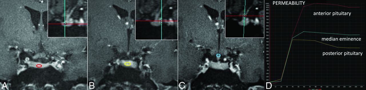
  • Fig 2.
    • Download figure
    • Open in new tab
    • Download powerpoint
    Fig 2.

    GRASP images in a healthy volunteer show the ROIs in the anterior pituitary gland (A), posterior pituitary gland (B), and median eminence (C). Sagittal reference planes are shown top right. Corresponding signal-time curves from the ROIs (D) demonstrate a different pattern of enhancement in the anterior (red) and posterior pituitary gland (yellow) and the median eminence (light blue).

  • Fig 3.
    • Download figure
    • Open in new tab
    • Download powerpoint
    Fig 3.

    A, GRASP image demonstrates placement of ROIs within the normal-appearing pituitary gland and microadenoma. B, Corresponding signal-time curves are shown. Peak enhancement from the anterior pituitary gland is seen at approximately 80 seconds with subsequent gradual washout. Peak enhancement from the microadenoma is seen at approximately 100 seconds with a subsequent plateau. There is a significant difference in the enhancement between the anterior pituitary gland and the microadenoma from 60 seconds onward.

  • Fig 4.
    • Download figure
    • Open in new tab
    • Download powerpoint
    Fig 4.

    Signal-time curve derived from an ROI applied to a cyst demonstrates a flat STC compared with the normal STCs from the anterior pituitary gland, posterior pituitary gland, and median eminence evaluated in the same patient.

Tables

  • Figures
    • View popup
    Table 1:

    Mean, SD, median, maximum, and lower and upper limits of a 95% confidence interval of the mean for the percentage change in the peak enhancement estimate from time T0 = 60 seconds to time T8 = 140 seconds in the normal-appearing anterior pituitary gland of group 2 patients

    TimeMeanSDMedianMaximumLowerUpper
    19.829.975.2530.995.1614.49
    26.116.654.5221.693.009.23
    32.002.521.058.530.823.18
    42.172.551.188.980.973.36
    51.410.871.062.761.011.82
    61.380.871.043.820.981.79
    70.940.870.713.280.531.35
    80.570.330.621.300.420.73
    • View popup
    Table 2:

    Values of perfusion parameters obtained from ROI analysis in different regions of the normal-appearing pituitary glanda

    ParameterAnteriorPosteriorMedian Eminence
    MeanSDMeanSDMeanSD
    AUC69980.6520169.3237500.1017701.7340246.3916626.70
    Peak754.94242.04439.61205.67446.62192.66
    TME (sec)86.5519.2358.9616.2362.6018.70
    Wash-in10.923.899.144.208.543.71
    Washout1.521.341.901.481.561.27
    • Note:—TME indicates time of maximum enhancement.

    • ↵a All parameters other than TME are dimensionless.

    • View popup
    Table 3:

    Mean, minimum, and maximum peak enhancement values measured at different times in the microadenoma of group 2 patients

    Time (sec)Mean PEMin PEMax PE
    6032790500
    70365120600
    80400170630
    90420200660
    100417180650
    110342165625
    120345172620
    130346171613
    140344170613
    • Note:—PE indicates peak enhancement; Min, minimum; Max, maximum.

PreviousNext
Back to top

In this issue

American Journal of Neuroradiology: 36 (8)
American Journal of Neuroradiology
Vol. 36, Issue 8
1 Aug 2015
  • Table of Contents
  • Index by author
  • Complete Issue (PDF)
Advertisement
Print
Download PDF
Email Article

Thank you for your interest in spreading the word on American Journal of Neuroradiology.

NOTE: We only request your email address so that the person you are recommending the page to knows that you wanted them to see it, and that it is not junk mail. We do not capture any email address.

Enter multiple addresses on separate lines or separate them with commas.
High-Resolution DCE-MRI of the Pituitary Gland Using Radial k-Space Acquisition with Compressed Sensing Reconstruction
(Your Name) has sent you a message from American Journal of Neuroradiology
(Your Name) thought you would like to see the American Journal of Neuroradiology web site.
CAPTCHA
This question is for testing whether or not you are a human visitor and to prevent automated spam submissions.
Cite this article
M.C. Rossi Espagnet, L. Bangiyev, M. Haber, K.T. Block, J. Babb, V. Ruggiero, F. Boada, O. Gonen, G.M. Fatterpekar
High-Resolution DCE-MRI of the Pituitary Gland Using Radial k-Space Acquisition with Compressed Sensing Reconstruction
American Journal of Neuroradiology Aug 2015, 36 (8) 1444-1449; DOI: 10.3174/ajnr.A4324

Citation Manager Formats

  • BibTeX
  • Bookends
  • EasyBib
  • EndNote (tagged)
  • EndNote 8 (xml)
  • Medlars
  • Mendeley
  • Papers
  • RefWorks Tagged
  • Ref Manager
  • RIS
  • Zotero
0 Responses
Respond to this article
Share
Bookmark this article
High-Resolution DCE-MRI of the Pituitary Gland Using Radial k-Space Acquisition with Compressed Sensing Reconstruction
M.C. Rossi Espagnet, L. Bangiyev, M. Haber, K.T. Block, J. Babb, V. Ruggiero, F. Boada, O. Gonen, G.M. Fatterpekar
American Journal of Neuroradiology Aug 2015, 36 (8) 1444-1449; DOI: 10.3174/ajnr.A4324
del.icio.us logo Twitter logo Facebook logo Mendeley logo
  • Tweet Widget
  • Facebook Like
  • Google Plus One
Purchase

Jump to section

  • Article
    • Abstract
    • ABBREVIATIONS:
    • Materials and Methods
    • Results
    • Discussion
    • Conclusions
    • Footnotes
    • References
  • Figures & Data
  • Info & Metrics
  • Responses
  • References
  • PDF

Related Articles

  • No related articles found.
  • PubMed
  • Google Scholar

Cited By...

  • Differentiation of Jugular Foramen Paragangliomas versus Schwannomas Using Golden-Angle Radial Sparse Parallel Dynamic Contrast-Enhanced MRI
  • Dynamic Contrast-Enhanced MRI to Differentiate Parotid Neoplasms Using Golden-Angle Radial Sparse Parallel Imaging
  • Role of High-Resolution Dynamic Contrast-Enhanced MRI with Golden-Angle Radial Sparse Parallel Reconstruction to Identify the Normal Pituitary Gland in Patients with Macroadenomas
  • Crossref
  • Google Scholar

This article has not yet been cited by articles in journals that are participating in Crossref Cited-by Linking.

More in this TOC Section

  • Diagnostic Neuroradiology of Monoclonal Antibodies
  • Clinical Outcomes After Chiari I Decompression
  • Segmentation of Brain Metastases with BLAST
Show more Adult Brain

Similar Articles

Advertisement

Indexed Content

  • Current Issue
  • Accepted Manuscripts
  • Article Preview
  • Past Issues
  • Editorials
  • Editor's Choice
  • Fellows' Journal Club
  • Letters to the Editor
  • Video Articles

Cases

  • Case Collection
  • Archive - Case of the Week
  • Archive - Case of the Month
  • Archive - Classic Case

More from AJNR

  • Trainee Corner
  • Imaging Protocols
  • MRI Safety Corner
  • Book Reviews

Multimedia

  • AJNR Podcasts
  • AJNR Scantastics

Resources

  • Turnaround Time
  • Submit a Manuscript
  • Submit a Video Article
  • Submit an eLetter to the Editor/Response
  • Manuscript Submission Guidelines
  • Statistical Tips
  • Fast Publishing of Accepted Manuscripts
  • Graphical Abstract Preparation
  • Imaging Protocol Submission
  • Evidence-Based Medicine Level Guide
  • Publishing Checklists
  • Author Policies
  • Become a Reviewer/Academy of Reviewers
  • News and Updates

About Us

  • About AJNR
  • Editorial Board
  • Editorial Board Alumni
  • Alerts
  • Permissions
  • Not an AJNR Subscriber? Join Now
  • Advertise with Us
  • Librarian Resources
  • Feedback
  • Terms and Conditions
  • AJNR Editorial Board Alumni

American Society of Neuroradiology

  • Not an ASNR Member? Join Now

© 2025 by the American Society of Neuroradiology All rights, including for text and data mining, AI training, and similar technologies, are reserved.
Print ISSN: 0195-6108 Online ISSN: 1936-959X

Powered by HighWire