
Article Figures & Data
Figures
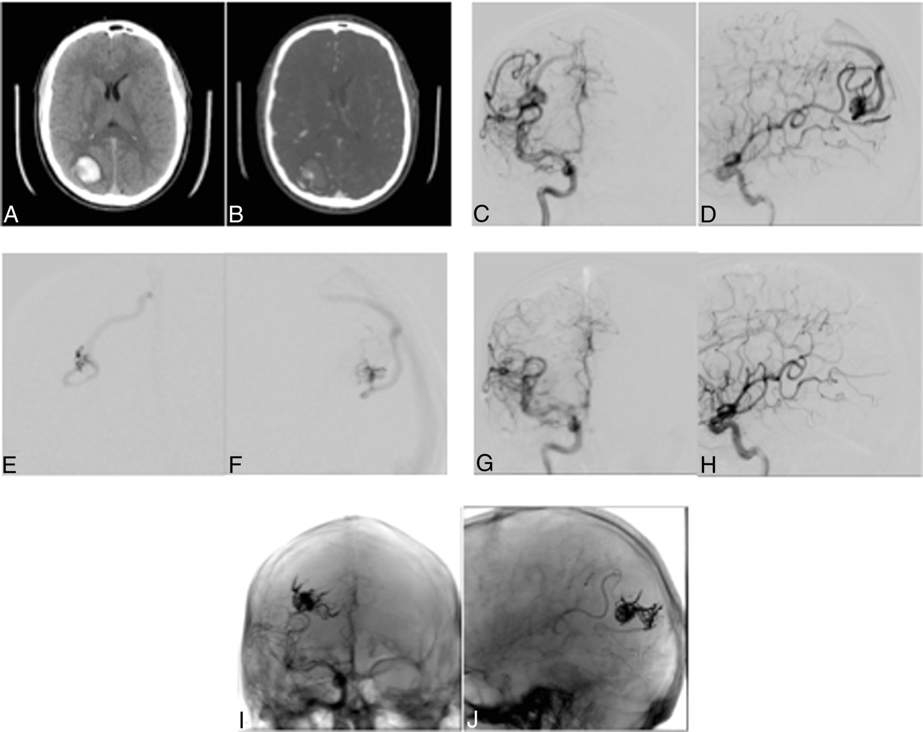
- Fig 1.
A 35-year-old man presenting with acute onset of dizziness and headache. A and B, noncontrast head CT and head CTA demonstrate a 3.4-cm right parietal hematoma and a 1.9-cm bAVM, respectively. C and D, Diagnostic right internal carotid artery anteroposterior and lateral angiograms demonstrate a Spetzler-Martin grade II (S1, E1, V0) and endovascular grade II (A2, E0, F0) bAVM supplied via angular branches of the right middle cerebral artery (supplementary right posterior cerebral artery supply not shown) with superficial venous drainage via mildly dilated cortical veins. E and F, Intraprocedural right middle cerebral artery angular branch microcatheter anteroposterior and lateral angiograms demonstrate a distinct plexiform nidus and no arteriovenous fistulous component. G and H, Right internal carotid artery anteroposterior and lateral angiograms following Onyx embolization via the right middle cerebral artery angular branch demonstrate resolution of arteriovenous shunting (left vertebral artery injection also documented resolution of arteriovenous shunting from a supplementary right posterior cerebral artery supply, not shown). I and J, Skull anteroposterior and lateral radiographs demonstrate the Onyx cast; note penetration of the Onyx material into a posterior component supplied by the right posterior cerebral artery on the lateral projection. Final mRS = 0. S indicates size; E, eloquence; V, vein; A, artery; F, fistula.
- Fig 2.
A 31-year-old man presenting with seizures found to have a 3.8-cm left frontoparietal bAVM, Spetzler-Martin grade III (S2, E1, V0), and endovascular grade V (A3, E1, F1). A and B, Diagnostic left internal carotid anteroposterior and lateral angiograms demonstrate predominant arterial supply to the lesion via posterior frontal and parietal branches of the left middle cerebral artery with superficial venous drainage via moderately dilated cortical veins. Supplemental supply via the left anterior and posterior cerebral arteries was also present (not shown) C, Diagnostic left middle cerebral artery lateral magnification angiogram demonstrates a dilated angular branch >2 mm with a direct venous connection consistent with an arteriovenous fistula component (arrow). D, Intraprocedural left middle cerebral artery angular branch superselective microcatheter injection oblique anteroposterior angiogram again demonstrates an arteriovenous fistula component (arrow). E, Following stage I Onyx embolization, the medial portion of the Onyx cast (Onyx subtracted, seen as absence of contrast enhancement within the previous nidus compared to image A) can be seen within a dilated medial cortical vein (arrow), and the previously seen dilated cortical veins coursing inferiorly in image A are no longer visualized consistent with venous occlusion (ellipse). F, Postprocedural day 2 emergent noncontrast head CT performed for neurologic deterioration and a dilated left pupil demonstrates a large 3.8 × 7.4 cm left frontal hemorrhage anterior to the Onyx cast with surrounding cerebral edema, causing moderate-to-severe rightward midline shift. The hematoma and residual bAVM underwent emergent surgical resection. Final mRS was 6 due to neurologic decline and ventilator-associated pneumonia. S indicates size; E, eloquence; V, vein; A, artery; F, fistula.


Tables
Feature and Value Points No. of feeding arteriesb <3 1 ≥3, <6 2 ≥6 3 Eloquence of adjacent areas Noneloquent 0 Eloquent 1 Presence of arteriovenous fistula componentc No AVF 0 AVF 1 ↵a Modified from Feliciano et al.12
↵b An arterial feeder was defined as a separate arterial pedicle or a pedicle arising ≥1.5 cm from another arterial pedicle. En passage arterial feeders were given a score of 3.
↵c The presence of an arteriovenous fistula was determined by criteria described by Yuki et al,13 including an abnormally dilated feeding artery, a direct arteriovenous connection to a dilated venous component or varix, the absence of a plexiform component between the 2 structures, and a diameter of the feeding artery more than twice as large as that of the arteries supplying the comparable areas not supplying the AVM (eg, the corresponding contralateral cerebral artery) or feeding artery diameter of >2 mm.
All Patients (n = 126) Endovascular (n = 8) Radiotherapya (n = 39) Surgical (n = 44) Multimodala (n = 31) Endovascular Complications (n = 4) Age (yr) (mean) (SD) 44.9 (±16.9) 42.8 (16.9) 45.6 (17.2) 48.7 (16.3) 37.9 (14.7) 41.3 (20.5) Female sex (%) 52 50 53 63 32 25 Endovascular grade (median) (IQR) 3 (2–4) 2 (1.75–2) 4 (2–4) 3 (2–3) 3 (2–4) 4 (3.5–4.25) Spetzler-Martin grade (median) (IQR) 2 (1–3) 1 (1.5–2) 3 (2–3) 1 (1–2) 3 (2–4) 3.5 (3–4) bAVM volume (mL) (mean) (SD) 9.8 (±17.2) 2.1 (±1.8) 9.7 (±13.4) 1.7 (±2.4) 17.4 (±19.2) 54.1 (±45.2) No. of feeding arteries (score) (median) (IQR) 2 (2–3) 1 (1–1.25) 3 (2–3) 3 (1–3) 2 (2–3) 3 (2.75–3) AVF component (%) 9% 0 10% 2% 9% 25% Eloquent (%) 53% 63% 74% 31% 54% 75% Final mRS score (median) (IQR) 0 (0–1) 0 (0–0) 0 (0–1) 0 (0–1) 1 (0–1) 5 (3.5–6) Note:—IQR indicates interquartile range.
↵a Cured or significantly reduced lesions without complications (in the “Multimodal” category, this includes embolization plus radiosurgery).


Jump to section
Related Articles
Cited By...
- Endovascular treatment in the multimodality management of brain arteriovenous malformations: report of the Society of NeuroInterventional Surgery Standards and Guidelines Committee
- Complications of Endovascular Treatments for Brain Arteriovenous Malformations: A Nationwide Surveillance
- Arteriovenous malformation embocure score (AVMES): response