Advertisement

Main menu

  • Home
  • Content
    • Current Issue
    • Accepted Manuscripts
    • Article Preview
    • Past Issue Archive
    • Video Articles
    • AJNR Case Collection
    • Case of the Week Archive
    • Case of the Month Archive
    • Classic Case Archive
  • Special Collections
    • AJNR Awards
    • Low-Field MRI
    • Alzheimer Disease
    • ASNR Foundation Special Collection
    • Photon-Counting CT
    • View All
  • Multimedia
    • AJNR Podcasts
    • AJNR SCANtastic
    • Trainee Corner
    • MRI Safety Corner
    • Imaging Protocols
  • For Authors
    • Submit a Manuscript
    • Submit a Video Article
    • Submit an eLetter to the Editor/Response
    • Manuscript Submission Guidelines
    • Statistical Tips
    • Fast Publishing of Accepted Manuscripts
    • Graphical Abstract Preparation
    • Imaging Protocol Submission
    • Author Policies
  • About Us
    • About AJNR
    • Editorial Board
    • Editorial Board Alumni
  • More
    • Become a Reviewer/Academy of Reviewers
    • Subscribers
    • Permissions
    • Alerts
    • Feedback
    • Advertisers
    • ASNR Home

User menu

  • Alerts
  • Log in

Search

  • Advanced search
American Journal of Neuroradiology

American Journal of Neuroradiology

ASHNR American Society of Functional Neuroradiology ASHNR American Society of Pediatric Neuroradiology ASSR
  • Alerts
  • Log in

Advanced Search

  • Home
  • Content
    • Current Issue
    • Accepted Manuscripts
    • Article Preview
    • Past Issue Archive
    • Video Articles
    • AJNR Case Collection
    • Case of the Week Archive
    • Case of the Month Archive
    • Classic Case Archive
  • Special Collections
    • AJNR Awards
    • Low-Field MRI
    • Alzheimer Disease
    • ASNR Foundation Special Collection
    • Photon-Counting CT
    • View All
  • Multimedia
    • AJNR Podcasts
    • AJNR SCANtastic
    • Trainee Corner
    • MRI Safety Corner
    • Imaging Protocols
  • For Authors
    • Submit a Manuscript
    • Submit a Video Article
    • Submit an eLetter to the Editor/Response
    • Manuscript Submission Guidelines
    • Statistical Tips
    • Fast Publishing of Accepted Manuscripts
    • Graphical Abstract Preparation
    • Imaging Protocol Submission
    • Author Policies
  • About Us
    • About AJNR
    • Editorial Board
    • Editorial Board Alumni
  • More
    • Become a Reviewer/Academy of Reviewers
    • Subscribers
    • Permissions
    • Alerts
    • Feedback
    • Advertisers
    • ASNR Home
  • Follow AJNR on Twitter
  • Visit AJNR on Facebook
  • Follow AJNR on Instagram
  • Join AJNR on LinkedIn
  • RSS Feeds

AJNR Awards, New Junior Editors, and more. Read the latest AJNR updates

Getting new auth cookie, if you see this message a lot, tell someone!
Research ArticleAdult Brain

Degree of Collaterals and Not Time Is the Determining Factor of Core Infarct Volume within 6 Hours of Stroke Onset

E. Cheng-Ching, J.A. Frontera, S. Man, J. Aoki, Y. Tateishi, F.K. Hui, D. Wisco, P. Ruggieri, M.S. Hussain and K. Uchino
American Journal of Neuroradiology July 2015, 36 (7) 1272-1276; DOI: https://doi.org/10.3174/ajnr.A4274
E. Cheng-Ching
aFrom the Cerebrovascular Center (E.C.-C., J.A.F., S.M., F.K.H., D.W., M.S.H., K.U.)
  • Find this author on Google Scholar
  • Find this author on PubMed
  • Search for this author on this site
J.A. Frontera
aFrom the Cerebrovascular Center (E.C.-C., J.A.F., S.M., F.K.H., D.W., M.S.H., K.U.)
  • Find this author on Google Scholar
  • Find this author on PubMed
  • Search for this author on this site
S. Man
aFrom the Cerebrovascular Center (E.C.-C., J.A.F., S.M., F.K.H., D.W., M.S.H., K.U.)
  • Find this author on Google Scholar
  • Find this author on PubMed
  • Search for this author on this site
J. Aoki
cDepartment of Neurology and Cerebrovascular Disease (J.A.), Nippon Medical School, Tokyo, Japan
  • Find this author on Google Scholar
  • Find this author on PubMed
  • Search for this author on this site
Y. Tateishi
dCerebrovascular Center (Y.T.), Department of Neurology and Strokology, Nagasaki University Hospital, Nagasaki, Japan.
  • Find this author on Google Scholar
  • Find this author on PubMed
  • Search for this author on this site
F.K. Hui
aFrom the Cerebrovascular Center (E.C.-C., J.A.F., S.M., F.K.H., D.W., M.S.H., K.U.)
  • Find this author on Google Scholar
  • Find this author on PubMed
  • Search for this author on this site
D. Wisco
aFrom the Cerebrovascular Center (E.C.-C., J.A.F., S.M., F.K.H., D.W., M.S.H., K.U.)
  • Find this author on Google Scholar
  • Find this author on PubMed
  • Search for this author on this site
P. Ruggieri
bDepartment of Neuroradiology (P.R.), Imaging Institute, Cleveland Clinic, Cleveland, Ohio
  • Find this author on Google Scholar
  • Find this author on PubMed
  • Search for this author on this site
M.S. Hussain
aFrom the Cerebrovascular Center (E.C.-C., J.A.F., S.M., F.K.H., D.W., M.S.H., K.U.)
  • Find this author on Google Scholar
  • Find this author on PubMed
  • Search for this author on this site
K. Uchino
aFrom the Cerebrovascular Center (E.C.-C., J.A.F., S.M., F.K.H., D.W., M.S.H., K.U.)
  • Find this author on Google Scholar
  • Find this author on PubMed
  • Search for this author on this site
  • Article
  • Figures & Data
  • Supplemental
  • Info & Metrics
  • Responses
  • References
  • PDF
Loading

Article Figures & Data

Figures

  • Tables
  • Fig 1.
    • Download figure
    • Open in new tab
    • Download powerpoint
    Fig 1.

    CTA collateral status in the region of the occluded vessel. A, Absent collaterals. B, Minimal (<50%) collaterals. C, Partial (>50%) collaterals. D, Full presence of collaterals.
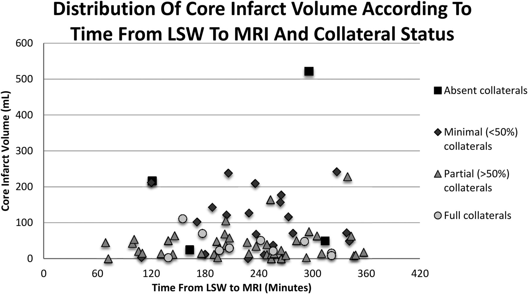
  • Fig 2.
    • Download figure
    • Open in new tab
    • Download powerpoint
    Fig 2.

    Distribution of DWI infarct volume of each patient according to the time from last seen well to MR imaging and collateral status.

  • Fig 3.
    • Download figure
    • Open in new tab
    • Download powerpoint
    Fig 3.

    Receiver operating characteristic curve demonstrating no association between the time from LSW to MR imaging (0–3 hours versus 3–6 hours) and DWI infarct volume.

Tables

  • Figures
  • Imaging characteristics by time from last seen well to MRI

    0 to <3 Hours (n = 21)3–6 Hours (n = 70)0–6 Hours (Entire Cohort) (n = 91)P Value (Difference, 0–3 vs 3–6)
    Age (yr) (median) (IQR)58 (54–77)73 (63.25–83)70 (59–82).034
    NIHSS (median) (IQR)13 (11–19)17 (13–22.75)17 (12–21.5).050
    CT ASPECTS (median) (IQR)9 (7–10)8 (I6–9)8 (6–9).202
    CT ASPECTS ≥7 (No.) (%)17/20 (85%)42/61 (69%)59/81 (72%).159
    CTA collaterals >50% (No.) (%)14/19 (74%)37/57 (65%)51/76 (67%).481
    DWI volume (mL) (median) (IQR)24.7 (13.9–63.8)29.4 (12.1–68.5)29.2 (12.3–68.5).906
    MRI DWI volume ≥70 mL of DWI volume (No.) (%)5 (23.8%)16 (22.8%)21 (22.8%).928
    MRI DWI volume ≥100 mL of DWI volume (No.) (%)4 (19%)13 (18.6%)17 (18.7%).961
    Terminal ICA occlusion (No.) (%)5 (23.8%)18 (25.7%)23 (25.3%).961
    Proximal M1 occlusion (No.) (%)5 (23.8%)21 (30%)26 (28.6%).706
    Distal M1 occlusion (No.) (%)11 (52.4%)31 (44.3%)42 (46.1%).393
PreviousNext
Back to top

In this issue

American Journal of Neuroradiology: 36 (7)
American Journal of Neuroradiology
Vol. 36, Issue 7
1 Jul 2015
  • Table of Contents
  • Index by author
  • Complete Issue (PDF)
Advertisement
Print
Download PDF
Email Article

Thank you for your interest in spreading the word on American Journal of Neuroradiology.

NOTE: We only request your email address so that the person you are recommending the page to knows that you wanted them to see it, and that it is not junk mail. We do not capture any email address.

Enter multiple addresses on separate lines or separate them with commas.
Degree of Collaterals and Not Time Is the Determining Factor of Core Infarct Volume within 6 Hours of Stroke Onset
(Your Name) has sent you a message from American Journal of Neuroradiology
(Your Name) thought you would like to see the American Journal of Neuroradiology web site.
CAPTCHA
This question is for testing whether or not you are a human visitor and to prevent automated spam submissions.
Cite this article
E. Cheng-Ching, J.A. Frontera, S. Man, J. Aoki, Y. Tateishi, F.K. Hui, D. Wisco, P. Ruggieri, M.S. Hussain, K. Uchino
Degree of Collaterals and Not Time Is the Determining Factor of Core Infarct Volume within 6 Hours of Stroke Onset
American Journal of Neuroradiology Jul 2015, 36 (7) 1272-1276; DOI: 10.3174/ajnr.A4274

Citation Manager Formats

  • BibTeX
  • Bookends
  • EasyBib
  • EndNote (tagged)
  • EndNote 8 (xml)
  • Medlars
  • Mendeley
  • Papers
  • RefWorks Tagged
  • Ref Manager
  • RIS
  • Zotero
0 Responses
Respond to this article
Share
Bookmark this article
Degree of Collaterals and Not Time Is the Determining Factor of Core Infarct Volume within 6 Hours of Stroke Onset
E. Cheng-Ching, J.A. Frontera, S. Man, J. Aoki, Y. Tateishi, F.K. Hui, D. Wisco, P. Ruggieri, M.S. Hussain, K. Uchino
American Journal of Neuroradiology Jul 2015, 36 (7) 1272-1276; DOI: 10.3174/ajnr.A4274
del.icio.us logo Twitter logo Facebook logo Mendeley logo
  • Tweet Widget
  • Facebook Like
  • Google Plus One
Purchase

Jump to section

  • Article
    • Abstract
    • ABBREVIATIONS:
    • Materials and Methods
    • Results
    • Discussion
    • Conclusions
    • Footnotes
    • REFERENCES
  • Figures & Data
  • Supplemental
  • Info & Metrics
  • Responses
  • References
  • PDF

Related Articles

  • PubMed
  • Google Scholar

Cited By...

  • Value of Contrast-Enhanced MRA versus Time-of-Flight MRA in Acute Ischemic Stroke MRI
  • Impact of time to endovascular reperfusion on outcome differs according to the involvement of the proximal MCA territory
  • ASPECTS decay during inter-facility transfer in patients with large vessel occlusion strokes
  • Initial hospital management of patients with emergent large vessel occlusion (ELVO): report of the standards and guidelines committee of the Society of NeuroInterventional Surgery
  • Crossref
  • Google Scholar

This article has not yet been cited by articles in journals that are participating in Crossref Cited-by Linking.

More in this TOC Section

  • Diagnostic Neuroradiology of Monoclonal Antibodies
  • Clinical Outcomes After Chiari I Decompression
  • Segmentation of Brain Metastases with BLAST
Show more Adult Brain

Similar Articles

Advertisement

Indexed Content

  • Current Issue
  • Accepted Manuscripts
  • Article Preview
  • Past Issues
  • Editorials
  • Editor's Choice
  • Fellows' Journal Club
  • Letters to the Editor
  • Video Articles

Cases

  • Case Collection
  • Archive - Case of the Week
  • Archive - Case of the Month
  • Archive - Classic Case

More from AJNR

  • Trainee Corner
  • Imaging Protocols
  • MRI Safety Corner
  • Book Reviews

Multimedia

  • AJNR Podcasts
  • AJNR Scantastics

Resources

  • Turnaround Time
  • Submit a Manuscript
  • Submit a Video Article
  • Submit an eLetter to the Editor/Response
  • Manuscript Submission Guidelines
  • Statistical Tips
  • Fast Publishing of Accepted Manuscripts
  • Graphical Abstract Preparation
  • Imaging Protocol Submission
  • Evidence-Based Medicine Level Guide
  • Publishing Checklists
  • Author Policies
  • Become a Reviewer/Academy of Reviewers
  • News and Updates

About Us

  • About AJNR
  • Editorial Board
  • Editorial Board Alumni
  • Alerts
  • Permissions
  • Not an AJNR Subscriber? Join Now
  • Advertise with Us
  • Librarian Resources
  • Feedback
  • Terms and Conditions
  • AJNR Editorial Board Alumni

American Society of Neuroradiology

  • Not an ASNR Member? Join Now

© 2025 by the American Society of Neuroradiology All rights, including for text and data mining, AI training, and similar technologies, are reserved.
Print ISSN: 0195-6108 Online ISSN: 1936-959X

Powered by HighWire