
Article Figures & Data
Figures
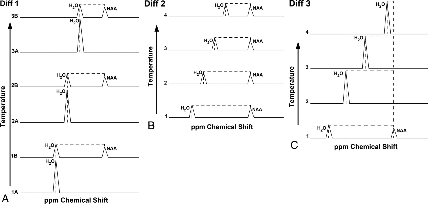
- Fig 1.
Schematic representation of methodologies for PRF thermometry: DIFF 1, DIFF 2, and DIFF 3 (see text). With increasing temperatures, the water resonance frequency is observed to shift at approximately 0.01 ppm/°C toward the lower resonance. A, DIFF 1, representing the initial-approach interleaving pairs of non-water-suppressed and water-suppressed proton spectra during increasing temperatures. Alphanumeric combinations denote the pair number, followed by either A for non-water-suppressed or B for water-suppressed spectra. The shifting water-resonance frequency is thus measured against the static NAA resonance, the latter obtained from B in each pair. B, DIFF 2 partially water-suppressed spectrum used for the simultaneous measurement of both water and NAA frequencies. C, DIFF 3, newly proposed accelerated methodology, wherein the NAA reference frequency is obtained from an initial water-suppressed spectrum and is followed by a high-temporal-resolution series of non-water-suppressed spectra, measured against the initial NAA reference frequency.
- Fig 2.
Linear regression from the DIFF 1 experiment. The y-axis represents the water-NAA chemical shift difference, plotted against continuous fiber optic measured temperatures from approximately 18 to 45°C. The slope of regression, 0.01 ppm/°C, is in agreement with the expected temperature dependency of the temperature-dependent proton chemical shift.
- Fig 3.
Linear regression from the DIFF 2 experiment. Similar correlation as noted in DIFF 1. By comparison with DIFF 1, approximately 15%–20% time savings is achieved by eliminating the paired approach used in Fig 2.
- Fig 4.
DIFF 3 versus temperature change. Excellent correlation is again noted between measured temperatures and the water-NAA chemical shift. Frequency shifts are measured from sequential, non-water-suppressed spectra against an initial NAA reference frequency. Elimination of the repeated metabolite spectra in DIFF 1 and DIFF 2 permits increasing temporal resolution, with collection of 102 data points between approximately 18 and 35°C, compared with <15 measures across a similar experimental session.
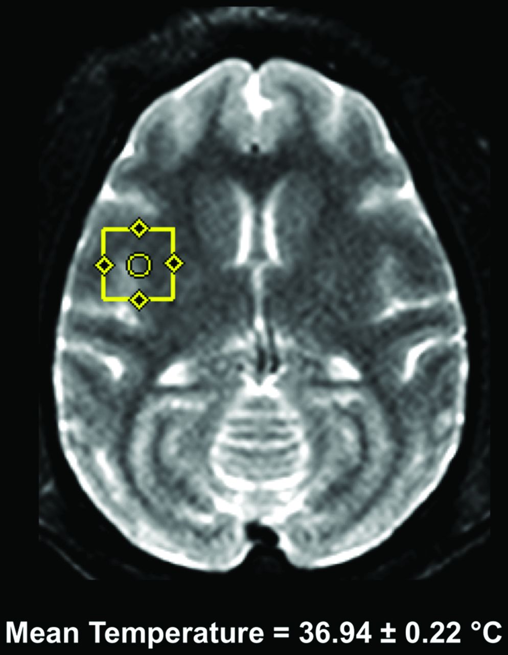
- Fig 5.
Single-voxel MR spectroscopy in a healthy nonhuman primate during physiologic monitoring under general anesthesia, with maintenance of rectal temperatures at approximately 37°C. Six repeated MR spectroscopy scans were acquired from the right operculum by using a standard point-resolved spectroscopy sequence across an approximately 3-hour imaging session. Temperatures were computed from the temperature-dependent water-NAA chemical shift difference (see text), with mean computed temperatures of 36.94 ± 0.22°C. Spectral fitted full width at half maximum varies between 4 and 5 Hz.
- Fig 6.
Multivoxel MR thermometry in a healthy nonhuman primate during physiologic monitoring under general anesthesia, with maintenance of rectal temperatures at approximately 37°C. Six repeated MR spectroscopy scans were acquired by using 2D chemical shift imaging spectroscopy (see text) across an approximately 3-hour imaging session. Computed temperatures are derived from the water-NAA proton chemical shift and color thermal grid overlaid on the axial T2-weighted reference image for display purposes. A relatively symmetric, zonal distribution of temperatures is noted across repeated studies; a representative peripheral voxel demonstrates the repeatability of in vivo imaging, with mean computed temperatures of 37.6 ± 0.02°C. Spectral-fitted full widths at half maximum vary between 4 and 5 Hz across voxels.
- Fig 7.
A, Single-voxel MR thermometry in an adult nonhuman primate 6 hours following endovascular MCA occlusion. Axial T2-weighted image (right) with symmetric 1.5-mL voxels placed in the bilateral opercular region. B, Area of infarction in the right hemisphere demonstrated on diffusion-weighted images (yellow arrows). Computed temperatures within the region of infarction are noted approaching 0.5°C less than those in the contralateral hemisphere.
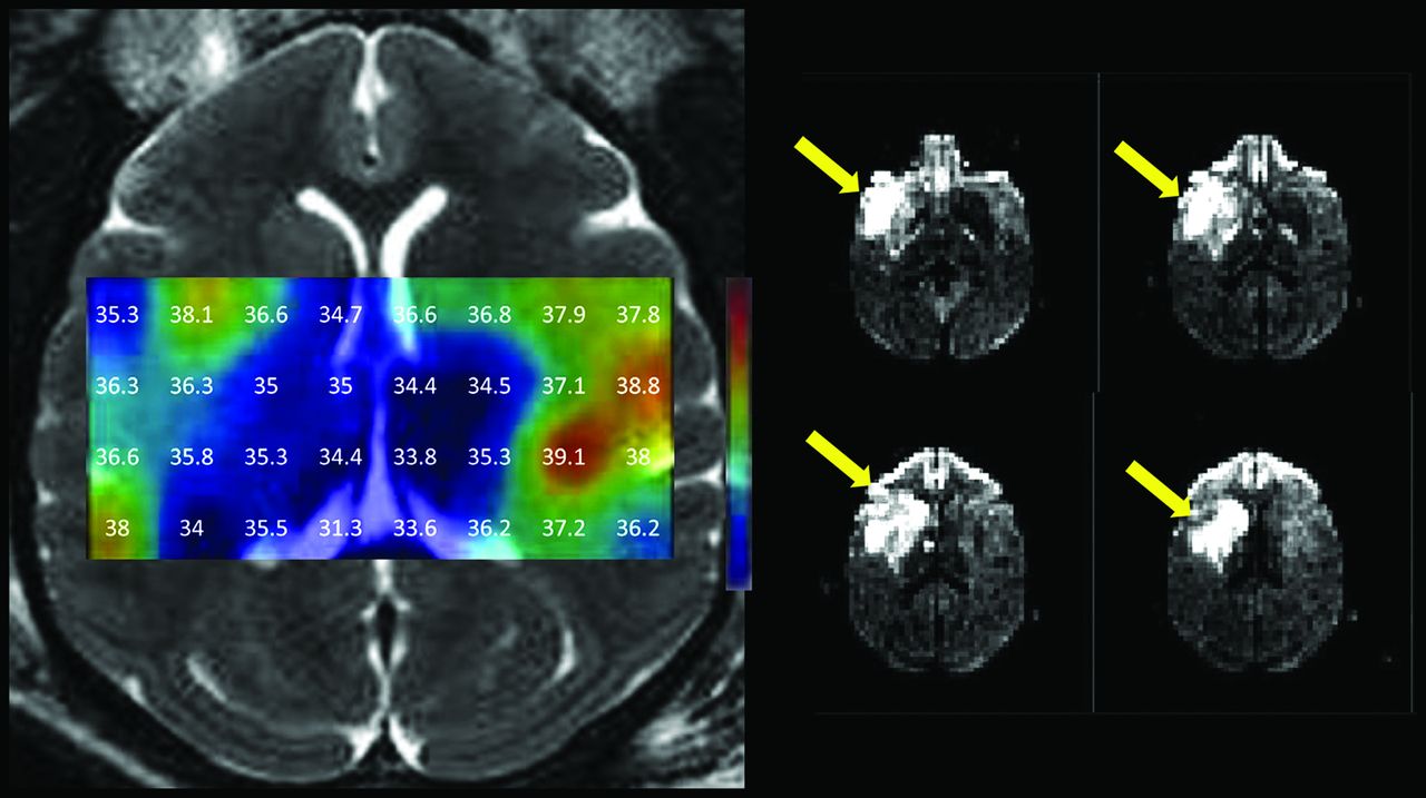
- Fig 8.
Multivoxel MR thermometry in an adult rhesus macaque obtained approximately 6 hours following endovascular MCA occlusion. Symmetric gradients observed under physiologic, preischemic conditions in Fig 6 were replaced by generally lower temperatures throughout the infarction territory of the right MCA seen on diffusion-weighted imaging (yellow arrows). Temperatures throughout the right hemispheric infarct territory generally diminish by comparison with the contralateral normal left hemisphere.


Jump to section
Related Articles
Cited By...
- Laser Interstitial Thermal Therapy for Intra-Axial Brain Tumors: Everything the Neuroradiologist Should Know
- Improving reproducibility of proton MRS brain thermometry: theoretical and empirical approaches
- MR Thermometry in Cerebrovascular Disease: Physiologic Basis, Hemodynamic Dependence, and a New Frontier in Stroke Imaging
- The Brain Thermal Response as a Potential Neuroimaging Biomarker of Cerebrovascular Impairment
- Cerebral Temperature Dysregulation: MR Thermographic Monitoring in a Nonhuman Primate Study of Acute Ischemic Stroke
- Body Temperature Modulates Infarction Growth following Endovascular Reperfusion