Advertisement

Main menu

  • Home
  • Content
    • Current Issue
    • Accepted Manuscripts
    • Article Preview
    • Past Issue Archive
    • Video Articles
    • AJNR Case Collection
    • Case of the Week Archive
    • Case of the Month Archive
    • Classic Case Archive
  • Special Collections
    • AJNR Awards
    • Low-Field MRI
    • Alzheimer Disease
    • ASNR Foundation Special Collection
    • Photon-Counting CT
    • View All
  • Multimedia
    • AJNR Podcasts
    • AJNR SCANtastic
    • Trainee Corner
    • MRI Safety Corner
    • Imaging Protocols
  • For Authors
    • Submit a Manuscript
    • Submit a Video Article
    • Submit an eLetter to the Editor/Response
    • Manuscript Submission Guidelines
    • Statistical Tips
    • Fast Publishing of Accepted Manuscripts
    • Graphical Abstract Preparation
    • Imaging Protocol Submission
    • Author Policies
  • About Us
    • About AJNR
    • Editorial Board
    • Editorial Board Alumni
  • More
    • Become a Reviewer/Academy of Reviewers
    • Subscribers
    • Permissions
    • Alerts
    • Feedback
    • Advertisers
    • ASNR Home

User menu

  • Alerts
  • Log in

Search

  • Advanced search
American Journal of Neuroradiology

American Journal of Neuroradiology

ASHNR American Society of Functional Neuroradiology ASHNR American Society of Pediatric Neuroradiology ASSR
  • Alerts
  • Log in

Advanced Search

  • Home
  • Content
    • Current Issue
    • Accepted Manuscripts
    • Article Preview
    • Past Issue Archive
    • Video Articles
    • AJNR Case Collection
    • Case of the Week Archive
    • Case of the Month Archive
    • Classic Case Archive
  • Special Collections
    • AJNR Awards
    • Low-Field MRI
    • Alzheimer Disease
    • ASNR Foundation Special Collection
    • Photon-Counting CT
    • View All
  • Multimedia
    • AJNR Podcasts
    • AJNR SCANtastic
    • Trainee Corner
    • MRI Safety Corner
    • Imaging Protocols
  • For Authors
    • Submit a Manuscript
    • Submit a Video Article
    • Submit an eLetter to the Editor/Response
    • Manuscript Submission Guidelines
    • Statistical Tips
    • Fast Publishing of Accepted Manuscripts
    • Graphical Abstract Preparation
    • Imaging Protocol Submission
    • Author Policies
  • About Us
    • About AJNR
    • Editorial Board
    • Editorial Board Alumni
  • More
    • Become a Reviewer/Academy of Reviewers
    • Subscribers
    • Permissions
    • Alerts
    • Feedback
    • Advertisers
    • ASNR Home
  • Follow AJNR on Twitter
  • Visit AJNR on Facebook
  • Follow AJNR on Instagram
  • Join AJNR on LinkedIn
  • RSS Feeds

AJNR Awards, New Junior Editors, and more. Read the latest AJNR updates

Review ArticleReview Articles
Open Access

4D-CTA in Neurovascular Disease: A Review

H.G.J. Kortman, E.J. Smit, M.T.H. Oei, R. Manniesing, M. Prokop and F.J.A. Meijer
American Journal of Neuroradiology June 2015, 36 (6) 1026-1033; DOI: https://doi.org/10.3174/ajnr.A4162
H.G.J. Kortman
aFrom the Department of Radiology and Nuclear Medicine, Radboud University Nijmegen Medical Center, Nijmegen, the Netherlands.
  • Find this author on Google Scholar
  • Find this author on PubMed
  • Search for this author on this site
E.J. Smit
aFrom the Department of Radiology and Nuclear Medicine, Radboud University Nijmegen Medical Center, Nijmegen, the Netherlands.
  • Find this author on Google Scholar
  • Find this author on PubMed
  • Search for this author on this site
M.T.H. Oei
aFrom the Department of Radiology and Nuclear Medicine, Radboud University Nijmegen Medical Center, Nijmegen, the Netherlands.
  • Find this author on Google Scholar
  • Find this author on PubMed
  • Search for this author on this site
R. Manniesing
aFrom the Department of Radiology and Nuclear Medicine, Radboud University Nijmegen Medical Center, Nijmegen, the Netherlands.
  • Find this author on Google Scholar
  • Find this author on PubMed
  • Search for this author on this site
M. Prokop
aFrom the Department of Radiology and Nuclear Medicine, Radboud University Nijmegen Medical Center, Nijmegen, the Netherlands.
  • Find this author on Google Scholar
  • Find this author on PubMed
  • Search for this author on this site
F.J.A. Meijer
aFrom the Department of Radiology and Nuclear Medicine, Radboud University Nijmegen Medical Center, Nijmegen, the Netherlands.
  • Find this author on Google Scholar
  • Find this author on PubMed
  • Search for this author on this site
  • ORCID record for F.J.A. Meijer
  • Article
  • Figures & Data
  • Supplemental
  • Info & Metrics
  • Responses
  • References
  • PDF
Loading

Article Figures & Data

Figures

  • Tables
  • Fig 1.
    • Download figure
    • Open in new tab
    • Download powerpoint
    Fig 1.

    Schematic diagram of 4D-CTA imaging techniques. The x-axis represents the time domain. The y-axis represents the z-dimension. The light-gray horizontal bar represents the area that needs to be covered in the z-dimension. A, Shuttle mode: the sinus represents continuous table movement back and forth in the z-axis dimension to provide adequate coverage of the region of interest at multiple points in time. Notice that the temporal resolution is a function of the speed of table movement, typically 2–4 seconds depending on coverage. B, Toggling-table technique: the bars represent table repositioning in the z-axis dimension to provide adequate coverage of the region of interest at multiple points in time. Notice that the temporal resolution is a function of the speed of table repositioning, typically 3–4 seconds. C, Volume scanning: complete coverage of the region of interest (horizontal bar) with 1 gantry rotation. Notice that the temporal resolution is a function of the scanning interval settings because each rotation provides full coverage. Volume CT scanning enables (D) continuous volume scanning. Temporal resolution is limited by the gantry rotation speed.

  • Fig 2.
    • Download figure
    • Open in new tab
    • Download powerpoint
    Fig 2.

    4D-CTA demonstrating a Borden type I dural arteriovenous fistula of the left sigmoid sinus. Selected 4D-CTA subtraction MIP images of a continuous 4D-CTA volume acquisition (320–detector row CT) in lateral (A) and oblique (B) projections in a patient presenting with left-sided tinnitus. Branches of the occipital artery are identified as arterial feeders of the dAVF. There is normal antegrade venous return. The 3D image (C) demonstrates the advantage of 4D-CTA to study vessels in relation to surrounding structures.

  • Fig 3.
    • Download figure
    • Open in new tab
    • Download powerpoint
    Fig 3.

    Timing-invariant CTA better estimates the extent of collateral circulation in a patient with right middle cerebral artery occlusion. The left image is a conventional CTA showing poor collateral circulation and suggests a poor prognosis. The right image is a TI-CTA image from a 4D-CTA acquisition (ie, temporal MIP), which shows good collateral filling and suggests a good prognosis. In this case, the patient had a good recovery.
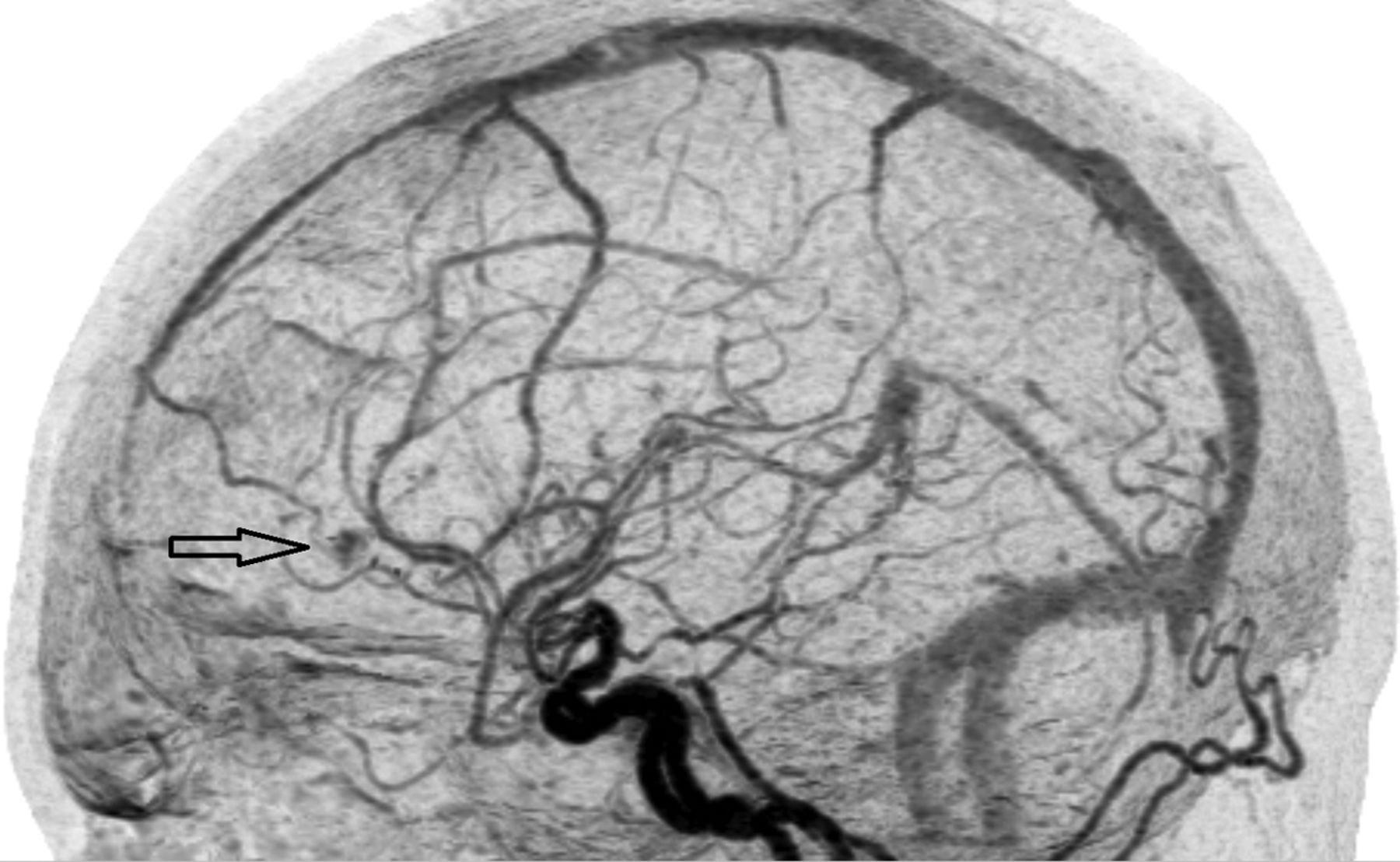
  • Fig 4.
    • Download figure
    • Open in new tab
    • Download powerpoint
    Fig 4.

    4D-CTA image demonstrating recurrence of an arteriovenous malformation. The arrow indicates the nidus, which is fed by arterial feeders from the anterior cerebral artery. There is cortical venous drainage toward the rostral superior sagittal sinus.

Tables

  • Figures
  • Classification of arteriovenous malformations and arteriovenous fistulas

    PathologyAVMAVF
    Distinguishing featurePresence of a nidusNo nidus
    Grading systemSpetzler-Martin classificationCognard classification
    Borden classification
    ClassificationSpetzler-Martin AVM grading scaleCognard classification:
    Size    Grade I: in sinus wall; normal antegrade venous drainage
        Small (<3 cm) = 1    Grade IIa: in sinus; reflux to sinus, not cortical veins
        Medium (3–6 cm) = 2    Grade IIb: retrograde drainage (reflux) to cortical veins
        Large (>6 cm) = 3    Grade III: direct cortical venous drainage; no venous ectasia
    Eloquence of adjacent brain    Grade IV: direct cortical venous drainage and venous ectasia
        Noneloquent = 0    Grade V: Spinal perimedullary venous drainage
        Eloquent = 1Borden classification:
    Venous drainage    Type I: dural arterial supply with antegrade venous drainage
        Superficial only = 0    Type Ia: simple dAVF with single meningeal arterial supply
        Deep component = 1    Type Ib: complex dAVF with multiple meningeal arteries
        Type II: retrograde cortical venous drainage
        Type III: dural arteries drain into cortical veins
PreviousNext
Back to top

In this issue

American Journal of Neuroradiology: 36 (6)
American Journal of Neuroradiology
Vol. 36, Issue 6
1 Jun 2015
  • Table of Contents
  • Index by author
  • Complete Issue (PDF)
Advertisement
Print
Download PDF
Email Article

Thank you for your interest in spreading the word on American Journal of Neuroradiology.

NOTE: We only request your email address so that the person you are recommending the page to knows that you wanted them to see it, and that it is not junk mail. We do not capture any email address.

Enter multiple addresses on separate lines or separate them with commas.
4D-CTA in Neurovascular Disease: A Review
(Your Name) has sent you a message from American Journal of Neuroradiology
(Your Name) thought you would like to see the American Journal of Neuroradiology web site.
CAPTCHA
This question is for testing whether or not you are a human visitor and to prevent automated spam submissions.
Cite this article
H.G.J. Kortman, E.J. Smit, M.T.H. Oei, R. Manniesing, M. Prokop, F.J.A. Meijer
4D-CTA in Neurovascular Disease: A Review
American Journal of Neuroradiology Jun 2015, 36 (6) 1026-1033; DOI: 10.3174/ajnr.A4162

Citation Manager Formats

  • BibTeX
  • Bookends
  • EasyBib
  • EndNote (tagged)
  • EndNote 8 (xml)
  • Medlars
  • Mendeley
  • Papers
  • RefWorks Tagged
  • Ref Manager
  • RIS
  • Zotero
0 Responses
Respond to this article
Share
Bookmark this article
4D-CTA in Neurovascular Disease: A Review
H.G.J. Kortman, E.J. Smit, M.T.H. Oei, R. Manniesing, M. Prokop, F.J.A. Meijer
American Journal of Neuroradiology Jun 2015, 36 (6) 1026-1033; DOI: 10.3174/ajnr.A4162
del.icio.us logo Twitter logo Facebook logo Mendeley logo
  • Tweet Widget
  • Facebook Like
  • Google Plus One
Purchase

Jump to section

  • Article
    • Abstract
    • ABBREVIATIONS:
    • Conclusions
    • Appendix: Search Strategy
    • Footnotes
    • References
  • Figures & Data
  • Supplemental
  • Info & Metrics
  • Responses
  • References
  • PDF

Related Articles

  • PubMed
  • Google Scholar

Cited By...

  • Management of a wake-up stroke
  • Color-Mapping of 4D-CTA for the Detection of Cranial Arteriovenous Shunts
  • Diagnostic accuracy of emergency CT angiography for presumed tandem internal carotid artery occlusion before acute endovascular therapy
  • Improved Detection of Anterior Circulation Occlusions: The "Delayed Vessel Sign" on Multiphase CT Angiography
  • Republished: Remote multifocal bleeding points producing a Sylvian subpial hematoma during endovascular coiling of an acutely ruptured cerebral aneurysm
  • Remote multifocal bleeding points producing a Sylvian subpial hematoma during endovascular coiling of an acutely ruptured cerebral aneurysm
  • Time-Resolved C-Arm Computed Tomographic Angiography Derived From Computed Tomographic Perfusion Acquisition: New Capability for One-Stop-Shop Acute Ischemic Stroke Treatment in the Angiosuite
  • Crossref
  • Google Scholar

This article has not yet been cited by articles in journals that are participating in Crossref Cited-by Linking.

More in this TOC Section

  • An Atlas of Neonatal Neurovascular Imaging Anatomy as Depicted with Microvascular Imaging: The Intracranial Arteries
  • An Atlas of Neonatal Neurovascular Imaging Anatomy as Depicted with Microvascular Imaging: The Intracranial Veins
  • Clinical Translation of Hyperpolarized 13C Metabolic Probes for Glioma Imaging
Show more Review Articles

Similar Articles

Advertisement

Indexed Content

  • Current Issue
  • Accepted Manuscripts
  • Article Preview
  • Past Issues
  • Editorials
  • Editor's Choice
  • Fellows' Journal Club
  • Letters to the Editor
  • Video Articles

Cases

  • Case Collection
  • Archive - Case of the Week
  • Archive - Case of the Month
  • Archive - Classic Case

More from AJNR

  • Trainee Corner
  • Imaging Protocols
  • MRI Safety Corner
  • Book Reviews

Multimedia

  • AJNR Podcasts
  • AJNR Scantastics

Resources

  • Turnaround Time
  • Submit a Manuscript
  • Submit a Video Article
  • Submit an eLetter to the Editor/Response
  • Manuscript Submission Guidelines
  • Statistical Tips
  • Fast Publishing of Accepted Manuscripts
  • Graphical Abstract Preparation
  • Imaging Protocol Submission
  • Evidence-Based Medicine Level Guide
  • Publishing Checklists
  • Author Policies
  • Become a Reviewer/Academy of Reviewers
  • News and Updates

About Us

  • About AJNR
  • Editorial Board
  • Editorial Board Alumni
  • Alerts
  • Permissions
  • Not an AJNR Subscriber? Join Now
  • Advertise with Us
  • Librarian Resources
  • Feedback
  • Terms and Conditions
  • AJNR Editorial Board Alumni

American Society of Neuroradiology

  • Not an ASNR Member? Join Now

© 2025 by the American Society of Neuroradiology All rights, including for text and data mining, AI training, and similar technologies, are reserved.
Print ISSN: 0195-6108 Online ISSN: 1936-959X

Powered by HighWire