
Article Figures & Data
Figures
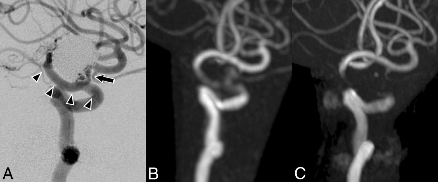
- Fig 1.
Case 1. A 65-year-old woman with a right paraclinoid internal carotid artery aneurysm was treated by stent-assisted coil embolization in March 2011. The latest DSA was performed in February 2013 (A). Silent MRA (B) and TOF MRA (C) were performed in March 2014. The clinoid-to-terminal segment of the internal carotid artery is covered by a stent (arrowheads). The anatomic outcome of the aneurysm in DSA is a neck remnant (arrow). Our subjective scores of Silent MRA are 4 and 3. The scores of TOF MRA are 1 and 1. In this case, the Silent MRA depiction of the neck remnant is also better than that of TOF MRA.
- Fig 2.
Case 2. A 75-year-old man with a right anterior choroidal artery aneurysm was treated in June 2010. The latest DSA was performed in December 2012 (A). Silent MRA (B) and TOF MRA (C) were performed in March 2014. The supraclinoid segment of the internal carotid artery to middle cerebral artery is covered by a stent (arrowheads). The anatomic outcome of the aneurysm in DSA is complete occlusion. Our subjective scores of Silent MRA are 4 and 3. The scores of TOF MRA are 2 and 2.
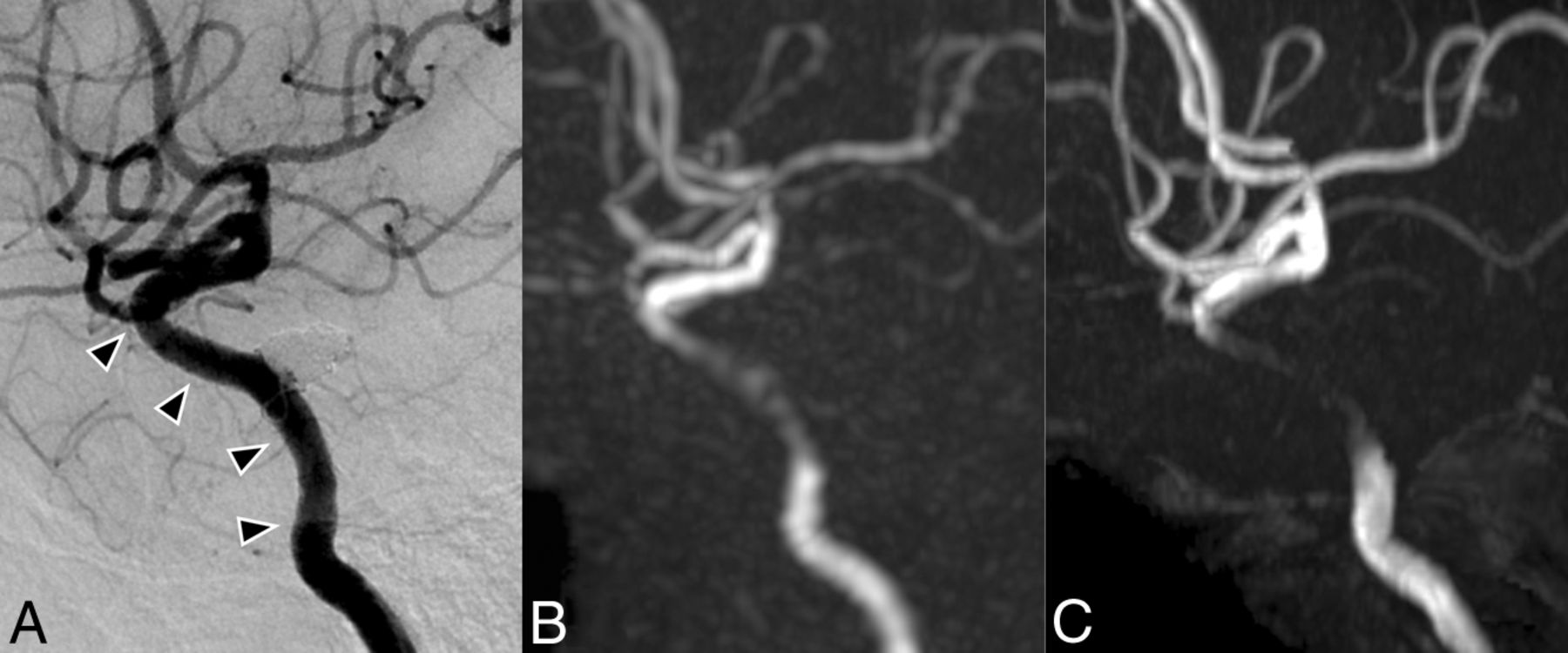
- Fig 3.
Case 3. A 74-year-old woman with a right paraclinoid internal carotid artery aneurysm was treated in October 2013. A DSA image (A) was obtained right after the treatment. Silent MRA (B) and TOF MRA (C) were performed in May 2014. The cavernous-to-supraclinoid segment of the internal carotid artery is covered by a stent (arrowheads). The anatomic outcome of the aneurysm in DSA is complete occlusion. Our subjective scores of Silent MRA are 4 and 4. The scores of TOF MRA are 1 and 1.
- Fig 4.
Case 6. A 50-year-old woman with a right paraclinoid internal carotid artery aneurysm was treated in June 2012. Silent MRA (B) and TOF MRA (C) were performed in June 2014, and DSA (A) was performed the next day. The cavernous-to-terminal segment of the internal carotid artery is covered by a stent (arrowheads). The anatomic outcome of the aneurysm in DSA is a small neck remnant (arrow). Our subjective scores of Silent MRA are 4 and 3. The scores of TOF MRA are 1 and 1.
- Fig 5.
Case 7. A 51-year-old woman with a left cavernous internal carotid artery aneurysm was treated in May 2012. The latest DSA was performed in June 2013 (A). Silent MRA (B) and TOF MRA (C) were performed in June 2014. The cavernous-to-clinoid segment of the internal carotid artery is covered by a stent (arrowheads). The anatomic outcome of the aneurysm in DSA is complete occlusion. In this case, the overall image quality of Silent MRA is not good, but visualization of the flow in a stent is still better than that of TOF MRA. Our subjective scores of Silent MRA are 2 and 3. The scores of TOF MRA are 1 and 1.


Tables
Patient data
Case No. Age (yr) Sex Aneurysm Location Stent Configurationa Interval between DSA and MRA Aneurysm Occlusion Status Scores of Silent MRAb Scores of TOF MRAb 1 65 F Rt. ICA paraclinoid Clinoid-terminal 13 mo NR 4/3 1/1 2 75 M Rt. ICA-AchoA Supraclinoid-MCA 15 mo CO 4/3 2/2 3 74 F Rt. ICA paraclinoid Cavernous-supraclinoid 7 mo CO 4/4 1/1 4 59 M Lt. ICA bifurcation Supraclinoid-MCA 1 day CO 3/3 2/1 5 45 F Rt. ICA paraclinoid Cavernous-terminal 7 mo CO 4/4 2/2 6 50 F Rt. ICA paraclinoid Cavernous-terminal 1 day NR 4/3 1/1 7 51 F Lt. ICA cavernous Cavernous-clinoid 12 mo CO 2/3 1/1 8 61 F Rt. ICA-PcomA Supraclinoid-terminal 2 days NR 4/4 2/2 9 64 M Lt. ICA paraclinoid Cavernous-supraclinoid 12 mo NR 3/3 2/1 Note:—AchoA indicates anterior choroidal artery; PcomA, posterior communicating artery; Clinoid, clinoid segment of ICA; terminal, terminal segment of ICA; supraclinoid, supraclinoid segment of ICA; Cavernous, cavernous segment of ICA; NR, neck remnant; CO, complete occlusion; Rt., right; Lt., left.
↵a Proximal-distal marker of a stent.
↵b Scores of observers 1/2.


Jump to section
Related Articles
Cited By...
- Visualization of Intracranial Aneurysms Treated with Woven EndoBridge Devices Using Ultrashort TE MR Imaging
- Validation of Zero TE-MRA in the Characterization of Cerebrovascular Diseases: A Feasibility Study
- Usefulness of Silent MR Angiography for Intracranial Aneurysms Treated with a Flow-Diverter Device
- Visualization of Aneurysmal Neck and Dome after Coiling with 3D Multifusion Imaging of Silent MRA and FSE-MR Cisternography
- Pointwise Encoding Time Reduction with Radial Acquisition with Subtraction-Based MRA during the Follow-Up of Stent-Assisted Coil Embolization of Anterior Circulation Aneurysms
- Usefulness of Vessel Wall MR Imaging for Follow-Up after Stent-Assisted Coil Embolization of Intracranial Aneurysms
- Non-Contrast-Enhanced Silent Scan MR Angiography of Intracranial Anterior Circulation Aneurysms Treated with a Low-Profile Visualized Intraluminal Support Device
- Usefulness of Non-Contrast-Enhanced MR Angiography Using a Silent Scan for Follow-Up after Y-Configuration Stent-Assisted Coil Embolization for Basilar Tip Aneurysms
- Contrast-Enhanced and Time-of-Flight MRA at 3T Compared with DSA for the Follow-Up of Intracranial Aneurysms Treated with the WEB Device