Advertisement

Main menu

  • Home
  • Content
    • Current Issue
    • Accepted Manuscripts
    • Article Preview
    • Past Issue Archive
    • Video Articles
    • AJNR Case Collection
    • Case of the Week Archive
    • Case of the Month Archive
    • Classic Case Archive
  • Special Collections
    • AJNR Awards
    • Low-Field MRI
    • Alzheimer Disease
    • ASNR Foundation Special Collection
    • Photon-Counting CT
    • View All
  • Multimedia
    • AJNR Podcasts
    • AJNR SCANtastic
    • Trainee Corner
    • MRI Safety Corner
    • Imaging Protocols
  • For Authors
    • Submit a Manuscript
    • Submit a Video Article
    • Submit an eLetter to the Editor/Response
    • Manuscript Submission Guidelines
    • Statistical Tips
    • Fast Publishing of Accepted Manuscripts
    • Graphical Abstract Preparation
    • Imaging Protocol Submission
    • Author Policies
  • About Us
    • About AJNR
    • Editorial Board
    • Editorial Board Alumni
  • More
    • Become a Reviewer/Academy of Reviewers
    • Subscribers
    • Permissions
    • Alerts
    • Feedback
    • Advertisers
    • ASNR Home

User menu

  • Alerts
  • Log in

Search

  • Advanced search
American Journal of Neuroradiology

American Journal of Neuroradiology

ASHNR American Society of Functional Neuroradiology ASHNR American Society of Pediatric Neuroradiology ASSR
  • Alerts
  • Log in

Advanced Search

  • Home
  • Content
    • Current Issue
    • Accepted Manuscripts
    • Article Preview
    • Past Issue Archive
    • Video Articles
    • AJNR Case Collection
    • Case of the Week Archive
    • Case of the Month Archive
    • Classic Case Archive
  • Special Collections
    • AJNR Awards
    • Low-Field MRI
    • Alzheimer Disease
    • ASNR Foundation Special Collection
    • Photon-Counting CT
    • View All
  • Multimedia
    • AJNR Podcasts
    • AJNR SCANtastic
    • Trainee Corner
    • MRI Safety Corner
    • Imaging Protocols
  • For Authors
    • Submit a Manuscript
    • Submit a Video Article
    • Submit an eLetter to the Editor/Response
    • Manuscript Submission Guidelines
    • Statistical Tips
    • Fast Publishing of Accepted Manuscripts
    • Graphical Abstract Preparation
    • Imaging Protocol Submission
    • Author Policies
  • About Us
    • About AJNR
    • Editorial Board
    • Editorial Board Alumni
  • More
    • Become a Reviewer/Academy of Reviewers
    • Subscribers
    • Permissions
    • Alerts
    • Feedback
    • Advertisers
    • ASNR Home
  • Follow AJNR on Twitter
  • Visit AJNR on Facebook
  • Follow AJNR on Instagram
  • Join AJNR on LinkedIn
  • RSS Feeds

AJNR Awards, New Junior Editors, and more. Read the latest AJNR updates

Research ArticleFunctional

Disrupted Resting-State Functional Connectivity in Progressive Supranuclear Palsy

M.C. Piattella, F. Tona, M. Bologna, E. Sbardella, A. Formica, N. Petsas, N. Filippini, A. Berardelli and P. Pantano
American Journal of Neuroradiology May 2015, 36 (5) 915-921; DOI: https://doi.org/10.3174/ajnr.A4229
M.C. Piattella
aFrom the Department of Neurology and Psychiatry (M.C.P., F.T., E.S., A.F., N.P., A.B., P.P.), Sapienza, University of Rome, Italy
  • Find this author on Google Scholar
  • Find this author on PubMed
  • Search for this author on this site
F. Tona
aFrom the Department of Neurology and Psychiatry (M.C.P., F.T., E.S., A.F., N.P., A.B., P.P.), Sapienza, University of Rome, Italy
  • Find this author on Google Scholar
  • Find this author on PubMed
  • Search for this author on this site
M. Bologna
bNeuromed Institute Istituto Di Ricovero e Cura a Carattere Scientifico (M.B., A.B., P.P.), Pozzilli, Italy
  • Find this author on Google Scholar
  • Find this author on PubMed
  • Search for this author on this site
E. Sbardella
aFrom the Department of Neurology and Psychiatry (M.C.P., F.T., E.S., A.F., N.P., A.B., P.P.), Sapienza, University of Rome, Italy
  • Find this author on Google Scholar
  • Find this author on PubMed
  • Search for this author on this site
A. Formica
aFrom the Department of Neurology and Psychiatry (M.C.P., F.T., E.S., A.F., N.P., A.B., P.P.), Sapienza, University of Rome, Italy
  • Find this author on Google Scholar
  • Find this author on PubMed
  • Search for this author on this site
N. Petsas
aFrom the Department of Neurology and Psychiatry (M.C.P., F.T., E.S., A.F., N.P., A.B., P.P.), Sapienza, University of Rome, Italy
  • Find this author on Google Scholar
  • Find this author on PubMed
  • Search for this author on this site
N. Filippini
cDepartment of Psychiatry and FMRIB Centre (N.F.), University of Oxford, United Kingdom.
  • Find this author on Google Scholar
  • Find this author on PubMed
  • Search for this author on this site
A. Berardelli
aFrom the Department of Neurology and Psychiatry (M.C.P., F.T., E.S., A.F., N.P., A.B., P.P.), Sapienza, University of Rome, Italy
bNeuromed Institute Istituto Di Ricovero e Cura a Carattere Scientifico (M.B., A.B., P.P.), Pozzilli, Italy
  • Find this author on Google Scholar
  • Find this author on PubMed
  • Search for this author on this site
P. Pantano
aFrom the Department of Neurology and Psychiatry (M.C.P., F.T., E.S., A.F., N.P., A.B., P.P.), Sapienza, University of Rome, Italy
bNeuromed Institute Istituto Di Ricovero e Cura a Carattere Scientifico (M.B., A.B., P.P.), Pozzilli, Italy
  • Find this author on Google Scholar
  • Find this author on PubMed
  • Search for this author on this site
  • Article
  • Figures & Data
  • Info & Metrics
  • Responses
  • References
  • PDF
Loading

Article Figures & Data

Figures

  • Tables
  • Fig 1.
    • Download figure
    • Open in new tab
    • Download powerpoint
    Fig 1.

    Maps of functional connectivity obtained from 5 seeds, ie, the dorsal midbrain tegmentum (yellow), thalamus (red), caudate (pink), putamen (green), and pallidum (blue)—in 12 healthy subjects (1-sample t test, P < .05, corrected for family-wise error). The images are presented according to radiologic orientation.

  • Fig 2.
    • Download figure
    • Open in new tab
    • Download powerpoint
    Fig 2.

    Differences between 19 patients with progressive supranuclear palsy and 12 healthy subjects in functional connectivity obtained from 5 seeds (2-sample t test, P < .05, corrected for family-wise error). Patients with PSP had significantly lower FC than healthy subjects in all 5 FC maps—that is, the dorsal midbrain tegmentum (yellow), thalamus (red), caudate (pink), putamen (green), and pallidum (blue). The images are presented according to radiologic orientation.
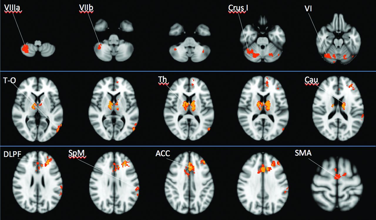
  • Fig 3.
    • Download figure
    • Open in new tab
    • Download powerpoint
    Fig 3.

    Images showing common areas of functional abnormalities shared by the 5 maps of FC. Different colors show the number of abnormal FC maps: in yellow, voxels of decreased FC in 4 maps; and in orange, voxels of decreased FC in 3 maps. Voxels of decreased FC in 2 maps are not shown. No focus of decreased FC in any of the 5 maps was identified. The images are presented according to radiologic orientation.

Tables

  • Figures
    • View popup
    Table 1:

    Clinical and radiologic characteristics of 19 patients with PSP and of 12 healthy subjects

    Healthy Subjectsa (n = 12)Patients with PSPa (n = 19)P Valueb
    Age (yr)69.172 ± 5.20170.933 ± 5.196.356
    Male/femalec3/910/9.158
    UPDRS–27.625 ± 17.952NA
    FAB–11.187 ± 3.799NA
    H&Y–2.9 ± 1.065NA
    MMSE29.135 ± 0.824.325 ± 3.886NA
    PSPRS–35.823 ± 16.994NA
        History–7.706 ± 3.820NA
        Mentation–3.647 ± 2.597NA
        Bulbar–3.117 ± 1.996NA
        Ocular–7.706 ± 2.932NA
        Limb–4.176 ± 3.486NA
        Gait–9.235 ± 5.750NA
    Thalamus V (mm3)9.483 ± 0.8408.005 ± 0.657<.0001d
    Caudate V (mm3)4.380 ± 0.3733.962 ± 0.477.015
    Putamen V (mm3)5.992 ± 0.4724.835 ± 0.575<.0001d
    Pallidum V (mm3)2.404 ± 0.4721.857 ± 0.293.0004d
    Brain stem V (mm3)14.906 ± 1.65212.618 ± 1.603.0006d
    Intracranial V (mm3)1620.661 ± 163.4821549.769 ± 114.184.165
    Cortical V (mm3)600.423 ± 41.923576.789 ± 49.823.167
    Mean FA0.508 ± 0.0190.441 ± 0.030<.0001d
    Mean MD (mm × sec−2) × 10−30.689 ± 0.0260.762 ± 0.030<.0001d
    Mean RD (mm × sec−2) × 10−30.478 ± 0.0290.561 ± 0.034<.0001d
    Mean AD (mm × sec−2) × 10−31.049 ± 0.0321.154 ± 0.056<.0001d
    • Note:—UPDRS indicates Unified Parkinson's Disease Rating Scale; FAB, Frontal Assessment Battery; H&Y, Hoehn and Yahr Scale; MMSE, Mini-Mental State Examination; PSPRS, PSP Rating Scale; V, volume (left and right values of subcortical volumes are averaged); RD, radial diffusivity; AD, axial diffusivity; –, not available; NA = not applicable.

    • ↵a Values are reported as mean ± SD.

    • ↵b Differences between groups were assessed by t test.

    • ↵c Differences between groups were assessed by χ2.

    • ↵d Statistically significant values after Bonferroni correction for multiple comparisons.

    • View popup
    Table 2:

    Significant correlations between parameter estimates of FC maps and clinical scores in patients with PSP

    Clinical ScalesaβP Value95% CIR2
    ThalamusBulbar−5.89.04−11.57 to −0.200.19
    ThalamusMentation−8.02.05−16.20 to −0.150.27
    ThalamusH&Y−3.51.03−6.60 to −0.400.53
    PallidumMMSE12.23.031.19–23.270.52
    dMTFAB42.16.042.24–82.080.26
    • Note:—FAB indicates Frontal Assessment Battery; H&Y, Hoehn and Yahr Scale; MMSE, Mini-Mental State Examination.

    • ↵a Bulbar and Mentation are subitems of the PSP Rating Scale.

PreviousNext
Back to top

In this issue

American Journal of Neuroradiology: 36 (5)
American Journal of Neuroradiology
Vol. 36, Issue 5
1 May 2015
  • Table of Contents
  • Index by author
  • Complete Issue (PDF)
Advertisement
Print
Download PDF
Email Article

Thank you for your interest in spreading the word on American Journal of Neuroradiology.

NOTE: We only request your email address so that the person you are recommending the page to knows that you wanted them to see it, and that it is not junk mail. We do not capture any email address.

Enter multiple addresses on separate lines or separate them with commas.
Disrupted Resting-State Functional Connectivity in Progressive Supranuclear Palsy
(Your Name) has sent you a message from American Journal of Neuroradiology
(Your Name) thought you would like to see the American Journal of Neuroradiology web site.
CAPTCHA
This question is for testing whether or not you are a human visitor and to prevent automated spam submissions.
Cite this article
M.C. Piattella, F. Tona, M. Bologna, E. Sbardella, A. Formica, N. Petsas, N. Filippini, A. Berardelli, P. Pantano
Disrupted Resting-State Functional Connectivity in Progressive Supranuclear Palsy
American Journal of Neuroradiology May 2015, 36 (5) 915-921; DOI: 10.3174/ajnr.A4229

Citation Manager Formats

  • BibTeX
  • Bookends
  • EasyBib
  • EndNote (tagged)
  • EndNote 8 (xml)
  • Medlars
  • Mendeley
  • Papers
  • RefWorks Tagged
  • Ref Manager
  • RIS
  • Zotero
0 Responses
Respond to this article
Share
Bookmark this article
Disrupted Resting-State Functional Connectivity in Progressive Supranuclear Palsy
M.C. Piattella, F. Tona, M. Bologna, E. Sbardella, A. Formica, N. Petsas, N. Filippini, A. Berardelli, P. Pantano
American Journal of Neuroradiology May 2015, 36 (5) 915-921; DOI: 10.3174/ajnr.A4229
del.icio.us logo Twitter logo Facebook logo Mendeley logo
  • Tweet Widget
  • Facebook Like
  • Google Plus One
Purchase

Jump to section

  • Article
    • Abstract
    • ABBREVIATIONS:
    • Materials and Methods
    • Results
    • Discussion
    • Conclusions
    • Footnotes
    • REFERENCES
  • Figures & Data
  • Info & Metrics
  • Responses
  • References
  • PDF

Related Articles

  • No related articles found.
  • PubMed
  • Google Scholar

Cited By...

  • Remote Cortical Network for Frontal Cognitive Symptoms derived from Tau Accumulation in Progressive Supranuclear Palsy
  • Crossref
  • Google Scholar

This article has not yet been cited by articles in journals that are participating in Crossref Cited-by Linking.

More in this TOC Section

  • Kurtosis and Epileptogenic Tubers: A Pilot Study
  • Glutaric Aciduria Type 1: DK vs. Conventional MRI
  • Brain Iron in Niemann-Pick Type C: 7T Study
Show more Functional

Similar Articles

Advertisement

Indexed Content

  • Current Issue
  • Accepted Manuscripts
  • Article Preview
  • Past Issues
  • Editorials
  • Editor's Choice
  • Fellows' Journal Club
  • Letters to the Editor
  • Video Articles

Cases

  • Case Collection
  • Archive - Case of the Week
  • Archive - Case of the Month
  • Archive - Classic Case

More from AJNR

  • Trainee Corner
  • Imaging Protocols
  • MRI Safety Corner
  • Book Reviews

Multimedia

  • AJNR Podcasts
  • AJNR Scantastics

Resources

  • Turnaround Time
  • Submit a Manuscript
  • Submit a Video Article
  • Submit an eLetter to the Editor/Response
  • Manuscript Submission Guidelines
  • Statistical Tips
  • Fast Publishing of Accepted Manuscripts
  • Graphical Abstract Preparation
  • Imaging Protocol Submission
  • Evidence-Based Medicine Level Guide
  • Publishing Checklists
  • Author Policies
  • Become a Reviewer/Academy of Reviewers
  • News and Updates

About Us

  • About AJNR
  • Editorial Board
  • Editorial Board Alumni
  • Alerts
  • Permissions
  • Not an AJNR Subscriber? Join Now
  • Advertise with Us
  • Librarian Resources
  • Feedback
  • Terms and Conditions
  • AJNR Editorial Board Alumni

American Society of Neuroradiology

  • Not an ASNR Member? Join Now

© 2025 by the American Society of Neuroradiology All rights, including for text and data mining, AI training, and similar technologies, are reserved.
Print ISSN: 0195-6108 Online ISSN: 1936-959X

Powered by HighWire