Advertisement

Main menu

  • Home
  • Content
    • Current Issue
    • Accepted Manuscripts
    • Article Preview
    • Past Issue Archive
    • Video Articles
    • AJNR Case Collection
    • Case of the Week Archive
    • Case of the Month Archive
    • Classic Case Archive
  • Special Collections
    • AJNR Awards
    • Low-Field MRI
    • Alzheimer Disease
    • ASNR Foundation Special Collection
    • Photon-Counting CT
    • View All
  • Multimedia
    • AJNR Podcasts
    • AJNR SCANtastic
    • Trainee Corner
    • MRI Safety Corner
    • Imaging Protocols
  • For Authors
    • Submit a Manuscript
    • Submit a Video Article
    • Submit an eLetter to the Editor/Response
    • Manuscript Submission Guidelines
    • Statistical Tips
    • Fast Publishing of Accepted Manuscripts
    • Graphical Abstract Preparation
    • Imaging Protocol Submission
    • Author Policies
  • About Us
    • About AJNR
    • Editorial Board
    • Editorial Board Alumni
  • More
    • Become a Reviewer/Academy of Reviewers
    • Subscribers
    • Permissions
    • Alerts
    • Feedback
    • Advertisers
    • ASNR Home

User menu

  • Alerts
  • Log in

Search

  • Advanced search
American Journal of Neuroradiology

American Journal of Neuroradiology

ASHNR American Society of Functional Neuroradiology ASHNR American Society of Pediatric Neuroradiology ASSR
  • Alerts
  • Log in

Advanced Search

  • Home
  • Content
    • Current Issue
    • Accepted Manuscripts
    • Article Preview
    • Past Issue Archive
    • Video Articles
    • AJNR Case Collection
    • Case of the Week Archive
    • Case of the Month Archive
    • Classic Case Archive
  • Special Collections
    • AJNR Awards
    • Low-Field MRI
    • Alzheimer Disease
    • ASNR Foundation Special Collection
    • Photon-Counting CT
    • View All
  • Multimedia
    • AJNR Podcasts
    • AJNR SCANtastic
    • Trainee Corner
    • MRI Safety Corner
    • Imaging Protocols
  • For Authors
    • Submit a Manuscript
    • Submit a Video Article
    • Submit an eLetter to the Editor/Response
    • Manuscript Submission Guidelines
    • Statistical Tips
    • Fast Publishing of Accepted Manuscripts
    • Graphical Abstract Preparation
    • Imaging Protocol Submission
    • Author Policies
  • About Us
    • About AJNR
    • Editorial Board
    • Editorial Board Alumni
  • More
    • Become a Reviewer/Academy of Reviewers
    • Subscribers
    • Permissions
    • Alerts
    • Feedback
    • Advertisers
    • ASNR Home
  • Follow AJNR on Twitter
  • Visit AJNR on Facebook
  • Follow AJNR on Instagram
  • Join AJNR on LinkedIn
  • RSS Feeds

AJNR Awards, New Junior Editors, and more. Read the latest AJNR updates

Review ArticleReview Articles
Open Access

Spinal Cord Ischemia: Practical Imaging Tips, Pearls, and Pitfalls

M.I. Vargas, J. Gariani, R. Sztajzel, I. Barnaure-Nachbar, B.M. Delattre, K.O. Lovblad and J.-L. Dietemann
American Journal of Neuroradiology May 2015, 36 (5) 825-830; DOI: https://doi.org/10.3174/ajnr.A4118
M.I. Vargas
aFrom the Divisions of Neuroradiology (M.I.V., I.B.-N., K.O.L.)
  • Find this author on Google Scholar
  • Find this author on PubMed
  • Search for this author on this site
J. Gariani
bRadiology (J.G., B.M.D.)
  • Find this author on Google Scholar
  • Find this author on PubMed
  • Search for this author on this site
R. Sztajzel
cNeurology (R.S.), Geneva University Hospitals, Geneva, Switzerland
  • Find this author on Google Scholar
  • Find this author on PubMed
  • Search for this author on this site
I. Barnaure-Nachbar
aFrom the Divisions of Neuroradiology (M.I.V., I.B.-N., K.O.L.)
  • Find this author on Google Scholar
  • Find this author on PubMed
  • Search for this author on this site
B.M. Delattre
bRadiology (J.G., B.M.D.)
  • Find this author on Google Scholar
  • Find this author on PubMed
  • Search for this author on this site
K.O. Lovblad
aFrom the Divisions of Neuroradiology (M.I.V., I.B.-N., K.O.L.)
  • Find this author on Google Scholar
  • Find this author on PubMed
  • Search for this author on this site
J.-L. Dietemann
dDivision of Radiology (J.-L.D.), Strasbourg University Hospitals, Strasbourg, France.
  • Find this author on Google Scholar
  • Find this author on PubMed
  • Search for this author on this site
  • Article
  • Figures & Data
  • Info & Metrics
  • Responses
  • References
  • PDF
Loading

Article Figures & Data

Figures

  • Tables
  • Fig 1.
    • Download figure
    • Open in new tab
    • Download powerpoint
    Fig 1.

    Localization of the artery of Adamkiewicz in a patient with aortic thrombus. MR angiography shows the thrombus in the abdominal aorta below the renal arteries (arrows, A). No ischemia is visible in the conus medullaris (B). The artery of Adamkiewicz is permeable (arrows, C).
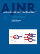
  • Fig 2.
    • Download figure
    • Open in new tab
    • Download powerpoint
    Fig 2.

    Ischemia provoked by an atheroma. Note the important atheromatosis of the abdominal aorta nicely shown by the volume-rendering reconstruction of CT angiography (A). Ischemia of the conus medullaris shown by MR imaging is hyperintense on T2 with a restriction of diffusion (arrows, B–E).
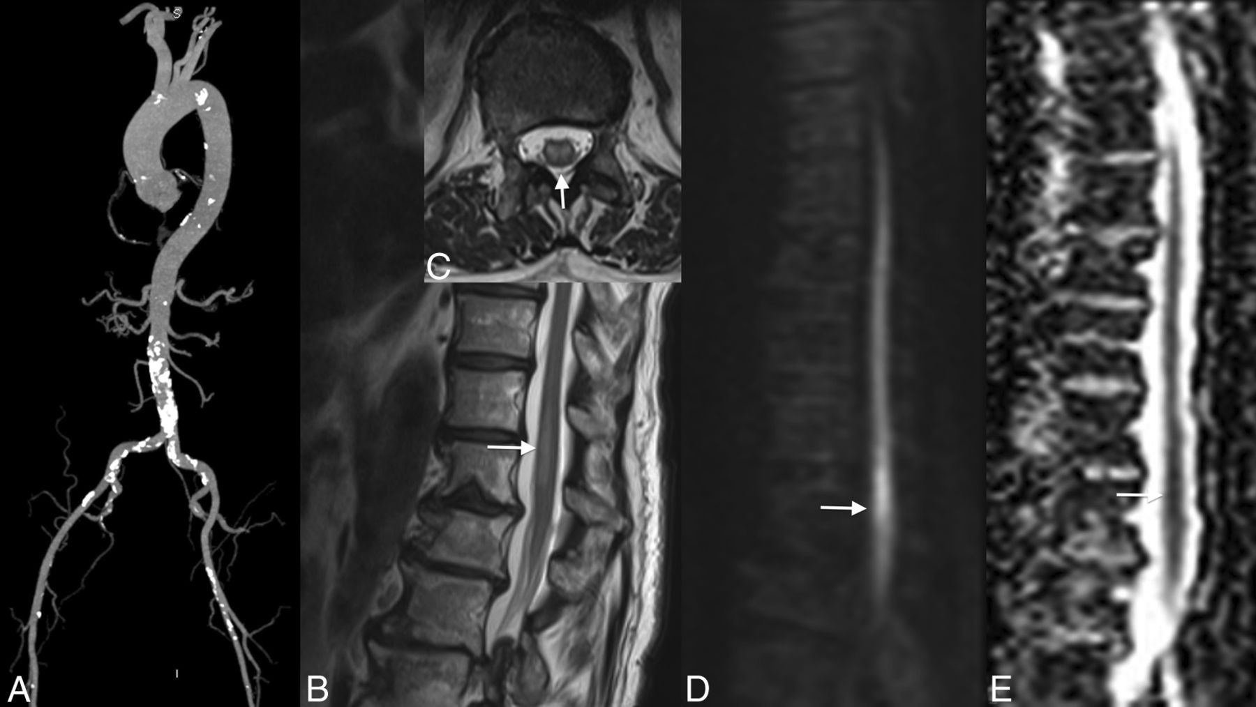
  • Fig 3.
    • Download figure
    • Open in new tab
    • Download powerpoint
    Fig 3.

    Evolution of ischemia. The first MR image shows the subtle signal anomaly on T2 and diffusion sequences (arrows, A–C). Follow-up 48 hours later shows an important tumefaction and high signal on T2WI associated with a restriction of diffusion of the cervical spinal cord at the C4–C7 levels (arrows, D–G).

  • Fig 4.
    • Download figure
    • Open in new tab
    • Download powerpoint
    Fig 4.

    Venous infarction in a patient with epidural and paraspinal abscesses. Note large intramedullary high signal on T2 of the cervical spinal cord (A). T1WI with contrast medium demonstrates an intramedullary enhancement (B and C), the anterior (arrows, B) and posterior epidural (white arrowhead, B), and paraspinal abscesses (black arrowhead, B). Note enhancement on axial T1 of both sides of the median line, reflecting venous ischemia.
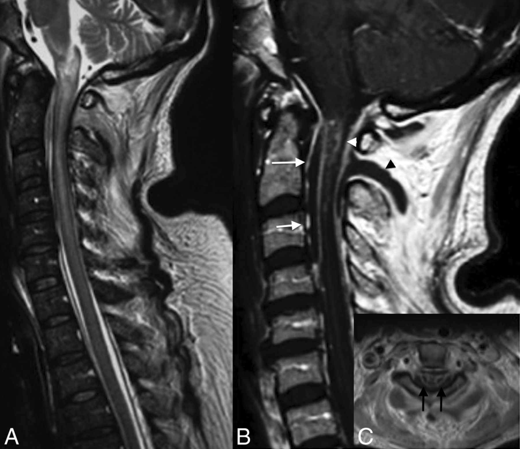
  • Fig 5.
    • Download figure
    • Open in new tab
    • Download powerpoint
    Fig 5.

    Cervical spinal canal stenosis and venous infarction. Note the cervical spinal canal stenosis from C4 to C6 due to cervical spondylosis (asterisks, A) and the intramedullary high signal on T2WI (arrow, B) at the same level with the “snake eye” appearance on axial T2WI (arrows, C).

  • Fig 6.
    • Download figure
    • Open in new tab
    • Download powerpoint
    Fig 6.

    Subacute ischemia. Note the slight hypersignal of the spinal anterior territory at the level of C4–C6 on T2WI (arrows, A and E), associated with a restriction of diffusion (arrows, C and D) and enhancement (arrows, D).

Tables

  • Figures
    • View popup
    Table 1:

    Technical MR imaging parameters of the spinal cord protocol at 3T

    SequencesTR (ms)TE (ms)Section Thickness (mm)B0
    SE T1670103
    SE T240001283
    STIR58601083
    Axial GE T2450173
    Axial SE T240001243
    Diffusion2600683b=500–700
    DTI2600732b=500–800, 20–25 directions
    • Note:—SE indicates spin-echo; GE, gradient-echo.

    • View popup
    Table 2:

    Technical MR imaging parameters of the spinal cord protocol at 1.5T

    SequencesTR (ms)TE (ms)Section Thickness (mm)B0
    SE T1590103
    SE T23270713
    STIR3000383
    Axial GE T2590243
    Axial SE T24640793
    Diffusion6000673b=500–700
    DTI3200673b=500–800, 20–25 directions
    • Note:—SE indicates spin-echo; GE, gradient-echo.

PreviousNext
Back to top

In this issue

American Journal of Neuroradiology: 36 (5)
American Journal of Neuroradiology
Vol. 36, Issue 5
1 May 2015
  • Table of Contents
  • Index by author
  • Complete Issue (PDF)
Advertisement
Print
Download PDF
Email Article

Thank you for your interest in spreading the word on American Journal of Neuroradiology.

NOTE: We only request your email address so that the person you are recommending the page to knows that you wanted them to see it, and that it is not junk mail. We do not capture any email address.

Enter multiple addresses on separate lines or separate them with commas.
Spinal Cord Ischemia: Practical Imaging Tips, Pearls, and Pitfalls
(Your Name) has sent you a message from American Journal of Neuroradiology
(Your Name) thought you would like to see the American Journal of Neuroradiology web site.
CAPTCHA
This question is for testing whether or not you are a human visitor and to prevent automated spam submissions.
Cite this article
M.I. Vargas, J. Gariani, R. Sztajzel, I. Barnaure-Nachbar, B.M. Delattre, K.O. Lovblad, J.-L. Dietemann
Spinal Cord Ischemia: Practical Imaging Tips, Pearls, and Pitfalls
American Journal of Neuroradiology May 2015, 36 (5) 825-830; DOI: 10.3174/ajnr.A4118

Citation Manager Formats

  • BibTeX
  • Bookends
  • EasyBib
  • EndNote (tagged)
  • EndNote 8 (xml)
  • Medlars
  • Mendeley
  • Papers
  • RefWorks Tagged
  • Ref Manager
  • RIS
  • Zotero
0 Responses
Respond to this article
Share
Bookmark this article
Spinal Cord Ischemia: Practical Imaging Tips, Pearls, and Pitfalls
M.I. Vargas, J. Gariani, R. Sztajzel, I. Barnaure-Nachbar, B.M. Delattre, K.O. Lovblad, J.-L. Dietemann
American Journal of Neuroradiology May 2015, 36 (5) 825-830; DOI: 10.3174/ajnr.A4118
del.icio.us logo Twitter logo Facebook logo Mendeley logo
  • Tweet Widget
  • Facebook Like
  • Google Plus One
Purchase

Jump to section

  • Article
    • Abstract
    • Footnotes
    • REFERENCES
  • Figures & Data
  • Info & Metrics
  • Responses
  • References
  • PDF

Related Articles

  • No related articles found.
  • PubMed
  • Google Scholar

Cited By...

  • Long-term Outcomes After Periprocedural and Spontaneous Spinal Cord Infarctions: A Population-Based Cohort Study
  • The Dominant Anterior Thoracic Artery of the Spinal Cord
  • Spontaneous spinal cord infarction: a practical approach
  • Reader Response: Teaching NeuroImage: Leber Hereditary Optic Neuropathy With Longitudinal Spinal Cord Lesion Mimicking Spinal Cord Infarction
  • Reversal of Acute Spinal Cord Ischemia by Intravenous Thrombolysis
  • Utility of MRI Enhancement Pattern in Myelopathies With Longitudinally Extensive T2 Lesions
  • Pediatric Spinal Cord Diseases
  • Spinal cord infarction in a young patient with methamphetamine abuse
  • Involvement of the Spinal Cord in Primary Mitochondrial Disorders: A Neuroimaging Mimicker of Inflammation and Ischemia in Children
  • Anatomy of the Great Posterior Radiculomedullary Artery
  • Acute extensive myelopathy after single heroin and cocaine exposure in a patient with toxicological evidence of long-term drug abstinence
  • Anterior spinal cord syndrome as a rare complication of acute bacterial meningitis in an adult
  • A practical approach to the diagnosis of spinal cord lesions
  • Is Catheter Angiography Still Necessary for the Follow-Up of Spinal Malformations after Treatment?
  • Clinical Reasoning: A young woman with respiratory failure, hearing loss, and paraplegia
  • Crossref
  • Google Scholar

This article has not yet been cited by articles in journals that are participating in Crossref Cited-by Linking.

More in this TOC Section

  • An Atlas of Neonatal Neurovascular Imaging Anatomy as Depicted with Microvascular Imaging: The Intracranial Arteries
  • An Atlas of Neonatal Neurovascular Imaging Anatomy as Depicted with Microvascular Imaging: The Intracranial Veins
  • Clinical Translation of Hyperpolarized 13C Metabolic Probes for Glioma Imaging
Show more Review Articles

Similar Articles

Advertisement

Indexed Content

  • Current Issue
  • Accepted Manuscripts
  • Article Preview
  • Past Issues
  • Editorials
  • Editor's Choice
  • Fellows' Journal Club
  • Letters to the Editor
  • Video Articles

Cases

  • Case Collection
  • Archive - Case of the Week
  • Archive - Case of the Month
  • Archive - Classic Case

More from AJNR

  • Trainee Corner
  • Imaging Protocols
  • MRI Safety Corner
  • Book Reviews

Multimedia

  • AJNR Podcasts
  • AJNR Scantastics

Resources

  • Turnaround Time
  • Submit a Manuscript
  • Submit a Video Article
  • Submit an eLetter to the Editor/Response
  • Manuscript Submission Guidelines
  • Statistical Tips
  • Fast Publishing of Accepted Manuscripts
  • Graphical Abstract Preparation
  • Imaging Protocol Submission
  • Evidence-Based Medicine Level Guide
  • Publishing Checklists
  • Author Policies
  • Become a Reviewer/Academy of Reviewers
  • News and Updates

About Us

  • About AJNR
  • Editorial Board
  • Editorial Board Alumni
  • Alerts
  • Permissions
  • Not an AJNR Subscriber? Join Now
  • Advertise with Us
  • Librarian Resources
  • Feedback
  • Terms and Conditions
  • AJNR Editorial Board Alumni

American Society of Neuroradiology

  • Not an ASNR Member? Join Now

© 2025 by the American Society of Neuroradiology All rights, including for text and data mining, AI training, and similar technologies, are reserved.
Print ISSN: 0195-6108 Online ISSN: 1936-959X

Powered by HighWire