
Article Figures & Data
Figures
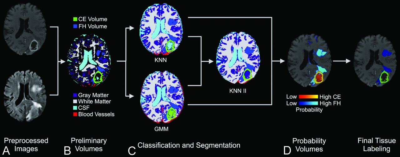
- Fig 1.
Work flow for iterative probabilistic voxel labeling. A, Downloaded TCIA images were preprocessed. B, Preliminary segmentation was performed to generate conservative yet highly specific preliminary volumes. C, In the classification step, these volumes were used to train the GMM and KNN probabilistic classifiers. The consensus of KNN and GMM classification was resampled and used to train a new classifier (KNN II), which assigned voxel tissue labels. The classifiers integrated their respective outputs to generate tissue-specific probability volumes. D, The voxels were assigned on the basis of their greatest probability of membership to a tissue label, and a voxel continuity filter was applied to eliminate clusters of less than 150 continuous voxels.
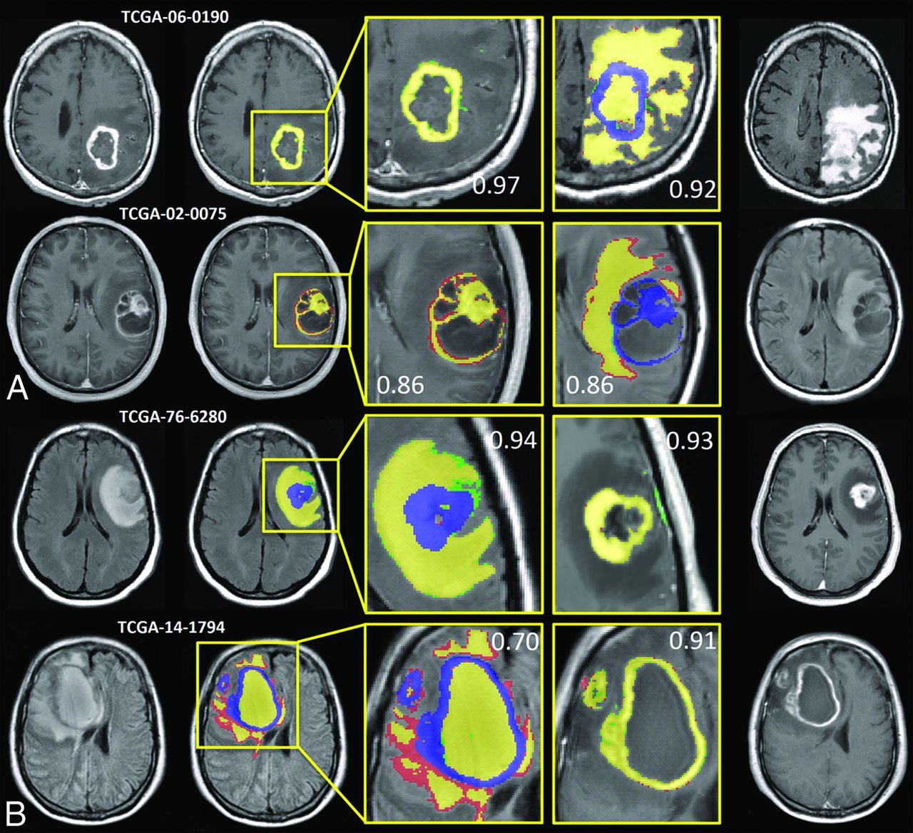
- Fig 2.
IPVL segment volumes that are highly analogous to operator-defined volumes. Results from 4 subjects representing the highest and lowest Dice similarity coefficient scores for CEV and FHV segmentations are shown. A, The highest DICE (top) and lowest DICE (bottom) examples of IPVL-segmented CEVs relative to operator-defined volumes are shown. The corresponding FHV segmentation results are shown (right) to demonstrate that CEV segmentations are independent of FHV segmentations. B, The highest DICE (top) and lowest DICE (bottom) examples of IPVL-segmented FHV relative to operator-defined volumes are shown. The corresponding CEV segmentation results are shown as well (right) to demonstrate that FHV segmentations are independent of CEV segmentations. Yellow indicates regions of intersection between operator and IPVL-defined volumes; red, operator-defined volume only; green, IPVL-defined volume only. Corresponding CEV segmentations are overlaid in blue on FLAIR images for clarity.
- Fig 3.
Quantitative comparison between IPVL-defined volumes and operator-derived volumes compared with interoperator comparisons. A and B, DICE comparisons for 2 sets of IPVL-defined and operator-defined CEVs are shown. DICE scores were calculated comparing CEVs generated by IPVL, operator 1, and operator 2. C and D, DICE score comparisons for IPVL-defined and operator-defined FHVs. DICE scores were calculated comparing FHVs generated by IPVL, operator 1, and operator 2.
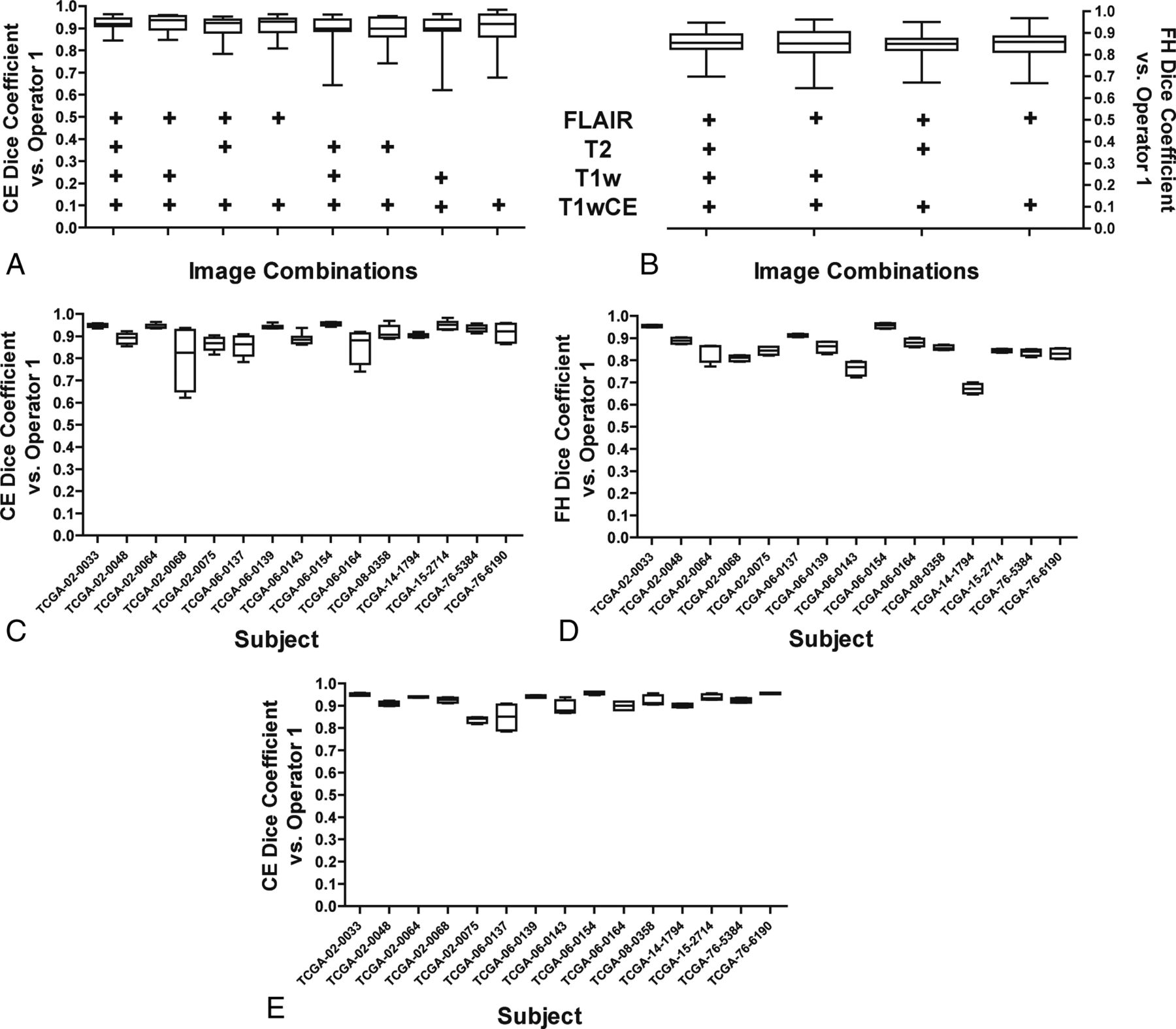
- Fig 4.
Effects of removing image sequences on IPVL segmentation. A, Select image sequences (such as T1WI) were removed before IPVL CEV segmentations for each subject. The image sequences that were available during IPVL segmentation are indicated by a plus sign. DICE scores were calculated for the resultant CEVs relative to operator 1–defined CEVs. The distribution of DICE scores across all subjects because of image sequence removal is shown as a boxplot. B, Select image sequences were removed before IPVL FHV segmentations. DICE scores were calculated for the resultant FHV segmentations relative to operator 1–defined FHVs. The distribution of DICE scores across all subjects due to image-sequence removal is shown as a boxplot. C, A boxplot demonstrates the range of DICE scores for IPVL-segmented CEVs relative to operator-defined CEVs per patient for all image combinations tested. D, A boxplot demonstrates the range of DICE scores for IPVL-segmented FHVs relative to operator-defined FHVs per patient for all image combinations tested. E, A boxplot demonstrates the range of DICE scores for IPVL-segmented CEVs relative to operator-defined CEVs per patient when only T1WI and FLAIR source images were used for IPVL segmentation.


