Advertisement

Main menu

  • Home
  • Content
    • Current Issue
    • Accepted Manuscripts
    • Article Preview
    • Past Issue Archive
    • Video Articles
    • AJNR Case Collection
    • Case of the Week Archive
    • Case of the Month Archive
    • Classic Case Archive
  • Special Collections
    • AJNR Awards
    • Low-Field MRI
    • Alzheimer Disease
    • ASNR Foundation Special Collection
    • Photon-Counting CT
    • View All
  • Multimedia
    • AJNR Podcasts
    • AJNR SCANtastic
    • Trainee Corner
    • MRI Safety Corner
    • Imaging Protocols
  • For Authors
    • Submit a Manuscript
    • Submit a Video Article
    • Submit an eLetter to the Editor/Response
    • Manuscript Submission Guidelines
    • Statistical Tips
    • Fast Publishing of Accepted Manuscripts
    • Graphical Abstract Preparation
    • Imaging Protocol Submission
    • Author Policies
  • About Us
    • About AJNR
    • Editorial Board
    • Editorial Board Alumni
  • More
    • Become a Reviewer/Academy of Reviewers
    • Subscribers
    • Permissions
    • Alerts
    • Feedback
    • Advertisers
    • ASNR Home

User menu

  • Alerts
  • Log in

Search

  • Advanced search
American Journal of Neuroradiology

American Journal of Neuroradiology

ASHNR American Society of Functional Neuroradiology ASHNR American Society of Pediatric Neuroradiology ASSR
  • Alerts
  • Log in

Advanced Search

  • Home
  • Content
    • Current Issue
    • Accepted Manuscripts
    • Article Preview
    • Past Issue Archive
    • Video Articles
    • AJNR Case Collection
    • Case of the Week Archive
    • Case of the Month Archive
    • Classic Case Archive
  • Special Collections
    • AJNR Awards
    • Low-Field MRI
    • Alzheimer Disease
    • ASNR Foundation Special Collection
    • Photon-Counting CT
    • View All
  • Multimedia
    • AJNR Podcasts
    • AJNR SCANtastic
    • Trainee Corner
    • MRI Safety Corner
    • Imaging Protocols
  • For Authors
    • Submit a Manuscript
    • Submit a Video Article
    • Submit an eLetter to the Editor/Response
    • Manuscript Submission Guidelines
    • Statistical Tips
    • Fast Publishing of Accepted Manuscripts
    • Graphical Abstract Preparation
    • Imaging Protocol Submission
    • Author Policies
  • About Us
    • About AJNR
    • Editorial Board
    • Editorial Board Alumni
  • More
    • Become a Reviewer/Academy of Reviewers
    • Subscribers
    • Permissions
    • Alerts
    • Feedback
    • Advertisers
    • ASNR Home
  • Follow AJNR on Twitter
  • Visit AJNR on Facebook
  • Follow AJNR on Instagram
  • Join AJNR on LinkedIn
  • RSS Feeds

AJNR Awards, New Junior Editors, and more. Read the latest AJNR updates

Review ArticleReview Article
Open Access

Current and Emerging MR Imaging Techniques for the Diagnosis and Management of CSF Flow Disorders: A Review of Phase-Contrast and Time–Spatial Labeling Inversion Pulse

S. Yamada, K. Tsuchiya, W.G. Bradley, M. Law, M.L. Winkler, M.T. Borzage, M. Miyazaki, E.J. Kelly and J.G. McComb
American Journal of Neuroradiology April 2015, 36 (4) 623-630; DOI: https://doi.org/10.3174/ajnr.A4030
S. Yamada
aFrom the Department of Neurosurgery (S.Y.), Toshiba Rinkan Hospital, Sagamihara, Kanagawa, Japan
  • Find this author on Google Scholar
  • Find this author on PubMed
  • Search for this author on this site
K. Tsuchiya
bDepartment of Radiology (K.T.), Kyorin University, Mitaka, Tokyo, Japan
  • Find this author on Google Scholar
  • Find this author on PubMed
  • Search for this author on this site
W.G. Bradley
cDepartment of Radiology (W.G.B.), University of California, San Diego, San Diego, California
  • Find this author on Google Scholar
  • Find this author on PubMed
  • Search for this author on this site
M. Law
dDepartment of Neuroradiology (M.L.), University of Southern California, Los Angeles, California
  • Find this author on Google Scholar
  • Find this author on PubMed
  • Search for this author on this site
M.L. Winkler
eSteinberg Diagnostic Imaging Center (M.L.W.), Las Vegas, Nevada
  • Find this author on Google Scholar
  • Find this author on PubMed
  • Search for this author on this site
M.T. Borzage
fDivision of Neuroradiology (M.T.B.), Department of Radiology, Institute for Maternal Fetal Health, Children's Hospital Los Angeles, Los Angeles, California
gDepartment of Biomedical Engineering (M.T.B.), USC Viterbi School of Engineering, University of Southern California, Los Angeles, California
  • Find this author on Google Scholar
  • Find this author on PubMed
  • Search for this author on this site
M. Miyazaki
hToshiba Medical Research Institute (M.M.), Vernon Hills, Illinois
  • Find this author on Google Scholar
  • Find this author on PubMed
  • Search for this author on this site
E.J. Kelly
iToshiba America Medical Systems Inc (E.J.K.), Tustin, California
  • Find this author on Google Scholar
  • Find this author on PubMed
  • Search for this author on this site
J.G. McComb
jDivision of Neurosurgery (J.G.M.), Children's Hospital Los Angeles, Los Angeles, California
kDepartment of Neurological Surgery (J.G.M.), Keck School of Medicine, University of Southern California, Los Angeles, California.
  • Find this author on Google Scholar
  • Find this author on PubMed
  • Search for this author on this site
  • Article
  • Figures & Data
  • Supplemental
  • Info & Metrics
  • Responses
  • References
  • PDF
Loading

Article Figures & Data

Figures

  • Fig 1.
    • Download figure
    • Open in new tab
    • Download powerpoint
    Fig 1.

    A 26-year-old healthy male volunteer. A, Geometry for the oblique-axial CSF phase-contrast scan. The section is positioned axially at a 90° angle through the aqueduct of Sylvius (rectangle, A). Aliasing (B) occurs if a phase value is greater than the maximum expected velocity, causing the phase to wrap back to a multiple of π, appearing black (black arrow) when it should appear white (or vice versa). Uncorrected (dotted line) and corrected (solid line) flow waveforms in milliliters per second represent bidirectional flow through the aqueduct (C). Aliasing can be corrected off-line by adding a multiple of 2 × π × Venc to aliased pixels.

  • Fig 2.
    • Download figure
    • Open in new tab
    • Download powerpoint
    Fig 2.

    An 18-year-old healthy female volunteer. A, A series of midline sagittal images depicting pulsatile CSF flow, where flow magnitude and direction are represented as gray-scale. Flow changes from positive to negative and back to positive (white indicates peak caudal flow; black, peak cranial flow). B, Depiction of a series of axial images at the level of the aqueduct (arrow). In both series, every odd phase of the 16 cardiac phases that were acquired is displayed.
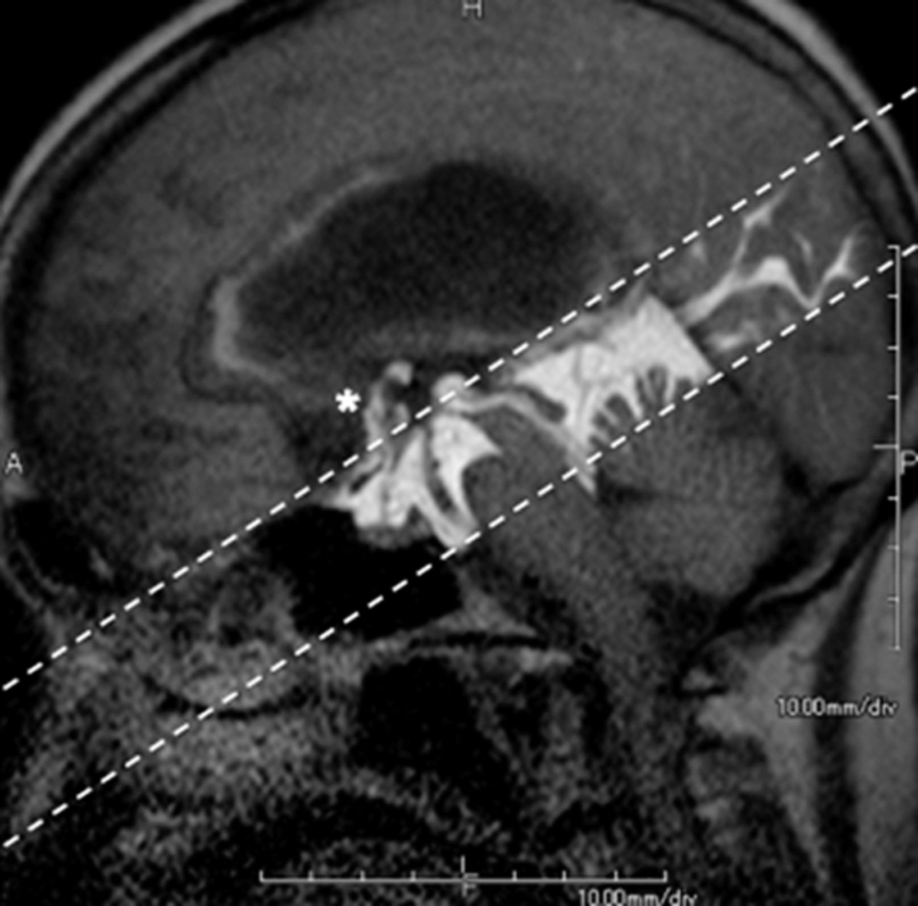
  • Fig 3.
    • Download figure
    • Open in new tab
    • Download powerpoint
    Fig 3.

    Hydrocephalus in a 74-year-old woman. The patient had undergone endoscopic third ventriculostomy. Patency of the fenestration on the floor of the third ventricle is readily and noninvasively confirmed postsurgery by the presence of CSF flow between the third ventricle and the basal cisterns (asterisk) emerging from the tagged region (dotted lines). (See On-line Video 1, which demonstrates postsurgical CSF flow between the third ventricle and the basal cisterns.)

  • Fig 4.
    • Download figure
    • Open in new tab
    • Download powerpoint
    Fig 4.

    Idiopathic normal pressure hydrocephalus in a 78-year-old man. Time-SLIP has consistently shown the presence of reflux flow from the third ventricle into the lateral ventricle in adult patients without hydrocephalus (A). This flow is shown to be restricted in NPH (B). Time-SLIP in the same patient confirms that this flow is restored after surgical intervention by inserting a CSF diverting shunt (C, artifacts on the right are from the shunt valve). (See On-line Videos 2 and 3, which demonstrate restricted flow and restored flow pre- and postsurgery, respectively.)

  • Fig 5.
    • Download figure
    • Open in new tab
    • Download powerpoint
    Fig 5.

    Chiari malformation in a 43-year-old woman pre- (top row) and post- (bottom row) surgery, shown at incremental TIs. Obliteration of the subarachnoid space at the craniocervical junction is associated with Chiari I malformation and a syrinx (top row, arrow). Following craniocervical decompression (bottom row, arrow), Time-SLIP shows CSF flow ventral to the brain stem and cervical spinal cord and a decrease in the size of the syrinx (bold arrow). (See On-line Videos 4 and 5, which demonstrate restricted flow and restored flow with a decrease in the size of the syrinx pre- and postsurgery, respectively.)

PreviousNext
Back to top

In this issue

American Journal of Neuroradiology: 36 (4)
American Journal of Neuroradiology
Vol. 36, Issue 4
1 Apr 2015
  • Table of Contents
  • Index by author
  • Complete Issue (PDF)
Advertisement
Print
Download PDF
Email Article

Thank you for your interest in spreading the word on American Journal of Neuroradiology.

NOTE: We only request your email address so that the person you are recommending the page to knows that you wanted them to see it, and that it is not junk mail. We do not capture any email address.

Enter multiple addresses on separate lines or separate them with commas.
Current and Emerging MR Imaging Techniques for the Diagnosis and Management of CSF Flow Disorders: A Review of Phase-Contrast and Time–Spatial Labeling Inversion Pulse
(Your Name) has sent you a message from American Journal of Neuroradiology
(Your Name) thought you would like to see the American Journal of Neuroradiology web site.
CAPTCHA
This question is for testing whether or not you are a human visitor and to prevent automated spam submissions.
Cite this article
S. Yamada, K. Tsuchiya, W.G. Bradley, M. Law, M.L. Winkler, M.T. Borzage, M. Miyazaki, E.J. Kelly, J.G. McComb
Current and Emerging MR Imaging Techniques for the Diagnosis and Management of CSF Flow Disorders: A Review of Phase-Contrast and Time–Spatial Labeling Inversion Pulse
American Journal of Neuroradiology Apr 2015, 36 (4) 623-630; DOI: 10.3174/ajnr.A4030

Citation Manager Formats

  • BibTeX
  • Bookends
  • EasyBib
  • EndNote (tagged)
  • EndNote 8 (xml)
  • Medlars
  • Mendeley
  • Papers
  • RefWorks Tagged
  • Ref Manager
  • RIS
  • Zotero
0 Responses
Respond to this article
Share
Bookmark this article
Current and Emerging MR Imaging Techniques for the Diagnosis and Management of CSF Flow Disorders: A Review of Phase-Contrast and Time–Spatial Labeling Inversion Pulse
S. Yamada, K. Tsuchiya, W.G. Bradley, M. Law, M.L. Winkler, M.T. Borzage, M. Miyazaki, E.J. Kelly, J.G. McComb
American Journal of Neuroradiology Apr 2015, 36 (4) 623-630; DOI: 10.3174/ajnr.A4030
del.icio.us logo Twitter logo Facebook logo Mendeley logo
  • Tweet Widget
  • Facebook Like
  • Google Plus One
Purchase

Jump to section

  • Article
    • Abstract
    • ABBREVIATIONS:
    • Footnotes
    • REFERENCES
  • Figures & Data
  • Supplemental
  • Info & Metrics
  • Responses
  • References
  • PDF

Related Articles

  • PubMed
  • Google Scholar

Cited By...

  • Optimizing scan efficiency of T1-weighted imaging for whole-brain intracranial vessel wall imaging
  • Advection versus diffusion in brain ventricular transport
  • The Spatial Patterns and Determinants of Cerebrospinal Fluid Circulation in the Human Brain
  • CSF Flow and Spinal Cord Motion in Patients With Spontaneous Intracranial Hypotension: A Phase Contrast MRI Study
  • Crossref
  • Google Scholar

This article has not yet been cited by articles in journals that are participating in Crossref Cited-by Linking.

More in this TOC Section

  • Giant Cell Arteritis: Important Imaging Findings
  • DCE MRI in Spinal Disease Assessment
  • Cerebrovascular Anomalies in Fetal Imaging
Show more Review Article

Similar Articles

Advertisement

Indexed Content

  • Current Issue
  • Accepted Manuscripts
  • Article Preview
  • Past Issues
  • Editorials
  • Editor's Choice
  • Fellows' Journal Club
  • Letters to the Editor
  • Video Articles

Cases

  • Case Collection
  • Archive - Case of the Week
  • Archive - Case of the Month
  • Archive - Classic Case

More from AJNR

  • Trainee Corner
  • Imaging Protocols
  • MRI Safety Corner
  • Book Reviews

Multimedia

  • AJNR Podcasts
  • AJNR Scantastics

Resources

  • Turnaround Time
  • Submit a Manuscript
  • Submit a Video Article
  • Submit an eLetter to the Editor/Response
  • Manuscript Submission Guidelines
  • Statistical Tips
  • Fast Publishing of Accepted Manuscripts
  • Graphical Abstract Preparation
  • Imaging Protocol Submission
  • Evidence-Based Medicine Level Guide
  • Publishing Checklists
  • Author Policies
  • Become a Reviewer/Academy of Reviewers
  • News and Updates

About Us

  • About AJNR
  • Editorial Board
  • Editorial Board Alumni
  • Alerts
  • Permissions
  • Not an AJNR Subscriber? Join Now
  • Advertise with Us
  • Librarian Resources
  • Feedback
  • Terms and Conditions
  • AJNR Editorial Board Alumni

American Society of Neuroradiology

  • Not an ASNR Member? Join Now

© 2025 by the American Society of Neuroradiology All rights, including for text and data mining, AI training, and similar technologies, are reserved.
Print ISSN: 0195-6108 Online ISSN: 1936-959X

Powered by HighWire