
Article Figures & Data
Figures
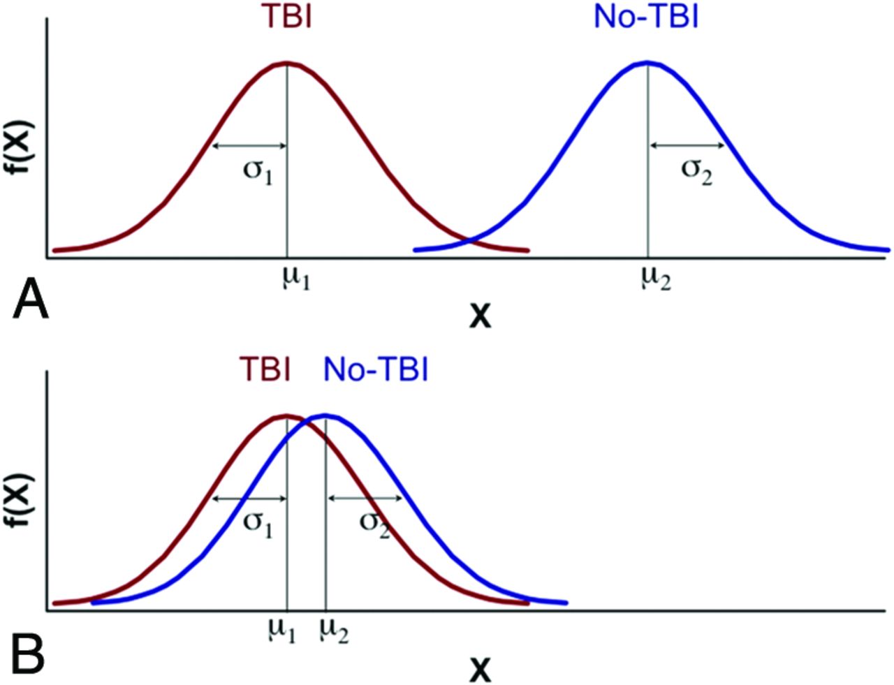
- Figure.
Hypothetic distribution of an advanced neuroimaging-dependent measure for TBI (red) and non-TBI (blue) groups. A, Idealized separation of distributions between the TBI and non-TBI groups due to a very selective “normal” non-TBI control group (ie, supernormal, with no history of TBI, and no neurologic, psychiatric, or other detectable abnormalities), which would be amenable to conventional statistical analyses based on the general linear model. B, Marked overlap of distributions between the TBI and non-TBI groups due to a non-TBI control group comprising subjects with pre-existing abnormalities present in the general population, which would be unlikely to yield a statistically significant differentiation by using the general linear model. Despite relatively marked overlap between distributions, classification approaches may be able to identify features unique to each group and therefore discriminate whether an individual belongs in the TBI or non-TBI group. To pursue implementation of such a binary classification, it will be necessary to characterize the variability associated with neuroimaging methods expected from the general population in the absence of TBI, which can be facilitated by constructing a large comprehensive normative data base.

Tables
NDAR FITBIR LORIS LONI IDA DART XNAT INDI/FCP COINS BIRN HID NIDB Core developers National Institutes of Health National Institutes of Health NINDS Montreal Neurological Institute University of Southern California American College of Radiology Washington University NITRC MIND Research Network National Institutes of Health NIGMS Olin Neuropsychiatry Research Center Types of data stored Neuroimaging Neuroimaging Neuroimaging Neuroimaging Imaging Neuroimaging Neuroimaging Neuroimaging Neuroimaging Neuroimaging Phenomic Phenomic Phenomic Phenomic Phenomic Phenomic Phenomic Phenomic Phenomic Phenomic Genetic Demographic Genetic Genetic Demographic Demographic Demographic Demographic Demographic Demographic Other: extensible Demographic Demographic Other: extensible Other: extensible Other: extensible Other: extensible Other: extensible Other: extensible Other: extensible Other: extensible Common data elements Required use Required use Flexible usage Flexible usage Flexible usage Flexible usage None None None None Workflow Yes Continuous project coordination No No Yes No Yes No No No No No Monitoring of data acquisition No No Yes No Yes No No No No Yes Data cleaning No No Yes Yes Yes Yes No No Yes Yes Data storage Developer hosting Developer hosting Developer hosting or local storage Developer hosting or local storage Developer hosting or local storage Local storage Developer hosting Developer hosting Local storage Local storage Data query Yes Yes Yes Yes Yes Yes No Yes Yes Yes Data download Yes Yes Yes Yes Yes Yes Yes Yes Yes Yes Interface with external data-processing No No Yes Yes Yes Yes No No Yes No Integrated data-processing tools No No No Yes Yes? Yes No No No No Disease-specific Yes: autism Yes: TBI No No No No No No No No Major datasets shared Data from 77,575 individuals related to autism In development ABIDE ABIDE Data from 150,000 individuals across a wide array of clinical domains HCP ABIDE ABIDE ABIDE ABIDE AddNeuroMed AIBL ADHD-200 MCIC Schizophrenic Brainscape CBRAIN ADNI CoRR NKI Rockland ELUDE CITA ADNIDOD NKI Rockland fBIRN GUSTO HCP MIRIAD IBIS ICBM OASIS MAVAN PPMI NeuGrid NeuroDevNet Access policies Government-defined with institutional agreement Government-defined with institutional agreement User-specified User-specified User-specified User-specified Unrestricted User-specified User-specified User-specified Data contribution Required as a condition of many NIH grants Required as a condition of many NIH grants Required for some multisite projects Required for some multisite projects Required for some multisite projects Voluntary Voluntary Voluntary Voluntary Voluntary Voluntary Voluntary Voluntary Note:—NDAR indicates National Database for Autism Research; NIGMS, National Institute of General Medical Sciences; INDI/FCP, International Neuroimaging Data Sharing Initiative/1000 Functional Connectome Project; IDA, Image Data Archive; COINS, Collaborative Informatics Neuroimaging Suite; NIDB, Neuroinformatics Database; HCP, Human Connectome Project; AIBL, Australian Imaging, Biomarker and Lifestyle Flagship Study of Ageing; ELUDE, Efficient Longitudinal Upload of Depression in the Elderly; CITA, Centro de Investigación y Terapias Avanzadas; ADNIDOD, Study of Brain Aging in Vietnam War Veterans; GUSTO, Singapore's birth cohort study; MIRIAD, Multisite Imaging Research In the Analysis of Depression; BIRN, Biomedical Informatics Research Network; fBIRN, Functional Bioinformatics Research Network; IBIS, Infant Imaging Brain Study; ICBM, International Consortium for Brain Mapping; OASIS, Open Access Structural Imaging Series; MAVAN, Maternal Adversity, Vulnerability, and Neurodevelopment; PPMI, Parkinson's Progressive Measures Initiative; XNAT, Extensible Neuroimaging Archive Toolkit; NINDS, National Institute of Neurologic Disorders and Stroke; ADHD, attention deficit/hyperactivity disorder; CoRR, Consortium for Reliability and Reproducibility; NKI Rockland, Nathan Kline Institute Rockland; MCIC, MIND Clinical Imaging Consortium.
- Table 2:
Core (required) demographic variables that should be collected for any normative data base
Recommended Core (Required) Variables CDE Variable Name Permissible Values Notes, Including Those from the CDE Web Site Age Age value 0–120 yr? For children younger than 1 year born at <36 weeks' gestation, it is recommended to also collect gestational age. Because of potential violation of privacy legislation and specifically HIPAA regulations, the calculated age should be recorded rather than using the actual date of birth. Gender Gender type Female, male, unknown, unspecified, not reported Note that “gender” refers to self-reported gender of the participant. Gender is the socially constructed identity of sex and is equated with phenotypic sex. Gender may differ from the sex of an individual determined genetically. The use of “gender” as opposed to “sex” is to facilitate the ease of self-report data collection. Handedness Hand preference type Left hand, right hand, both hands, unknown Hand which the participant/subject uses predominantly, not necessarily the hand he/she writes with exclusively Race Race USA category American Indian or Alaska native, Asian, black or African-American, Native Hawaiian or other Pacific Islander, white, unknown, not reported The patient's self-declared racial origination, independent of ethnic origination, using OMB-approved categories. We note that these may not be applicable to all non-USA regions. Ethnicity Ethnicity USA category Hispanic or Latino, not Hispanic or Latino, unknown, not reported Category of ethnicity the participant/subject most closely identifies with. We note that these may not be applicable to all non-USA regions. Socioeconomic status Education level USA-type Never attended/kindergarten only; 1st grade; 2nd grade; 3rd grade; 4th grade; 5th grade; 6th grade; 7th grade; 8th grade; 9th grade; 10th grade; 11th grade; 12th grade, no diploma; high school graduate; GED or equivalent; some college, no degree; associate degree: occupational/technical/vocational program; associate degree: academic program; bachelor's degree (eg, BA, AB, BS, BBA); master's degree (eg, MA, MS, MEng, MEd, MBA); professional school degree (eg, MD, DDS, DVM, JD); doctoral degree (eg, PhD, EdD); unknown Highest grade or level of school participant/subject has completed or the highest degree received. Note that for children, these should reflect the highest education level of the primary caregiver. Job classification category Official/manager, professional, technician, sales worker, administrative support worker, craft worker, operative, laborer/helper, service worker, social worker, unknown, none Category that classifies work performed by participant or, in the case of children, the work performed by the primary caregiver. Family income range $15,000 to $24,999, $25,000 to $34,999, $35,000 to $49,999, $50,000 to $74,999, $75,000 to $99,999, ≥$100,000, refused, unknown, <$15,000; Range, in US dollars, of the annual pretax, prededuction total income, of the household of which the participant/subject is a member Academic achievement Education service type Special education, regular education, none, unknown, early intervention Type of educational services received (as a child) Note:—GED indicates General Educational Development; HIPPA, Health Insurance Portability and Accountability Act; OBM, Office of Budget and Management.
Parameter “Core” “Preferred” No. 1 (High Angular Resolution) “Preferred” No. 2 (More b-Values) Orientation Axial Axial Axial Coil Any Phased array ≥8 channels Phased array ≥8 channels Readout EPI EPI EPI TR (ms) ∼9000 ∼9000 ∼9000 TE (ms) Min Min (<100) Min (<100) FOV (mm2) 256 × 256 or 350 (66% phase) 256 × 256 or 350 (66% phase) 256 × 256 or 350 (66% phase) Matrix size 128 × 128 128 × 128 128 × 128 Sections/thickness (mm) Any/≤3 59/2.7 59/2.7 Section gap (mm) 0 0 0 Voxel size (mm) ≤3 in all dimensions Isotropic 2.73 Isotropic 2.73 Directions ≥12 64 ≥12 Dual-echo Any No No Fat-suppression Any Yes Yes Phase-encode direction A to P A to P A to P BW (Hz/pixel) 1346 1346 1346 Parallel imaging factor Any 2 2 b-value (s/mm2) 2 (0, ∼1000) 2 (0, 1300) ≥3 (eg, 0, ∼1000, ∼2000) No. b=0 images 1 1 per 8 directions if allowed 1 per 8 directions if allowed Note:—Min indicates minimum; A to P, anterior to posterior; BW, bandwidth.


