Advertisement

Main menu

  • Home
  • Content
    • Current Issue
    • Accepted Manuscripts
    • Article Preview
    • Past Issue Archive
    • Video Articles
    • AJNR Case Collection
    • Case of the Week Archive
    • Case of the Month Archive
    • Classic Case Archive
  • Special Collections
    • AJNR Awards
    • Low-Field MRI
    • Alzheimer Disease
    • ASNR Foundation Special Collection
    • Photon-Counting CT
    • View All
  • Multimedia
    • AJNR Podcasts
    • AJNR SCANtastic
    • Trainee Corner
    • MRI Safety Corner
    • Imaging Protocols
  • For Authors
    • Submit a Manuscript
    • Submit a Video Article
    • Submit an eLetter to the Editor/Response
    • Manuscript Submission Guidelines
    • Statistical Tips
    • Fast Publishing of Accepted Manuscripts
    • Graphical Abstract Preparation
    • Imaging Protocol Submission
    • Author Policies
  • About Us
    • About AJNR
    • Editorial Board
    • Editorial Board Alumni
  • More
    • Become a Reviewer/Academy of Reviewers
    • Subscribers
    • Permissions
    • Alerts
    • Feedback
    • Advertisers
    • ASNR Home

User menu

  • Alerts
  • Log in

Search

  • Advanced search
American Journal of Neuroradiology

American Journal of Neuroradiology

ASHNR American Society of Functional Neuroradiology ASHNR American Society of Pediatric Neuroradiology ASSR
  • Alerts
  • Log in

Advanced Search

  • Home
  • Content
    • Current Issue
    • Accepted Manuscripts
    • Article Preview
    • Past Issue Archive
    • Video Articles
    • AJNR Case Collection
    • Case of the Week Archive
    • Case of the Month Archive
    • Classic Case Archive
  • Special Collections
    • AJNR Awards
    • Low-Field MRI
    • Alzheimer Disease
    • ASNR Foundation Special Collection
    • Photon-Counting CT
    • View All
  • Multimedia
    • AJNR Podcasts
    • AJNR SCANtastic
    • Trainee Corner
    • MRI Safety Corner
    • Imaging Protocols
  • For Authors
    • Submit a Manuscript
    • Submit a Video Article
    • Submit an eLetter to the Editor/Response
    • Manuscript Submission Guidelines
    • Statistical Tips
    • Fast Publishing of Accepted Manuscripts
    • Graphical Abstract Preparation
    • Imaging Protocol Submission
    • Author Policies
  • About Us
    • About AJNR
    • Editorial Board
    • Editorial Board Alumni
  • More
    • Become a Reviewer/Academy of Reviewers
    • Subscribers
    • Permissions
    • Alerts
    • Feedback
    • Advertisers
    • ASNR Home
  • Follow AJNR on Twitter
  • Visit AJNR on Facebook
  • Follow AJNR on Instagram
  • Join AJNR on LinkedIn
  • RSS Feeds

AJNR Awards, New Junior Editors, and more. Read the latest AJNR updates

Research ArticleSpine Imaging and Spine Image-Guided Interventions

Percutaneous Injection of Radiopaque Gelified Ethanol for the Treatment of Lumbar and Cervical Intervertebral Disk Herniations: Experience and Clinical Outcome in 80 Patients

M. Bellini, D.G. Romano, S. Leonini, I. Grazzini, C. Tabano, M. Ferrara, P. Piu, L. Monti and A. Cerase
American Journal of Neuroradiology March 2015, 36 (3) 600-605; DOI: https://doi.org/10.3174/ajnr.A4166
M. Bellini
aFrom the Neuroimaging and Neurointerventional Unit - NINT (M.B., D.G.R., S.L., M.F., L.M., A.C.)
  • Find this author on Google Scholar
  • Find this author on PubMed
  • Search for this author on this site
  • ORCID record for M. Bellini
D.G. Romano
aFrom the Neuroimaging and Neurointerventional Unit - NINT (M.B., D.G.R., S.L., M.F., L.M., A.C.)
  • Find this author on Google Scholar
  • Find this author on PubMed
  • Search for this author on this site
S. Leonini
aFrom the Neuroimaging and Neurointerventional Unit - NINT (M.B., D.G.R., S.L., M.F., L.M., A.C.)
  • Find this author on Google Scholar
  • Find this author on PubMed
  • Search for this author on this site
I. Grazzini
cDepartment of Medical, Surgical and Neuro Sciences, Diagnostic Imaging (I.G.), University of Siena, Siena, Italy
  • Find this author on Google Scholar
  • Find this author on PubMed
  • Search for this author on this site
C. Tabano
dUnit of Radiology (C.T.), Hospital of Arzignano, Vicenza, Italy.
  • Find this author on Google Scholar
  • Find this author on PubMed
  • Search for this author on this site
M. Ferrara
aFrom the Neuroimaging and Neurointerventional Unit - NINT (M.B., D.G.R., S.L., M.F., L.M., A.C.)
  • Find this author on Google Scholar
  • Find this author on PubMed
  • Search for this author on this site
P. Piu
bUnit of Neurology (P.P.), Department of Neurological and Neurosensorial Sciences, Hospital “Santa Maria alle Scotte,” Siena, Italy
  • Find this author on Google Scholar
  • Find this author on PubMed
  • Search for this author on this site
L. Monti
aFrom the Neuroimaging and Neurointerventional Unit - NINT (M.B., D.G.R., S.L., M.F., L.M., A.C.)
  • Find this author on Google Scholar
  • Find this author on PubMed
  • Search for this author on this site
A. Cerase
aFrom the Neuroimaging and Neurointerventional Unit - NINT (M.B., D.G.R., S.L., M.F., L.M., A.C.)
  • Find this author on Google Scholar
  • Find this author on PubMed
  • Search for this author on this site
  • Article
  • Figures & Data
  • Info & Metrics
  • Responses
  • References
  • PDF
Loading

Article Figures & Data

Figures

  • Fig 1.
    • Download figure
    • Open in new tab
    • Download powerpoint
    Fig 1.

    Progressive intervertebral disk filling of RGE. Real-time fluoroscopic images showing RGE inside the nucleus pulposus and in the herniated disk (arrows). Tungsten allows proper visualization of the material, including monitoring of possible leakage.

  • Fig 2.
    • Download figure
    • Open in new tab
    • Download powerpoint
    Fig 2.

    VAS scores recorded before and after percutaneous intradiskal injection of RGE. The left panel displays the boxplot of the data. Before treatment, the VAS scores were mostly concentrated on the upper values of the scale (median = 7; 25th percentile = 7; 75th percentile = 8); therefore, this distribution was negatively skewed (skewness = −0.29). The distribution of the VAS scores in the posttreatment survey (median = 3; 25th percentile = 2; 75th percentile = 4) became positively skewed (skewness = 0.91). No outliers were identified. The reduction of 4 points in the median of the VAS scores between pre- and posttreatment was significant for the nonparametric Wilcoxon matched-pairs signed rank test (W = −3076; P < .0001). The barplots in the right panel show the means and SDs of the data. The decrease of the means of the VAS scores from 7.16 ± 0.79 to 3.61 ± 1.72 was significant under the paired t test with 78 df (t = 17.61; P < .0001).
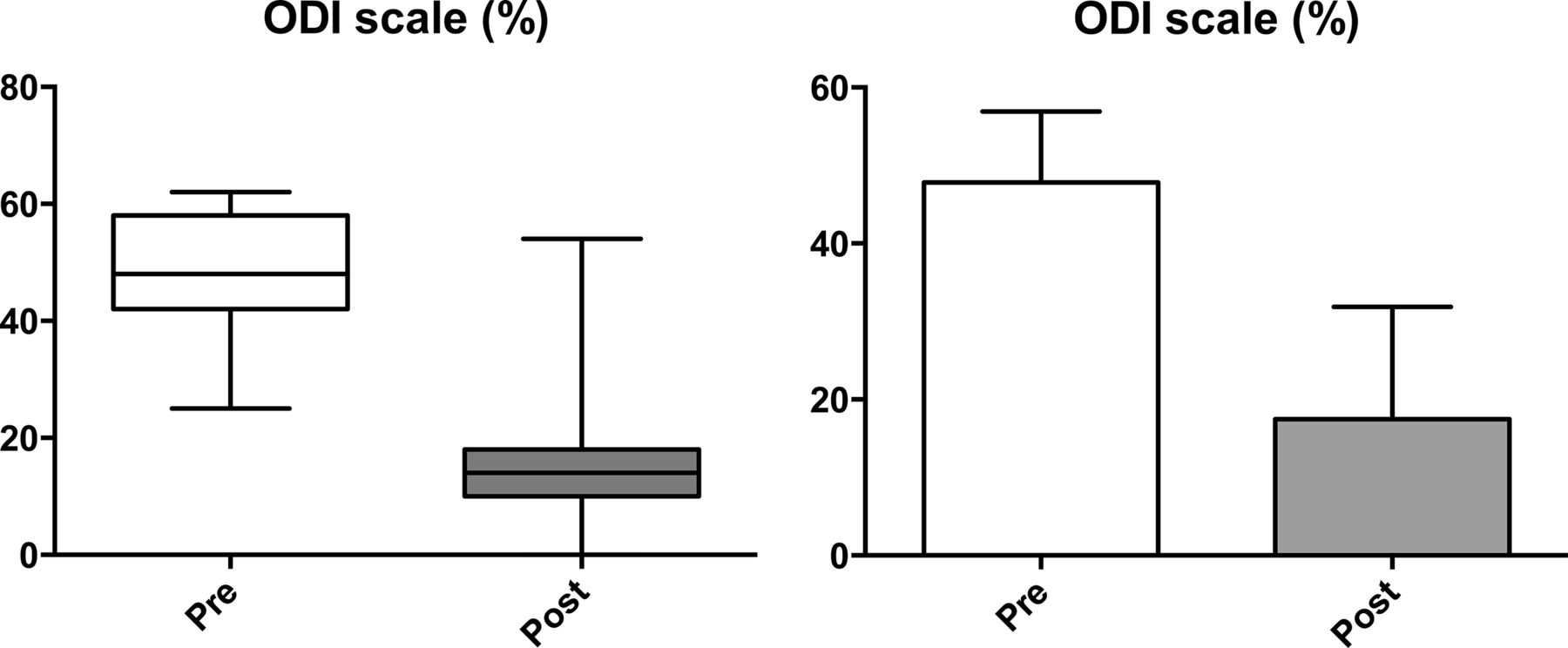
  • Fig 3.
    • Download figure
    • Open in new tab
    • Download powerpoint
    Fig 3.

    ODI scores recorded before and after percutaneous intradiskal injection of RGE. The index is expressed in percentage points and ranges from 0% to 100%. The lower limit corresponds to the absence of disability; the upper limit, to the maximum degree of disability (patients are bed-bound). The left panel displays the boxplot of the data. Before treatment, the distribution of ODI scores (median = 48%; 25th percentile = 42%; 75th percentile = 58%) was slightly negatively skewed (skewness = −0.05), and it became positively skewed (skewness = 1.21) in the posttreatment survey (median = 14%; 25th percentile = 10%; 75th percentile = 18%). No outliers were identified. Between the pre- and posttreatment periods, the medians of the VAS scores dropped by 34 percentage points. This decline was significant for the nonparametric Wilcoxon matched-pairs signed rank test (W = −3076; P < .0001). The barplots in the right panel show the means and SDs of the data. The decrease of the mean of the ODI scores from 47.85 ± 9.05 to 17.47 ± 14.41 was significant with the paired t test with 78 df (t = 17.71, P < .0001).

  • Fig 4.
    • Download figure
    • Open in new tab
    • Download powerpoint
    Fig 4.

    Treatment of a very large extruded LDH in a 58-year-old woman with left sciatica (VAS = 8). A, In a preoperative MR imaging study, sagittal and axial T2 sequences show a large uncontained and partially migrated disk herniation at L5–S1 (median left posterolateral), with considerable root involvement (white arrow). B, CT control shows the optimal distribution of RGE in a disk herniation without leakage (black arrow). The patient did not have significant symptoms (VAS = 3). C, MR imaging study obtained 6 months later shows complete dehydration and retraction of disk herniation. The patient was completely asymptomatic (VAS = 0).

  • Fig 5.
    • Download figure
    • Open in new tab
    • Download powerpoint
    Fig 5.

    Treatment of a CDH in a 45-year-old woman with left brachialgia (VAS = 7). A, T2-weighted sagittal and T2*-weighted axial MR images show a C4–C5 left posterolateral disk herniation. Note also other asymptomatic disk bulging at the C3–C4, C5–C6, and C6–C7 levels. Anteroposterior radiograph (B) and CT axial image (C) obtained at the end of the procedure confirm the proper distribution of RGE in the treated disk, especially in the herniated portion (arrows).

PreviousNext
Back to top

In this issue

American Journal of Neuroradiology: 36 (3)
American Journal of Neuroradiology
Vol. 36, Issue 3
1 Mar 2015
  • Table of Contents
  • Index by author
  • Complete Issue (PDF)
Advertisement
Print
Download PDF
Email Article

Thank you for your interest in spreading the word on American Journal of Neuroradiology.

NOTE: We only request your email address so that the person you are recommending the page to knows that you wanted them to see it, and that it is not junk mail. We do not capture any email address.

Enter multiple addresses on separate lines or separate them with commas.
Percutaneous Injection of Radiopaque Gelified Ethanol for the Treatment of Lumbar and Cervical Intervertebral Disk Herniations: Experience and Clinical Outcome in 80 Patients
(Your Name) has sent you a message from American Journal of Neuroradiology
(Your Name) thought you would like to see the American Journal of Neuroradiology web site.
CAPTCHA
This question is for testing whether or not you are a human visitor and to prevent automated spam submissions.
Cite this article
M. Bellini, D.G. Romano, S. Leonini, I. Grazzini, C. Tabano, M. Ferrara, P. Piu, L. Monti, A. Cerase
Percutaneous Injection of Radiopaque Gelified Ethanol for the Treatment of Lumbar and Cervical Intervertebral Disk Herniations: Experience and Clinical Outcome in 80 Patients
American Journal of Neuroradiology Mar 2015, 36 (3) 600-605; DOI: 10.3174/ajnr.A4166

Citation Manager Formats

  • BibTeX
  • Bookends
  • EasyBib
  • EndNote (tagged)
  • EndNote 8 (xml)
  • Medlars
  • Mendeley
  • Papers
  • RefWorks Tagged
  • Ref Manager
  • RIS
  • Zotero
0 Responses
Respond to this article
Share
Bookmark this article
Percutaneous Injection of Radiopaque Gelified Ethanol for the Treatment of Lumbar and Cervical Intervertebral Disk Herniations: Experience and Clinical Outcome in 80 Patients
M. Bellini, D.G. Romano, S. Leonini, I. Grazzini, C. Tabano, M. Ferrara, P. Piu, L. Monti, A. Cerase
American Journal of Neuroradiology Mar 2015, 36 (3) 600-605; DOI: 10.3174/ajnr.A4166
del.icio.us logo Twitter logo Facebook logo Mendeley logo
  • Tweet Widget
  • Facebook Like
  • Google Plus One
Purchase

Jump to section

  • Article
    • Abstract
    • ABBREVIATIONS:
    • Materials and Methods
    • Results
    • Discussion
    • Conclusions
    • Acknowledgments
    • Footnotes
    • REFERENCES
  • Figures & Data
  • Info & Metrics
  • Responses
  • References
  • PDF

Related Articles

  • No related articles found.
  • PubMed
  • Google Scholar

Cited By...

  • Polymer-assisted intratumoral delivery of ethanol: Preclinical investigation of safety and efficacy in a murine breast cancer model
  • Crossref
  • Google Scholar

This article has not yet been cited by articles in journals that are participating in Crossref Cited-by Linking.

More in this TOC Section

  • Optimization of Photon Counting CT Myelography
  • Characteristics of SIH Type I Culprit Lesions
  • Management Outcomes For VO Spine Biopsy
Show more Spine Imaging and Spine Image-Guided Interventions

Similar Articles

Advertisement

Indexed Content

  • Current Issue
  • Accepted Manuscripts
  • Article Preview
  • Past Issues
  • Editorials
  • Editor's Choice
  • Fellows' Journal Club
  • Letters to the Editor
  • Video Articles

Cases

  • Case Collection
  • Archive - Case of the Week
  • Archive - Case of the Month
  • Archive - Classic Case

More from AJNR

  • Trainee Corner
  • Imaging Protocols
  • MRI Safety Corner
  • Book Reviews

Multimedia

  • AJNR Podcasts
  • AJNR Scantastics

Resources

  • Turnaround Time
  • Submit a Manuscript
  • Submit a Video Article
  • Submit an eLetter to the Editor/Response
  • Manuscript Submission Guidelines
  • Statistical Tips
  • Fast Publishing of Accepted Manuscripts
  • Graphical Abstract Preparation
  • Imaging Protocol Submission
  • Evidence-Based Medicine Level Guide
  • Publishing Checklists
  • Author Policies
  • Become a Reviewer/Academy of Reviewers
  • News and Updates

About Us

  • About AJNR
  • Editorial Board
  • Editorial Board Alumni
  • Alerts
  • Permissions
  • Not an AJNR Subscriber? Join Now
  • Advertise with Us
  • Librarian Resources
  • Feedback
  • Terms and Conditions
  • AJNR Editorial Board Alumni

American Society of Neuroradiology

  • Not an ASNR Member? Join Now

© 2025 by the American Society of Neuroradiology All rights, including for text and data mining, AI training, and similar technologies, are reserved.
Print ISSN: 0195-6108 Online ISSN: 1936-959X

Powered by HighWire