Advertisement

Main menu

  • Home
  • Content
    • Current Issue
    • Accepted Manuscripts
    • Article Preview
    • Past Issue Archive
    • Video Articles
    • AJNR Case Collection
    • Case of the Week Archive
    • Case of the Month Archive
    • Classic Case Archive
  • Special Collections
    • AJNR Awards
    • Low-Field MRI
    • Alzheimer Disease
    • ASNR Foundation Special Collection
    • Photon-Counting CT
    • View All
  • Multimedia
    • AJNR Podcasts
    • AJNR SCANtastic
    • Trainee Corner
    • MRI Safety Corner
    • Imaging Protocols
  • For Authors
    • Submit a Manuscript
    • Submit a Video Article
    • Submit an eLetter to the Editor/Response
    • Manuscript Submission Guidelines
    • Statistical Tips
    • Fast Publishing of Accepted Manuscripts
    • Graphical Abstract Preparation
    • Imaging Protocol Submission
    • Author Policies
  • About Us
    • About AJNR
    • Editorial Board
    • Editorial Board Alumni
  • More
    • Become a Reviewer/Academy of Reviewers
    • Subscribers
    • Permissions
    • Alerts
    • Feedback
    • Advertisers
    • ASNR Home

User menu

  • Alerts
  • Log in

Search

  • Advanced search
American Journal of Neuroradiology

American Journal of Neuroradiology

ASHNR American Society of Functional Neuroradiology ASHNR American Society of Pediatric Neuroradiology ASSR
  • Alerts
  • Log in

Advanced Search

  • Home
  • Content
    • Current Issue
    • Accepted Manuscripts
    • Article Preview
    • Past Issue Archive
    • Video Articles
    • AJNR Case Collection
    • Case of the Week Archive
    • Case of the Month Archive
    • Classic Case Archive
  • Special Collections
    • AJNR Awards
    • Low-Field MRI
    • Alzheimer Disease
    • ASNR Foundation Special Collection
    • Photon-Counting CT
    • View All
  • Multimedia
    • AJNR Podcasts
    • AJNR SCANtastic
    • Trainee Corner
    • MRI Safety Corner
    • Imaging Protocols
  • For Authors
    • Submit a Manuscript
    • Submit a Video Article
    • Submit an eLetter to the Editor/Response
    • Manuscript Submission Guidelines
    • Statistical Tips
    • Fast Publishing of Accepted Manuscripts
    • Graphical Abstract Preparation
    • Imaging Protocol Submission
    • Author Policies
  • About Us
    • About AJNR
    • Editorial Board
    • Editorial Board Alumni
  • More
    • Become a Reviewer/Academy of Reviewers
    • Subscribers
    • Permissions
    • Alerts
    • Feedback
    • Advertisers
    • ASNR Home
  • Follow AJNR on Twitter
  • Visit AJNR on Facebook
  • Follow AJNR on Instagram
  • Join AJNR on LinkedIn
  • RSS Feeds

AJNR Awards, New Junior Editors, and more. Read the latest AJNR updates

Research ArticleBrain

Strategies of Collateral Blood Flow Assessment in Ischemic Stroke: Prediction of the Follow-Up Infarct Volume in Conventional and Dynamic CTA

S.E. Beyer, K.M. Thierfelder, L. von Baumgarten, M. Rottenkolber, F.G. Meinel, H. Janssen, B. Ertl-Wagner, M.F. Reiser and W.H. Sommer
American Journal of Neuroradiology March 2015, 36 (3) 488-494; DOI: https://doi.org/10.3174/ajnr.A4131
S.E. Beyer
aFrom the Institute for Clinical Radiology (S.E.B., K.M.T., F.G.M., B.E.-W., M.F.R., W.H.S.)
  • Find this author on Google Scholar
  • Find this author on PubMed
  • Search for this author on this site
K.M. Thierfelder
aFrom the Institute for Clinical Radiology (S.E.B., K.M.T., F.G.M., B.E.-W., M.F.R., W.H.S.)
  • Find this author on Google Scholar
  • Find this author on PubMed
  • Search for this author on this site
L. von Baumgarten
bDepartments of Neurology (L.v.B.)
  • Find this author on Google Scholar
  • Find this author on PubMed
  • Search for this author on this site
M. Rottenkolber
dDepartment of Medical Informatics, Biometry and Epidemiology (M.R.), Ludwig Maximilians University Munich, Munich, Germany.
  • Find this author on Google Scholar
  • Find this author on PubMed
  • Search for this author on this site
F.G. Meinel
aFrom the Institute for Clinical Radiology (S.E.B., K.M.T., F.G.M., B.E.-W., M.F.R., W.H.S.)
  • Find this author on Google Scholar
  • Find this author on PubMed
  • Search for this author on this site
H. Janssen
cNeuroradiology (H.J.), Ludwig Maximilians University Hospital Munich, Munich, Germany
  • Find this author on Google Scholar
  • Find this author on PubMed
  • Search for this author on this site
B. Ertl-Wagner
aFrom the Institute for Clinical Radiology (S.E.B., K.M.T., F.G.M., B.E.-W., M.F.R., W.H.S.)
  • Find this author on Google Scholar
  • Find this author on PubMed
  • Search for this author on this site
M.F. Reiser
aFrom the Institute for Clinical Radiology (S.E.B., K.M.T., F.G.M., B.E.-W., M.F.R., W.H.S.)
  • Find this author on Google Scholar
  • Find this author on PubMed
  • Search for this author on this site
W.H. Sommer
aFrom the Institute for Clinical Radiology (S.E.B., K.M.T., F.G.M., B.E.-W., M.F.R., W.H.S.)
  • Find this author on Google Scholar
  • Find this author on PubMed
  • Search for this author on this site
  • Article
  • Figures & Data
  • Supplemental
  • Info & Metrics
  • Responses
  • References
  • PDF
Loading

Article Figures & Data

Figures

  • Tables
  • Fig 1.
    • Download figure
    • Open in new tab
    • Download powerpoint
    Fig 1.

    Definition of arterial, arteriovenous, and venous phases. Example of the separation the 3 phases by using the time-to-peak contrast enhancement of the contralateral middle cerebral artery and the superior sagittal sinus in a typical patient.
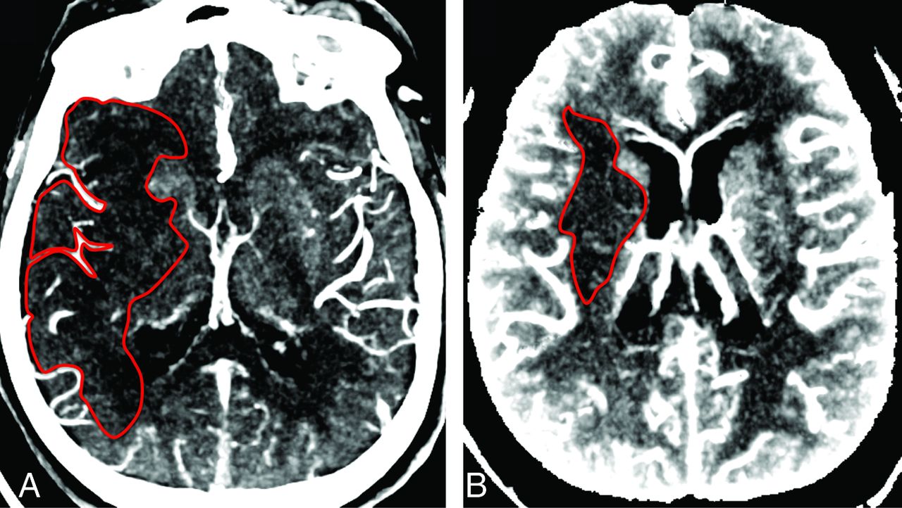
  • Fig 2.
    • Download figure
    • Open in new tab
    • Download powerpoint
    Fig 2.

    Comparison of conventional and dynamic CTA. A, MIP (10-mm) of conventional CTA source images. Volume of hypoattenuation (1 representative section outlined) is 90.5 mL. B, Temporal MIPs (10 mm) of dynamic CTA source images reconstructed for the arteriovenous phase. Volume of hypoattenuation (1 representative section outlined) is 7.5 mL. The follow-up infarct volume was 6.5 mL.
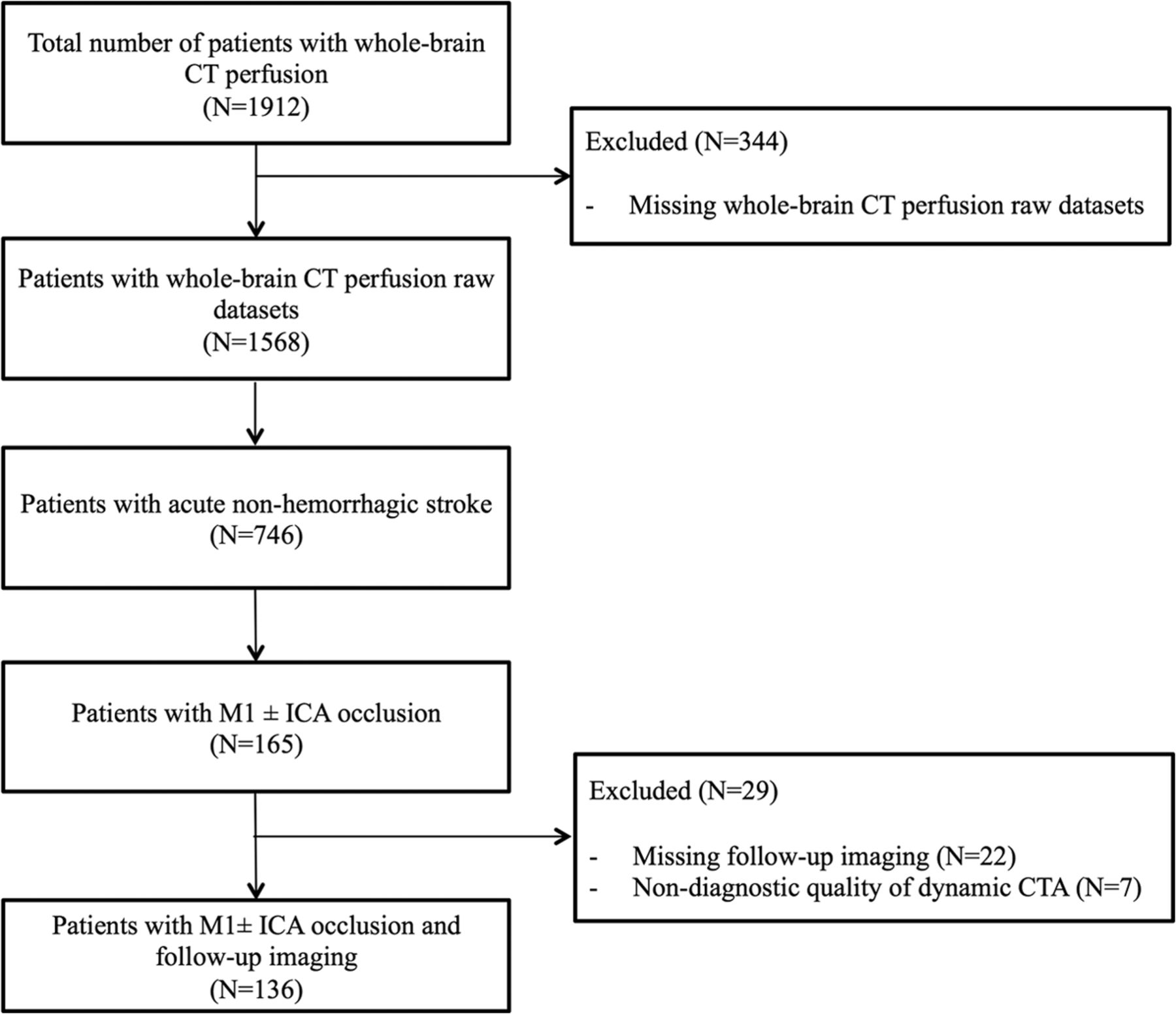
  • Fig 3.
    • Download figure
    • Open in new tab
    • Download powerpoint
    Fig 3.

    Inclusion and exclusion flow chart.

  • Fig 4.
    • Download figure
    • Open in new tab
    • Download powerpoint
    Fig 4.

    Comparison of collateral grading systems. The coefficient of determination is an indicator of model fit. Higher values indicate a better explanation of the variance of the outcome variable.

  • Fig 5.
    • Download figure
    • Open in new tab
    • Download powerpoint
    Fig 5.

    Examples of the volume of hypoattenuation and collateral vessels. A and B, Examples of 2 typical patients. The top rows show 10-mm MIPs of each phase in dynamic CTA. The areas of hypoattenuation of these MIPs are outlined, and the total lesion volumes of each phase are given beneath the images. The bottom rows show 20-mm MIPs of each phase in dynamic CTA, illustrating the collateral vessels. Collateral grades for each phase are given beneath the images and correspond to (in the order given) the 20-point relative score, the 5-point relative score, and the 4-point absolute score. The area of infarction on follow-up is shown on the right.

Tables

  • Figures
    • View popup
    Table 1:

    Patient characteristics and univariate linear regression analysis to determine the effect of patient characteristics on follow-up lesion size

    All Patients (N = 136)β-CoefficientaP Valuea
    Age (yr)b70.4 ± 13.8β = −0.043.235
    Male sex (No.) (%)56 (41.2)β = 1.434.157
    Time from symptom onset to initial imagingc (min)d139 (89–227)β = −0.001.897
    Time to follow-up imaging (days)d3 (1–5)β = −0.095.051
    Additional ICA occlusion (No.) (%)65 (47.8)β = 4.911<.001
    Visible early temporal branch (No.) (%)28 (20.6)β = −2.431.048
    IV thrombolysis (No.) (%)90 (66.2)β = −3.926<.001
    Mechanical recanalization (No.) (%)72 (52.9)β = −3.776<.001
    Follow-up lesion volumee (mL)d79 (19–218)N/AN/A
    • Note:—N/A indicates not available.

    • ↵a Results of univariate linear regression analysis with follow-up lesion volume (square-root-transformed) as a dependent variable. Positive β-values indicate an increase in follow-up lesion volume.

    • ↵b Mean.

    • ↵c Documented for 70 patients.

    • ↵d Median (first-third quartile).

    • ↵e As measured by DWI-MRI (b=1000) or NECT.

    • View popup
    Table 2:

    Model fit of multivariate linear regression for follow-up lesion volumea

    Conventional CTA (R2)Dynamic CT Angiography (R2)
    Arterial PhaseArteriovenous PhaseVenous Phase
    4-Point absolute0.3920.4390.4620.374
    5-Point relative0.3480.3490.4410.321
    20-Point relative0.4360.4210.4830.368
    Volume of hypoattenuation0.4830.5670.6140.586
    • Note:—R2 indicates the adjusted coefficient of determination corrected for sex, time to follow-up imaging, additional ICA occlusion, visible early temporal branch, IV thrombolysis, and mechanical recanalization. It is an indicator of model fit; higher values indicate a better explanation of the variance of the outcome variable.

    • ↵a Overall, dynamic CTA allows a better prediction of follow-up lesion volume than conventional CTA. Specifically, model fit was highest for reconstructions of dynamic CTA images within the arteriovenous phase. Models containing the volume of hypoattenuation performed better than models containing any of the collateral vessel grading scores.

PreviousNext
Back to top

In this issue

American Journal of Neuroradiology: 36 (3)
American Journal of Neuroradiology
Vol. 36, Issue 3
1 Mar 2015
  • Table of Contents
  • Index by author
  • Complete Issue (PDF)
Advertisement
Print
Download PDF
Email Article

Thank you for your interest in spreading the word on American Journal of Neuroradiology.

NOTE: We only request your email address so that the person you are recommending the page to knows that you wanted them to see it, and that it is not junk mail. We do not capture any email address.

Enter multiple addresses on separate lines or separate them with commas.
Strategies of Collateral Blood Flow Assessment in Ischemic Stroke: Prediction of the Follow-Up Infarct Volume in Conventional and Dynamic CTA
(Your Name) has sent you a message from American Journal of Neuroradiology
(Your Name) thought you would like to see the American Journal of Neuroradiology web site.
CAPTCHA
This question is for testing whether or not you are a human visitor and to prevent automated spam submissions.
Cite this article
S.E. Beyer, K.M. Thierfelder, L. von Baumgarten, M. Rottenkolber, F.G. Meinel, H. Janssen, B. Ertl-Wagner, M.F. Reiser, W.H. Sommer
Strategies of Collateral Blood Flow Assessment in Ischemic Stroke: Prediction of the Follow-Up Infarct Volume in Conventional and Dynamic CTA
American Journal of Neuroradiology Mar 2015, 36 (3) 488-494; DOI: 10.3174/ajnr.A4131

Citation Manager Formats

  • BibTeX
  • Bookends
  • EasyBib
  • EndNote (tagged)
  • EndNote 8 (xml)
  • Medlars
  • Mendeley
  • Papers
  • RefWorks Tagged
  • Ref Manager
  • RIS
  • Zotero
0 Responses
Respond to this article
Share
Bookmark this article
Strategies of Collateral Blood Flow Assessment in Ischemic Stroke: Prediction of the Follow-Up Infarct Volume in Conventional and Dynamic CTA
S.E. Beyer, K.M. Thierfelder, L. von Baumgarten, M. Rottenkolber, F.G. Meinel, H. Janssen, B. Ertl-Wagner, M.F. Reiser, W.H. Sommer
American Journal of Neuroradiology Mar 2015, 36 (3) 488-494; DOI: 10.3174/ajnr.A4131
del.icio.us logo Twitter logo Facebook logo Mendeley logo
  • Tweet Widget
  • Facebook Like
  • Google Plus One
Purchase

Jump to section

  • Article
    • Abstract
    • ABBREVIATIONS:
    • Materials and Methods
    • Results
    • Discussion
    • Conclusions
    • Footnotes
    • REFERENCES
  • Figures & Data
  • Supplemental
  • Info & Metrics
  • Responses
  • References
  • PDF

Related Articles

  • PubMed
  • Google Scholar

Cited By...

  • Guidelines for evaluation and management of cerebral collateral circulation in ischaemic stroke 2017
  • Value of Quantitative Collateral Scoring on CT Angiography in Patients with Acute Ischemic Stroke
  • Assessment of Collateral Status by Dynamic CT Angiography in Acute MCA Stroke: Timing of Acquisition and Relationship with Final Infarct Volume
  • Prediction of Stent-Retriever Thrombectomy Outcomes by Dynamic Multidetector CT Angiography in Patients with Acute Carotid T or MCA Occlusions
  • Cortical Venous Filling on Dynamic Computed Tomographic Angiography: A Novel Predictor of Clinical Outcome in Patients With Acute Middle Cerebral Artery Stroke
  • Impact of Collateral Status Evaluated by Dynamic Computed Tomographic Angiography on Clinical Outcome in Patients With Ischemic Stroke
  • Crossref
  • Google Scholar

This article has not yet been cited by articles in journals that are participating in Crossref Cited-by Linking.

More in this TOC Section

  • Predictors of Reperfusion in Patients with Acute Ischemic Stroke
  • Enhanced Axonal Metabolism during Early Natalizumab Treatment in Relapsing-Remitting Multiple Sclerosis
  • Progression of Microstructural Damage in Spinocerebellar Ataxia Type 2: A Longitudinal DTI Study
Show more Brain

Similar Articles

Advertisement

Indexed Content

  • Current Issue
  • Accepted Manuscripts
  • Article Preview
  • Past Issues
  • Editorials
  • Editor's Choice
  • Fellows' Journal Club
  • Letters to the Editor
  • Video Articles

Cases

  • Case Collection
  • Archive - Case of the Week
  • Archive - Case of the Month
  • Archive - Classic Case

More from AJNR

  • Trainee Corner
  • Imaging Protocols
  • MRI Safety Corner
  • Book Reviews

Multimedia

  • AJNR Podcasts
  • AJNR Scantastics

Resources

  • Turnaround Time
  • Submit a Manuscript
  • Submit a Video Article
  • Submit an eLetter to the Editor/Response
  • Manuscript Submission Guidelines
  • Statistical Tips
  • Fast Publishing of Accepted Manuscripts
  • Graphical Abstract Preparation
  • Imaging Protocol Submission
  • Evidence-Based Medicine Level Guide
  • Publishing Checklists
  • Author Policies
  • Become a Reviewer/Academy of Reviewers
  • News and Updates

About Us

  • About AJNR
  • Editorial Board
  • Editorial Board Alumni
  • Alerts
  • Permissions
  • Not an AJNR Subscriber? Join Now
  • Advertise with Us
  • Librarian Resources
  • Feedback
  • Terms and Conditions
  • AJNR Editorial Board Alumni

American Society of Neuroradiology

  • Not an ASNR Member? Join Now

© 2025 by the American Society of Neuroradiology All rights, including for text and data mining, AI training, and similar technologies, are reserved.
Print ISSN: 0195-6108 Online ISSN: 1936-959X

Powered by HighWire