Advertisement

Main menu

  • Home
  • Content
    • Current Issue
    • Accepted Manuscripts
    • Article Preview
    • Past Issue Archive
    • Video Articles
    • AJNR Case Collection
    • Case of the Week Archive
    • Case of the Month Archive
    • Classic Case Archive
  • Special Collections
    • AJNR Awards
    • Low-Field MRI
    • Alzheimer Disease
    • ASNR Foundation Special Collection
    • Photon-Counting CT
    • View All
  • Multimedia
    • AJNR Podcasts
    • AJNR SCANtastic
    • Trainee Corner
    • MRI Safety Corner
    • Imaging Protocols
  • For Authors
    • Submit a Manuscript
    • Submit a Video Article
    • Submit an eLetter to the Editor/Response
    • Manuscript Submission Guidelines
    • Statistical Tips
    • Fast Publishing of Accepted Manuscripts
    • Graphical Abstract Preparation
    • Imaging Protocol Submission
    • Author Policies
  • About Us
    • About AJNR
    • Editorial Board
    • Editorial Board Alumni
  • More
    • Become a Reviewer/Academy of Reviewers
    • Subscribers
    • Permissions
    • Alerts
    • Feedback
    • Advertisers
    • ASNR Home

User menu

  • Alerts
  • Log in

Search

  • Advanced search
American Journal of Neuroradiology

American Journal of Neuroradiology

ASHNR American Society of Functional Neuroradiology ASHNR American Society of Pediatric Neuroradiology ASSR
  • Alerts
  • Log in

Advanced Search

  • Home
  • Content
    • Current Issue
    • Accepted Manuscripts
    • Article Preview
    • Past Issue Archive
    • Video Articles
    • AJNR Case Collection
    • Case of the Week Archive
    • Case of the Month Archive
    • Classic Case Archive
  • Special Collections
    • AJNR Awards
    • Low-Field MRI
    • Alzheimer Disease
    • ASNR Foundation Special Collection
    • Photon-Counting CT
    • View All
  • Multimedia
    • AJNR Podcasts
    • AJNR SCANtastic
    • Trainee Corner
    • MRI Safety Corner
    • Imaging Protocols
  • For Authors
    • Submit a Manuscript
    • Submit a Video Article
    • Submit an eLetter to the Editor/Response
    • Manuscript Submission Guidelines
    • Statistical Tips
    • Fast Publishing of Accepted Manuscripts
    • Graphical Abstract Preparation
    • Imaging Protocol Submission
    • Author Policies
  • About Us
    • About AJNR
    • Editorial Board
    • Editorial Board Alumni
  • More
    • Become a Reviewer/Academy of Reviewers
    • Subscribers
    • Permissions
    • Alerts
    • Feedback
    • Advertisers
    • ASNR Home
  • Follow AJNR on Twitter
  • Visit AJNR on Facebook
  • Follow AJNR on Instagram
  • Join AJNR on LinkedIn
  • RSS Feeds

AJNR Awards, New Junior Editors, and more. Read the latest AJNR updates

Research ArticleBrain
Open Access

Combining MRI with NIHSS Thresholds to Predict Outcome in Acute Ischemic Stroke: Value for Patient Selection

P.W. Schaefer, B. Pulli, W.A. Copen, J.A. Hirsch, T. Leslie-Mazwi, L.H. Schwamm, O. Wu, R.G. González and A.J. Yoo
American Journal of Neuroradiology February 2015, 36 (2) 259-264; DOI: https://doi.org/10.3174/ajnr.A4103
P.W. Schaefer
aFrom the Departments of Radiology (P.W.S., B.P., W.A.C., J.A.H., T.L.-M., O.W., R.G.G., A.J.Y.)
  • Find this author on Google Scholar
  • Find this author on PubMed
  • Search for this author on this site
B. Pulli
aFrom the Departments of Radiology (P.W.S., B.P., W.A.C., J.A.H., T.L.-M., O.W., R.G.G., A.J.Y.)
  • Find this author on Google Scholar
  • Find this author on PubMed
  • Search for this author on this site
W.A. Copen
aFrom the Departments of Radiology (P.W.S., B.P., W.A.C., J.A.H., T.L.-M., O.W., R.G.G., A.J.Y.)
  • Find this author on Google Scholar
  • Find this author on PubMed
  • Search for this author on this site
J.A. Hirsch
aFrom the Departments of Radiology (P.W.S., B.P., W.A.C., J.A.H., T.L.-M., O.W., R.G.G., A.J.Y.)
  • Find this author on Google Scholar
  • Find this author on PubMed
  • Search for this author on this site
T. Leslie-Mazwi
aFrom the Departments of Radiology (P.W.S., B.P., W.A.C., J.A.H., T.L.-M., O.W., R.G.G., A.J.Y.)
  • Find this author on Google Scholar
  • Find this author on PubMed
  • Search for this author on this site
L.H. Schwamm
bNeurology (L.H.S.), Massachusetts General Hospital, Harvard Medical School, Boston, Massachusetts.
  • Find this author on Google Scholar
  • Find this author on PubMed
  • Search for this author on this site
O. Wu
aFrom the Departments of Radiology (P.W.S., B.P., W.A.C., J.A.H., T.L.-M., O.W., R.G.G., A.J.Y.)
  • Find this author on Google Scholar
  • Find this author on PubMed
  • Search for this author on this site
R.G. González
aFrom the Departments of Radiology (P.W.S., B.P., W.A.C., J.A.H., T.L.-M., O.W., R.G.G., A.J.Y.)
  • Find this author on Google Scholar
  • Find this author on PubMed
  • Search for this author on this site
A.J. Yoo
aFrom the Departments of Radiology (P.W.S., B.P., W.A.C., J.A.H., T.L.-M., O.W., R.G.G., A.J.Y.)
  • Find this author on Google Scholar
  • Find this author on PubMed
  • Search for this author on this site
  • Article
  • Figures & Data
  • Supplemental
  • Info & Metrics
  • Responses
  • References
  • PDF
Loading

Article Figures & Data

Figures

  • Tables
  • Fig 1.
    • Download figure
    • Open in new tab
    • Download powerpoint
    Fig 1.

    Baseline imaging volumes and NIHSS scores of patients with good (mRS 0–2) and poor (mRS 3–6) clinical outcome at 90 days after stroke. Horizontal lines indicate the thresholds used to predict outcome with DWI (A) (>70 mL MTT predicts poor outcome), NIHSS score (B) (<8 or >20 predicts good or poor outcome, respectively), MTT (C) (<50 mL predicts good outcome), and Tmax (D) (<50 mL predicts good outcome). The filled circles mark patients for whom prediction was incorrect.

  • Fig 2.
    • Download figure
    • Open in new tab
    • Download powerpoint
    Fig 2.

    Baseline infarct volumes, clinical status, and probability for poor outcome in treated-versus-untreated patients. A, Mean DWI volume (left) and NIHSS score (right) in patients with good outcome stratified by treatment; asterisks indicate a significant difference. B, Probability (determined by logistic regression) for poor outcome versus DWI plots between untreated and treated patients (black circles represent untreated; gray circles, treated).
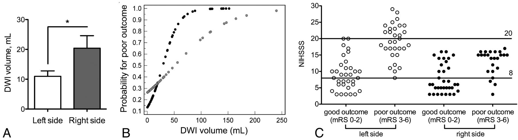
  • Fig 3.
    • Download figure
    • Open in new tab
    • Download powerpoint
    Fig 3.

    Baseline infarct volumes, probability for poor outcome, and NIHSS score thresholds in left- versus right-sided strokes. A, Mean DWI volume in patients with good outcome stratified by side of involvement. Asterisks indicate a significant difference. B, Probability (determined by logistic regression) for poor outcome versus DWI plots between left- and right-sided strokes (left = black circles, right = gray circles). C, NIHSS score scatterplots and thresholds stratified by side of involvement.

Tables

  • Figures
    • View popup
    Table 1:

    Baseline clinical and imaging characteristics, treatment, and predictors of 3-month mRS

    All Patients (n = 123)mRS 0–2 (n = 68)mRS 3–6 (n = 55)P Value
    Age (yr)69.8 ± 15.968.9 ± 16.970.8 ± 14.7.727a
    Baseline NIHSS score11 (6–16)7 (5–11.5)16 (14–20.75)<.0001a
    Female sex55 (44.7%)31 (45.6%)24 (43.6%).973b
    Right hemisphere74 (47.2%)34 (50.0%)24 (43.6%).602b
    Time to MRI3:57 ± 1:263:45 ± 1:294:12 ± 1:20.085c
    DWI volume (mL)37.4 ± 47.716.3 ± 20.163.4 ± 58.2<.0001a
    MTT volume (mL)129.5 ± 101.380.6 ± 78.3189.9 ± 94.1<.0001a
    Tmax volume (mL)148.1 ± 114.989.5 ± 86.5217.3 ± 106.0<.0001a
    Absolute mismatch (mL)92.7 ± 81.464.9 ± 69.1127.1 ± 82.9<.0001a
    Relative mismatch8.7 ± 21.610.9 ± 27.86.0 ± 8.8.541a
    Reperfusion therapyNone: 37 (30.1%)22 (32.4%)15 (25.4%)
    IV-tPA: 61 (49.6%)36 (52.9%)25 (45.5%).200b
    IAT ± IV-tPA: 25 (20.3%)10 (14.7%)15 (27.3%)
    Occlusion levelICA: 24 (19.5%)6 (8.8%)18 (32.7%)
    MCA M1: 39 (31.7%)15 (22.1%)24 (43.6%)<.0001b
    MCA M2/3: 36 (29.3%)24 (35.3%)12 (21.8%)
    None: 24 (19.5%)23 (33.8%)1 (1.8%)
    • Note:—IAT indicates intra-arterial thrombolysis.

    • ↵a Mann-Whitney U test.

    • ↵b χ2 test.

    • ↵c Student t test.

    • View popup
    Table 2:

    Validation of the previously described model combining clinical and imaging thresholds to predict outcome in acute ischemic stroke

    ThresholdPositive Predictive Value (%)Prognostic Yield (%)
    Poor outcome
        DWI > 70 mL86.4a17.9
        NIHSS score > 20100.0a11.4
    Good outcome
        MTT < 0 mL89.5a30.9
        Tmax < 50 mL88.9a29.3
        NIHSS score < 892.3a31.7
    Combined clinical thresholds (NIHSS score)94.3a43.1
    Combined imaging thresholds (DWI + MTT)88.3a48.8
    Combined imaging thresholds (DWI + Tmax > 6 seconds)87.9a47.2
    Combined clinical and imaging thresholds88.8a65.0b
    • ↵a No significant difference in PPV between any of the applied thresholds.

    • ↵b Significantly increased over use of single parameters (P < .0001) and combined clinical or imaging thresholds (P = .01).

    • View popup
    Table 3:

    Differences of clinical and imaging thresholds in patients receiving thrombolysis versus untreated patients

    No Reperfusion TherapyIV and/or IA TherapyP Value
    DWI
        AUC ± SE for predicting poor outcome0.935 ± 0.060.724 ± 0.05.011
        90% Specificity for poor outcome (prognostic yield) (mL)>19.7 (42.3%)>51.8 (19.5%).016a
        95% Specificity for poor outcome (prognostic yield) (mL)>20.5 (40.5%)>74.8 (16.1%).007a
    >30.6 (35.1%)>103.1 (10.4%).003a
    NIHSS score
        AUC ± SE for predicting poor outcome0.903 ± 0.060.827 ± 0.04.314
        90% Specificity for poor outcome (prognostic yield)>12 (37.8%)>16 (23.0%).142a
        95% Specificity for poor outcome (prognostic yield)>15 (27.0%)>19 (14.9%).182a
        100% Specificity for poor outcome (prognostic yield)>17 (16.2%)>20 (9.2%).414a
    • ↵a P value for comparison among prognostic yields.

    • View popup
    Table 4:

    Differences of clinical and imaging thresholds in patients stratified by affected cerebral hemisphere

    Right SideLeft SideP Value
    DWI
        AUC ± SE0.706 ± 0.070.875 ± 0.04.043
        90% Specificity for poor outcome (prognostic yield) (mL)>39.6 (20.7%)>23.4 (40.0%).034a
        95% Specificity for poor outcome (prognostic yield) (mL)>98.5 (12.1%)>51.8 (26.2%).082a
        100% Specificity for poor outcome (prognostic yield) (mL)>103.1 (10.4%)>74.8 (16.9%).435a
    NIHSS score
        AUC ± SE0.808 ± 0.060.911 ± 0.03.077
        90% Specificity for poor or good outcome (prognostic yield)>15 or <6 (29.3%)>17 or <10 (66.2%)<.001a
        95% Specificity for poor or good outcome (prognostic yield)>16 or <5 (17.2%)>20 or <9 (49.2%)<.001a
        100% Specificity for poor or good outcome (prognostic yield)>16 or <3 (1.7%)>20 or <7 (38.5%)<.001a
    • ↵a P value for comparison between prognostic yields.

PreviousNext
Back to top

In this issue

American Journal of Neuroradiology: 36 (2)
American Journal of Neuroradiology
Vol. 36, Issue 2
1 Feb 2015
  • Table of Contents
  • Index by author
  • Complete Issue (PDF)
Advertisement
Print
Download PDF
Email Article

Thank you for your interest in spreading the word on American Journal of Neuroradiology.

NOTE: We only request your email address so that the person you are recommending the page to knows that you wanted them to see it, and that it is not junk mail. We do not capture any email address.

Enter multiple addresses on separate lines or separate them with commas.
Combining MRI with NIHSS Thresholds to Predict Outcome in Acute Ischemic Stroke: Value for Patient Selection
(Your Name) has sent you a message from American Journal of Neuroradiology
(Your Name) thought you would like to see the American Journal of Neuroradiology web site.
CAPTCHA
This question is for testing whether or not you are a human visitor and to prevent automated spam submissions.
Cite this article
P.W. Schaefer, B. Pulli, W.A. Copen, J.A. Hirsch, T. Leslie-Mazwi, L.H. Schwamm, O. Wu, R.G. González, A.J. Yoo
Combining MRI with NIHSS Thresholds to Predict Outcome in Acute Ischemic Stroke: Value for Patient Selection
American Journal of Neuroradiology Feb 2015, 36 (2) 259-264; DOI: 10.3174/ajnr.A4103

Citation Manager Formats

  • BibTeX
  • Bookends
  • EasyBib
  • EndNote (tagged)
  • EndNote 8 (xml)
  • Medlars
  • Mendeley
  • Papers
  • RefWorks Tagged
  • Ref Manager
  • RIS
  • Zotero
0 Responses
Respond to this article
Share
Bookmark this article
Combining MRI with NIHSS Thresholds to Predict Outcome in Acute Ischemic Stroke: Value for Patient Selection
P.W. Schaefer, B. Pulli, W.A. Copen, J.A. Hirsch, T. Leslie-Mazwi, L.H. Schwamm, O. Wu, R.G. González, A.J. Yoo
American Journal of Neuroradiology Feb 2015, 36 (2) 259-264; DOI: 10.3174/ajnr.A4103
del.icio.us logo Twitter logo Facebook logo Mendeley logo
  • Tweet Widget
  • Facebook Like
  • Google Plus One
Purchase

Jump to section

  • Article
    • Abstract
    • ABBREVIATIONS:
    • Materials and Methods
    • Results
    • Discussion
    • Conclusions
    • Footnotes
    • REFERENCES
  • Figures & Data
  • Supplemental
  • Info & Metrics
  • Responses
  • References
  • PDF

Related Articles

  • PubMed
  • Google Scholar

Cited By...

  • Risk factors of unexplained early neurological deterioration after treatment for ischemic stroke due to large vessel occlusion: a post hoc analysis of the HERMES study
  • Effect of IV alteplase on the ischemic brain lesion at 24-48 hours after ischemic stroke
  • Outcome After Reperfusion Therapies in Patients With Large Baseline Diffusion-Weighted Imaging Stroke Lesions: A THRACE Trial (Mechanical Thrombectomy After Intravenous Alteplase Versus Alteplase Alone After Stroke) Subgroup Analysis
  • Impact of Leukoaraiosis Burden on Hemispheric Lateralization of the National Institutes of Health Stroke Scale Deficit in Acute Ischemic Stroke
  • Crossref
  • Google Scholar

This article has not yet been cited by articles in journals that are participating in Crossref Cited-by Linking.

More in this TOC Section

  • Multimodal CT Provides Improved Performance for Lacunar Infarct Detection
  • Optimal MRI Sequence for Identifying Occlusion Location in Acute Stroke: Which Value of Time-Resolved Contrast-Enhanced MRA?
  • Evaluating the Effects of White Matter Multiple Sclerosis Lesions on the Volume Estimation of 6 Brain Tissue Segmentation Methods
Show more Brain

Similar Articles

Advertisement

Indexed Content

  • Current Issue
  • Accepted Manuscripts
  • Article Preview
  • Past Issues
  • Editorials
  • Editor's Choice
  • Fellows' Journal Club
  • Letters to the Editor
  • Video Articles

Cases

  • Case Collection
  • Archive - Case of the Week
  • Archive - Case of the Month
  • Archive - Classic Case

More from AJNR

  • Trainee Corner
  • Imaging Protocols
  • MRI Safety Corner
  • Book Reviews

Multimedia

  • AJNR Podcasts
  • AJNR Scantastics

Resources

  • Turnaround Time
  • Submit a Manuscript
  • Submit a Video Article
  • Submit an eLetter to the Editor/Response
  • Manuscript Submission Guidelines
  • Statistical Tips
  • Fast Publishing of Accepted Manuscripts
  • Graphical Abstract Preparation
  • Imaging Protocol Submission
  • Evidence-Based Medicine Level Guide
  • Publishing Checklists
  • Author Policies
  • Become a Reviewer/Academy of Reviewers
  • News and Updates

About Us

  • About AJNR
  • Editorial Board
  • Editorial Board Alumni
  • Alerts
  • Permissions
  • Not an AJNR Subscriber? Join Now
  • Advertise with Us
  • Librarian Resources
  • Feedback
  • Terms and Conditions
  • AJNR Editorial Board Alumni

American Society of Neuroradiology

  • Not an ASNR Member? Join Now

© 2025 by the American Society of Neuroradiology All rights, including for text and data mining, AI training, and similar technologies, are reserved.
Print ISSN: 0195-6108 Online ISSN: 1936-959X

Powered by HighWire