Advertisement

Main menu

  • Home
  • Content
    • Current Issue
    • Accepted Manuscripts
    • Article Preview
    • Past Issue Archive
    • Video Articles
    • AJNR Case Collection
    • Case of the Week Archive
    • Case of the Month Archive
    • Classic Case Archive
  • Special Collections
    • AJNR Awards
    • Low-Field MRI
    • Alzheimer Disease
    • ASNR Foundation Special Collection
    • Photon-Counting CT
    • View All
  • Multimedia
    • AJNR Podcasts
    • AJNR SCANtastic
    • Trainee Corner
    • MRI Safety Corner
    • Imaging Protocols
  • For Authors
    • Submit a Manuscript
    • Submit a Video Article
    • Submit an eLetter to the Editor/Response
    • Manuscript Submission Guidelines
    • Statistical Tips
    • Fast Publishing of Accepted Manuscripts
    • Graphical Abstract Preparation
    • Imaging Protocol Submission
    • Author Policies
  • About Us
    • About AJNR
    • Editorial Board
    • Editorial Board Alumni
  • More
    • Become a Reviewer/Academy of Reviewers
    • Subscribers
    • Permissions
    • Alerts
    • Feedback
    • Advertisers
    • ASNR Home

User menu

  • Alerts
  • Log in

Search

  • Advanced search
American Journal of Neuroradiology

American Journal of Neuroradiology

ASHNR American Society of Functional Neuroradiology ASHNR American Society of Pediatric Neuroradiology ASSR
  • Alerts
  • Log in

Advanced Search

  • Home
  • Content
    • Current Issue
    • Accepted Manuscripts
    • Article Preview
    • Past Issue Archive
    • Video Articles
    • AJNR Case Collection
    • Case of the Week Archive
    • Case of the Month Archive
    • Classic Case Archive
  • Special Collections
    • AJNR Awards
    • Low-Field MRI
    • Alzheimer Disease
    • ASNR Foundation Special Collection
    • Photon-Counting CT
    • View All
  • Multimedia
    • AJNR Podcasts
    • AJNR SCANtastic
    • Trainee Corner
    • MRI Safety Corner
    • Imaging Protocols
  • For Authors
    • Submit a Manuscript
    • Submit a Video Article
    • Submit an eLetter to the Editor/Response
    • Manuscript Submission Guidelines
    • Statistical Tips
    • Fast Publishing of Accepted Manuscripts
    • Graphical Abstract Preparation
    • Imaging Protocol Submission
    • Author Policies
  • About Us
    • About AJNR
    • Editorial Board
    • Editorial Board Alumni
  • More
    • Become a Reviewer/Academy of Reviewers
    • Subscribers
    • Permissions
    • Alerts
    • Feedback
    • Advertisers
    • ASNR Home
  • Follow AJNR on Twitter
  • Visit AJNR on Facebook
  • Follow AJNR on Instagram
  • Join AJNR on LinkedIn
  • RSS Feeds

AJNR Awards, New Junior Editors, and more. Read the latest AJNR updates

Research ArticleBrain

Double Inversion Recovery MR Sequence for the Detection of Subacute Subarachnoid Hemorrhage

J. Hodel, R. Aboukais, B. Dutouquet, E. Kalsoum, M.A. Benadjaoud, D. Chechin, M. Zins, A. Rahmouni, A. Luciani, J.-P. Pruvo, J.-P. Lejeune and X. Leclerc
American Journal of Neuroradiology February 2015, 36 (2) 251-258; DOI: https://doi.org/10.3174/ajnr.A4102
J. Hodel
aFrom the Departments of Neuroradiology (J.H., B.D., E.K., J.-P.P., X.L.)
eDepartment of Radiology (J.H., M.Z.), Hôpital Saint Joseph, Paris, France
  • Find this author on Google Scholar
  • Find this author on PubMed
  • Search for this author on this site
R. Aboukais
bNeurosurgery (R.A., J.-P.L.), Hôpital Roger Salengro, Lille, France
  • Find this author on Google Scholar
  • Find this author on PubMed
  • Search for this author on this site
B. Dutouquet
aFrom the Departments of Neuroradiology (J.H., B.D., E.K., J.-P.P., X.L.)
  • Find this author on Google Scholar
  • Find this author on PubMed
  • Search for this author on this site
E. Kalsoum
aFrom the Departments of Neuroradiology (J.H., B.D., E.K., J.-P.P., X.L.)
  • Find this author on Google Scholar
  • Find this author on PubMed
  • Search for this author on this site
M.A. Benadjaoud
cInstitut National De La Santé et De La Recherche Médicale (M.A.B.), Centre for Research in Epidemiology and Population Health, Villejuif, France
  • Find this author on Google Scholar
  • Find this author on PubMed
  • Search for this author on this site
D. Chechin
dPhilips Medical Systems (D.C.), Suresnes, France
  • Find this author on Google Scholar
  • Find this author on PubMed
  • Search for this author on this site
M. Zins
eDepartment of Radiology (J.H., M.Z.), Hôpital Saint Joseph, Paris, France
  • Find this author on Google Scholar
  • Find this author on PubMed
  • Search for this author on this site
A. Rahmouni
fDepartment of Radiology (A.R., A.L.), Centre Hospitalier Universitaire, Henri Mondor, Créteil, France.
  • Find this author on Google Scholar
  • Find this author on PubMed
  • Search for this author on this site
A. Luciani
fDepartment of Radiology (A.R., A.L.), Centre Hospitalier Universitaire, Henri Mondor, Créteil, France.
  • Find this author on Google Scholar
  • Find this author on PubMed
  • Search for this author on this site
J.-P. Pruvo
aFrom the Departments of Neuroradiology (J.H., B.D., E.K., J.-P.P., X.L.)
  • Find this author on Google Scholar
  • Find this author on PubMed
  • Search for this author on this site
J.-P. Lejeune
bNeurosurgery (R.A., J.-P.L.), Hôpital Roger Salengro, Lille, France
  • Find this author on Google Scholar
  • Find this author on PubMed
  • Search for this author on this site
X. Leclerc
aFrom the Departments of Neuroradiology (J.H., B.D., E.K., J.-P.P., X.L.)
  • Find this author on Google Scholar
  • Find this author on PubMed
  • Search for this author on this site
  • Article
  • Figures & Data
  • Info & Metrics
  • Responses
  • References
  • PDF
Loading

Article Figures & Data

Figures

  • Tables
  • Fig 1.
    • Download figure
    • Open in new tab
    • Download powerpoint
    Fig 1.

    Axial DIR images in a 37-year-old healthy volunteer showing the basal cisterns (A and B) and the subarachnoid spaces at the brain convexity (C). In all the healthy volunteers scanned with the DIR sequence, the CSF appeared hypointense without CSF flow-related artifacts (B, arrowheads). Note the regional variation of gray matter signal intensity by using DIR, such as the difference between the prefrontal (C, arrows) and motor (C, arrowheads) cortices. The absence of CSF signal abnormality on DIR images in all the healthy volunteers and the strong agreement among readers suggest that the hyperintensities observed in patients with SAH by using DIR were not linked to artifacts.

  • Fig 2.
    • Download figure
    • Open in new tab
    • Download powerpoint
    Fig 2.

    Subacute SAH related to an aneurysm of the anterior communicating artery in a 42-year-old woman. No signal abnormality is visible on 3D FLAIR (A). SAH is visible by using axial SWI minimum-intensity-projection reformations in the right Sylvian fissure (B, arrows). The hypointensity visible in the left Sylvian fissure on SWI was considered a cortical vein by the 3 blinded readers by using both average and minimum-intensity-projection reformations (B, arrowhead). On the axial T2* image, SAH is bilateral, involving the Sylvian fissures (C, arrows). The DIR sequence reveals extensive SAH prevailing in the Sylvian fissures (D, arrows) and interhemispheric and occipital sulci (D, arrowheads).

  • Fig 3.
    • Download figure
    • Open in new tab
    • Download powerpoint
    Fig 3.

    Subacute SAH related to an aneurysm of the anterior communicating artery in a 63-year-old man. With 2D FLAIR (A), 3D FLAIR (B), or T2* (C) images, no subacute hemorrhage is visible. Conversely, marked subarachnoid signal abnormalities along the anterior and posterior interhemispheric sulci are observed by using DIR (D, arrows). SAH involving the parietal lobes is also visible (D, arrowheads).
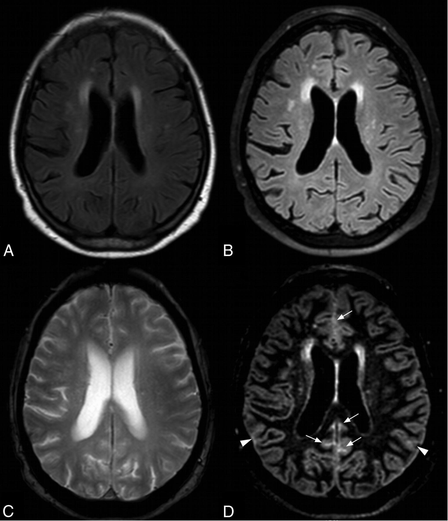
  • Fig 4.
    • Download figure
    • Open in new tab
    • Download powerpoint
    Fig 4.

    Subacute SAH related to an aneurysm of the left internal carotid artery in a 25-year-old woman. With 2D FLAIR images, the detection of SAH is challenging due to potential CSF flow-related artifacts (A, arrow). With an axial average SWI reformat, a slight rim of hemorrhage is visible within the interpeduncular fossa (B, arrow). On the 3D FLAIR image, SAH is subtle due to a lack of contrast (C, arrows). With DIR images, SAH is obvious, with a marked hyperintensity within the interpeduncular fossa, vermis, and left Sylvian fissure (D, arrows). Note the marked signal intensity of SAH with DIR compared with the other MR images.

Tables

  • Figures
    • View popup
    Table 1:

    Demographic and clinical data at admission

    PatientsControls
    No.2520
    Sex16 M/9 F13 M/7 F
    Age (yr) (mean) (range)52 (28–71)50 (26–69)
    Fisher grading (mean)3
        Grade 1 (No.)5
        Grade 2 (No.)2
        Grade 3 (No.)6
        Grade 4 (No.)12
    GCS (mean) (range)14.1 (10–15)
    WFNS score (mean) (range)1.5 (1–4)
        No vascular lesion (No.)10
        Brain aneurysm (No.)15
    Etiologic work-up (CTA and DSA)
        ACA (No.)6
        MCA (No.)(right 2, left 2)
        Right PcomA (No.)2
        Left vertebral artery (No.)1
        Left ICA (No.)1
        Basilar artery (No.)1
    • Note:—GCS indicates Glasgow Coma Scale; WFNS, World Federation of Neurosurgical Societies; ACA, anterior communicating artery; PcomA, posterior communicating artery.

    • View popup
    Table 2:

    MR imaging sequence parameters

    2D FLAIR2D T2*3D FLAIR3D SWI3D DIR
    Acquisition planeAxialAxialSagittalSagittalSagittal
    TR/TE (ms)11,000/1251077/164800/26713/195500/255
    TI (ms)2800–1650–2600/625
    Acquired voxel size (mm)0.8 × 1.4 × 40.9 × 1.2 × 41.2 × 1.2 × 1.21.2 × 1.2 × 1.21.2 × 1.2 × 1.2
    Bandwidth (Hz)27121614331721433
    Echo-train length31–182–173
    No. of sections3636280280280
    SENSE1.4–2.52.52.5
    CLEARYesYesYesYesYes
    Fat suppressionNoNoSPIRNoSPIR
    Acquisition time3 min3 min3 min 20 sec3 min4 min
    • Note:—SENSE indicates sensitivity encoding; CLEAR, inhomogeneity correction; SPIR, spectral presaturation with inversion recovery.

    • View popup
    Table 3:

    Interobserver agreement among readers 1, 2, and 3 for each set of images according to the area considered

    CTMRI
    2D FLAIR3D FLAIR2D T2*3D SWI3D DIR
    Total
        R1 vs R2 (95% CI)1 (1–1)0.85 (0.77–0.93)0.79 (0.69–0.89)0.84 (0.76–0.91)0.79 (0.72–0.87)0.94 (0.91–0.97)
        R1 vs R3 (95% CI)0.94 (0.85–1)0.68 (0.57–0.79)0.65 (0.53–0.76)0.74 (0.65–0.83)0.64 (0.55–0.73)0.90 (0.87–0.93)
        R2 vs R3 (95% CI)0.94 (0.85–1.02)0.70 (0.60–0.81)0.70 (0.59–0.80)0.72 (0.63–0.81)0.73 (0.65–0.81)0.93 (0.91–0.96)
    Subarachnoid
        Interhemispheric
            R1 vs R2 (95% CI)1 (1–1)0.91 (0.74–1)0.83 (0.60–1)0.76 (0.54–0.98)0.68 (0.44–0.92)1 (1–1)
            R1 vs R3 (95% CI)1 (1–1)0.83 (0.60–1.06)0.76 (0.50–1.02)0.87 (0.70–1.05)0.44 (0.11–0.77)1 (1–1)
            R2 vs R3 (95% CI)1 (1–1)0.91 (0.74–1.08)0.76 (0.50–1.02)0.77 (0.56–0.99)0.63 (0.39–0.88)1 (1–1)
        Sylvian fissures
            R1 vs R2 (95% CI)1 (1–1)1 (1–1)1 (1–1)0.94 (0.83–1)0.82 (0.63–1)0.95 (0.88–1)
            R1 vs R3 (95% CI)1 (1–1)0.88 (0.66–1.11)1 (1–1)1 (1–1)0.59 (0.31–0.86)0.90 (0.81–1)
            R2 vs R3 (95% CI)1 (1–1)0.88 (0.66–1.11)1 (1–1)0.94 (0.83–1.06)0.73 (0.50–0.96)0.95 (0.89–1.02)
        Convexity
            R1 vs R2 (95% CI)1 (1–1)0.83 (0.68–0.98)0.56 (0.27–0.84)0.73 (0.57–0.89)0.63 (0.43–0.84)0.90 (0.85–0.95)
            R1 vs R3 (95% CI)0.83 (0.60–1.07)0.61 (0.42–0.81)0.48 (0.23–0.73)0.60 (0.42–0.78)0.63 (0.44–0.83)0.87 (0.81–0.92)
            R2 vs R3 (95% CI)0.83 (0.60–1.07)0.60 (0.40–0.79)0.56 (0.33–0.80)0.56 (0.37–0.74)0.70 (0.55–0.86)0.90 (0.85–0.95)
        Basal cisterns
            R1 vs R2 (95% CI)1 (1–1)0.33 (−0.33–0.98)0.70 (0.41–0.99)0.74 (0.39–1)0.64 (0.36–0.92)0.94 (0.88–1)
            R1 vs R3 (95% CI)1 (1–1)0.43 (−0.07–0.92)0.25 (−0.20–0.70)0.45 (−0.01–0.98)0.25 (−0.13–0.62)0.89 (0.82–0.97)
            R2 vs R3 (95% CI)1 (1–1)0.39 (−0.29–1.07)0.30 (−0.17–0.77)0.74 (0.39–1.10)0.41 (0.09–0.73)0.95 (0.90–1)
    Intraventricular
        R1 vs R2 (95% CI)1 (1–1)1 (1–1)1 (1–1)1 (1–1)0.95 (0.85–1)1 (1–1)
        R1 vs R3 (95% CI)1 (1–1)0 (−1.12–1.12)0 (−1.95–1.95)0.66 (0.19–1.13)0.86 (0.70–1.02)0 (−1.38–1.38)
        R2 vs R3 (95% CI)1 (1–1)0 (−1.12–1.12)0 (−-1.95–1.95)0.66 (0.19–1.13)0.82 (0.64–1)0 (−1.38–1.38)
    • Note:—R1–R3 indicate readers 1–3; Total, all the subarachnoid and ventricular areas; Convexity, bilateral frontal, parietal, temporal, and occipital convexity areas; Basal cisterns, perimesencephalic and prepontine cisterns and cisterna magna.

    • View popup
    Table 4:

    Number of patients with at least 1 subarachnoid and/or ventricular signal abnormality (diagnosis of SAH and IVH, respectively) for each imaging modality after consensus among readers

    CTMRI
    2D FLAIR3D FLAIR2D T2*3D SWI3D DIR
    Patients with SAH71215141525
    Interhemispherica067131525
    Sylvian fissuresa4449817
    Convexitya388101119
    Basal cisternsa0154723
    Patients with IVH000350
    • ↵a Number of patients with at least 1 subarachnoid signal abnormality.

PreviousNext
Back to top

In this issue

American Journal of Neuroradiology: 36 (2)
American Journal of Neuroradiology
Vol. 36, Issue 2
1 Feb 2015
  • Table of Contents
  • Index by author
  • Complete Issue (PDF)
Advertisement
Print
Download PDF
Email Article

Thank you for your interest in spreading the word on American Journal of Neuroradiology.

NOTE: We only request your email address so that the person you are recommending the page to knows that you wanted them to see it, and that it is not junk mail. We do not capture any email address.

Enter multiple addresses on separate lines or separate them with commas.
Double Inversion Recovery MR Sequence for the Detection of Subacute Subarachnoid Hemorrhage
(Your Name) has sent you a message from American Journal of Neuroradiology
(Your Name) thought you would like to see the American Journal of Neuroradiology web site.
CAPTCHA
This question is for testing whether or not you are a human visitor and to prevent automated spam submissions.
Cite this article
J. Hodel, R. Aboukais, B. Dutouquet, E. Kalsoum, M.A. Benadjaoud, D. Chechin, M. Zins, A. Rahmouni, A. Luciani, J.-P. Pruvo, J.-P. Lejeune, X. Leclerc
Double Inversion Recovery MR Sequence for the Detection of Subacute Subarachnoid Hemorrhage
American Journal of Neuroradiology Feb 2015, 36 (2) 251-258; DOI: 10.3174/ajnr.A4102

Citation Manager Formats

  • BibTeX
  • Bookends
  • EasyBib
  • EndNote (tagged)
  • EndNote 8 (xml)
  • Medlars
  • Mendeley
  • Papers
  • RefWorks Tagged
  • Ref Manager
  • RIS
  • Zotero
0 Responses
Respond to this article
Share
Bookmark this article
Double Inversion Recovery MR Sequence for the Detection of Subacute Subarachnoid Hemorrhage
J. Hodel, R. Aboukais, B. Dutouquet, E. Kalsoum, M.A. Benadjaoud, D. Chechin, M. Zins, A. Rahmouni, A. Luciani, J.-P. Pruvo, J.-P. Lejeune, X. Leclerc
American Journal of Neuroradiology Feb 2015, 36 (2) 251-258; DOI: 10.3174/ajnr.A4102
del.icio.us logo Twitter logo Facebook logo Mendeley logo
  • Tweet Widget
  • Facebook Like
  • Google Plus One
Purchase

Jump to section

  • Article
    • Abstract
    • ABBREVIATIONS:
    • Materials and Methods
    • Results
    • Discussion
    • Conclusions
    • Footnotes
    • REFERENCES
  • Figures & Data
  • Info & Metrics
  • Responses
  • References
  • PDF

Related Articles

  • No related articles found.
  • PubMed
  • Google Scholar

Cited By...

  • Detection of aneurysmal subarachnoid hemorrhage 3 months after initial bleeding: evaluation of T2* and FLAIR MR sequences at 3 T in comparison with initial non-enhanced CT as a gold standard
  • Crossref
  • Google Scholar

This article has not yet been cited by articles in journals that are participating in Crossref Cited-by Linking.

More in this TOC Section

  • Multimodal CT Provides Improved Performance for Lacunar Infarct Detection
  • Optimal MRI Sequence for Identifying Occlusion Location in Acute Stroke: Which Value of Time-Resolved Contrast-Enhanced MRA?
  • Evaluating the Effects of White Matter Multiple Sclerosis Lesions on the Volume Estimation of 6 Brain Tissue Segmentation Methods
Show more Brain

Similar Articles

Advertisement

Indexed Content

  • Current Issue
  • Accepted Manuscripts
  • Article Preview
  • Past Issues
  • Editorials
  • Editor's Choice
  • Fellows' Journal Club
  • Letters to the Editor
  • Video Articles

Cases

  • Case Collection
  • Archive - Case of the Week
  • Archive - Case of the Month
  • Archive - Classic Case

More from AJNR

  • Trainee Corner
  • Imaging Protocols
  • MRI Safety Corner
  • Book Reviews

Multimedia

  • AJNR Podcasts
  • AJNR Scantastics

Resources

  • Turnaround Time
  • Submit a Manuscript
  • Submit a Video Article
  • Submit an eLetter to the Editor/Response
  • Manuscript Submission Guidelines
  • Statistical Tips
  • Fast Publishing of Accepted Manuscripts
  • Graphical Abstract Preparation
  • Imaging Protocol Submission
  • Evidence-Based Medicine Level Guide
  • Publishing Checklists
  • Author Policies
  • Become a Reviewer/Academy of Reviewers
  • News and Updates

About Us

  • About AJNR
  • Editorial Board
  • Editorial Board Alumni
  • Alerts
  • Permissions
  • Not an AJNR Subscriber? Join Now
  • Advertise with Us
  • Librarian Resources
  • Feedback
  • Terms and Conditions
  • AJNR Editorial Board Alumni

American Society of Neuroradiology

  • Not an ASNR Member? Join Now

© 2025 by the American Society of Neuroradiology All rights, including for text and data mining, AI training, and similar technologies, are reserved.
Print ISSN: 0195-6108 Online ISSN: 1936-959X

Powered by HighWire