
Article Figures & Data
Figures
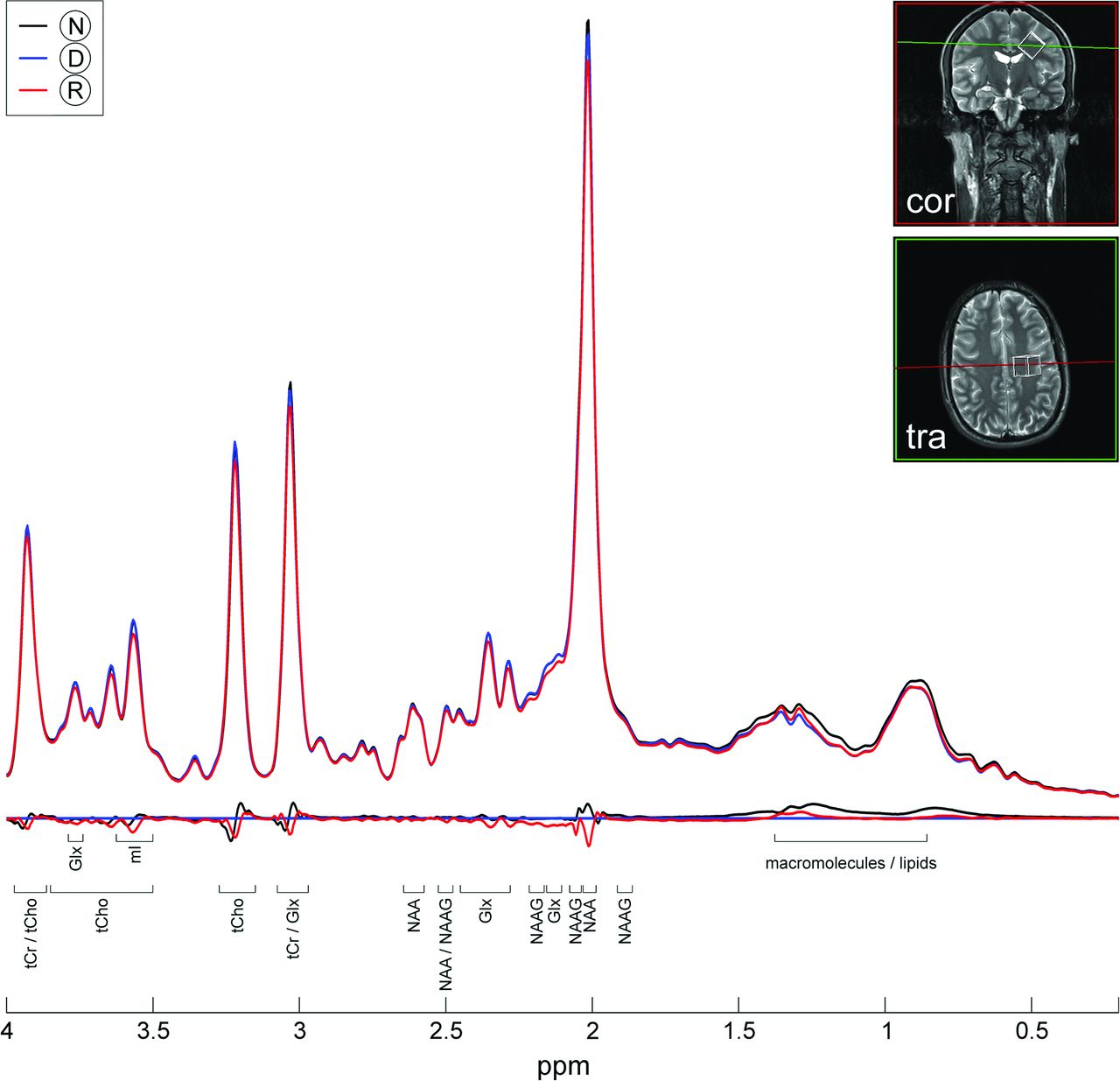
- Fig 1.
Representative 1H-MR spectroscopy spectral data fits. The voxel was placed in the left paracentral lobule. Spectral data fits (upper row) and subtracted spectra (lower row) for visualization of metabolite resonances at normo- (black line, N), de- (blue line, D), and rehydration (red line, R). tCr indicates total creatine, mI, myo-inositol; tCho, total choline; NAA+NAAG, N-acetylaspartate and N-acetyl aspartylglutamate; Glx, glutamine + glutamate.
- Fig 2.
Serial data of brain tissue fluid H2Obrain. Twelve hours of thirsting (dehydration) increased serum osmolality by 0.67% and decreased H2Obrain by 1.63%. Subsequent oral fluid intake (rehydration) during 1 hour lowered serum osmolality by 0.96% and led to an increase of H2Obrain by 0.43%. This finding shows that even subtle changes in H2Obrain on minor osmotic challenges are detectable by 1H-MR spectroscopy.
- Fig 3.
Volumetric morphometry at normo-, de-, and rehydration illustrated as percentage volume change normalized to session 2 (dehydration). Volume changes of the entire brain, cerebral cortex, white matter, and hypothalamus/thalamus were significant in repeated measures ANOVA and compatible with cell shrinking during hyperosmolality and cell swelling during hypo-osmolality. Subject-specific pair-wise differences are plotted with respect to dehydration. Post hoc tests revealed hydration states with significant volume changes between each one (indicated by asterisks; for details see On-line Table 3). Box indicates upper and lower quartiles; thick black line, median; whiskers, most extreme values of the interquartile range; crosses, outliers; D, dehydration; N, normohydration; R, rehydration; asterisk, significant difference.
- Fig 4.
Cortical thickness analysis. Local changes of cortical thickness (A, red-to-yellow: for thinning on dehydration; blue-to-light blue: for thickening on rehydration). Dehydration primarily induces cortical thinning (upper row), which reverses on rehydration (bottom row). Note that these prevailing changes are not uniformly distributed over the cerebral surface. Changes on the mesial surface (not shown) were slightly less pronounced but similar. The corresponding statistical significance (B, red-to-yellow: for thinning on dehydration; blue-to-light blue: for thickening upon rehydration) is expressed by increasingly lower false-positive probabilities across subjects.
- Fig 5.
T1-relaxometry. Quantitative T1-relaxation times of the cerebral cortex (upper row), the subcortical gray (middle row), and white matter (bottom row) during normo-, de-, and rehydration. Statistical analyses revealed no longitudinal changes in T1 relaxation times. Box indicates upper and lower quartiles; thick black line, median; whiskers, most extreme values of the interquartile range; crosses, outliers.


Tables
Blood/serum parameters during normo-, de-, and rehydration
Parameter Normohyd Dehyd Rehyd Na+ (mmol/L) 137.1 ± 2.7 138.5 ± 2.2 136.9 ± 2.3 K+ (mmol/L) 4.6 ± 0.4a 5.2 ± 0.6a 4.9 ± 0.4a HCT (%) 41.9 ± 2.9 43.8 ± 2.9 42.5 ± 2.9 MCHC (g/dL) 34.3 ± 0.9 34.3 ± 0.8 34.3 ± 0.8 Urea (mg/dL) 27.4 ± 6.2 26.8 ± 5.9 25.5 ± 5.6 OSMserum (mOsm/kg) 312.7 ± 4.8a 314.8 ± 4.1a 311.8 ± 4.7a Glucose (mg/dL) 92.1 ± 6.8a 84.4 ± 6.9a 85.5 ± 5.9a Note:—Normohyd indicates normohydration; Dehyd, dehydration; Rehyd, rehydration; MCHC, mean corpuscular hemoglobin concentration.
↵a Significant changes among different levels of hydration (normo-, de-, and rehydration) at type I error probabilities of P < .05 (1-sided t tests; df = 14).


Jump to section
Related Articles
Cited By...
- Association between brain imaging biomarkers and continuous glucose monitoring-derived glycemic control indices in Japanese patients with type 2 diabetes mellitus
- Cortical microstructure and hemispheric specialization - a diffusion-imaging analysis in younger and older adults
- Reader response: Gray matter volume modifications in migraine: A cross-sectional and longitudinal study
- Large Changes in Brain Volume Observed in an Asymptomatic Young Child With Type 1 Diabetes