Advertisement

Main menu

  • Home
  • Content
    • Current Issue
    • Accepted Manuscripts
    • Article Preview
    • Past Issue Archive
    • Video Articles
    • AJNR Case Collection
    • Case of the Week Archive
    • Case of the Month Archive
    • Classic Case Archive
  • Special Collections
    • AJNR Awards
    • Low-Field MRI
    • Alzheimer Disease
    • ASNR Foundation Special Collection
    • Photon-Counting CT
    • View All
  • Multimedia
    • AJNR Podcasts
    • AJNR SCANtastic
    • Trainee Corner
    • MRI Safety Corner
    • Imaging Protocols
  • For Authors
    • Submit a Manuscript
    • Submit a Video Article
    • Submit an eLetter to the Editor/Response
    • Manuscript Submission Guidelines
    • Statistical Tips
    • Fast Publishing of Accepted Manuscripts
    • Graphical Abstract Preparation
    • Imaging Protocol Submission
    • Author Policies
  • About Us
    • About AJNR
    • Editorial Board
    • Editorial Board Alumni
  • More
    • Become a Reviewer/Academy of Reviewers
    • Subscribers
    • Permissions
    • Alerts
    • Feedback
    • Advertisers
    • ASNR Home

User menu

  • Alerts
  • Log in

Search

  • Advanced search
American Journal of Neuroradiology

American Journal of Neuroradiology

ASHNR American Society of Functional Neuroradiology ASHNR American Society of Pediatric Neuroradiology ASSR
  • Alerts
  • Log in

Advanced Search

  • Home
  • Content
    • Current Issue
    • Accepted Manuscripts
    • Article Preview
    • Past Issue Archive
    • Video Articles
    • AJNR Case Collection
    • Case of the Week Archive
    • Case of the Month Archive
    • Classic Case Archive
  • Special Collections
    • AJNR Awards
    • Low-Field MRI
    • Alzheimer Disease
    • ASNR Foundation Special Collection
    • Photon-Counting CT
    • View All
  • Multimedia
    • AJNR Podcasts
    • AJNR SCANtastic
    • Trainee Corner
    • MRI Safety Corner
    • Imaging Protocols
  • For Authors
    • Submit a Manuscript
    • Submit a Video Article
    • Submit an eLetter to the Editor/Response
    • Manuscript Submission Guidelines
    • Statistical Tips
    • Fast Publishing of Accepted Manuscripts
    • Graphical Abstract Preparation
    • Imaging Protocol Submission
    • Author Policies
  • About Us
    • About AJNR
    • Editorial Board
    • Editorial Board Alumni
  • More
    • Become a Reviewer/Academy of Reviewers
    • Subscribers
    • Permissions
    • Alerts
    • Feedback
    • Advertisers
    • ASNR Home
  • Follow AJNR on Twitter
  • Visit AJNR on Facebook
  • Follow AJNR on Instagram
  • Join AJNR on LinkedIn
  • RSS Feeds

AJNR Awards, New Junior Editors, and more. Read the latest AJNR updates

Research ArticleAdult Brain

Differentiation of Parkinsonism-Predominant Multiple System Atrophy from Idiopathic Parkinson Disease Using 3T Susceptibility-Weighted MR Imaging, Focusing on Putaminal Change and Lesion Asymmetry

I. Hwang, C.-H. Sohn, K.M. Kang, B.S. Jeon, H.-J. Kim, S.H. Choi, T.J. Yun and J.-h. Kim
American Journal of Neuroradiology December 2015, 36 (12) 2227-2234; DOI: https://doi.org/10.3174/ajnr.A4442
I. Hwang
aFrom the Departments of Radiology (I.H., C.-H.S., K.M.K, S.H.C., T.J.Y., J.-h.K.)
  • Find this author on Google Scholar
  • Find this author on PubMed
  • Search for this author on this site
  • ORCID record for I. Hwang
C.-H. Sohn
aFrom the Departments of Radiology (I.H., C.-H.S., K.M.K, S.H.C., T.J.Y., J.-h.K.)
cDepartment of Radiology (C.-H.S.), Seoul National University College of Medicine, Seoul, Korea
dInstitute of Radiation Medicine (C.-H.S.), Seoul National University Medical Research Center, Seoul, Korea.
  • Find this author on Google Scholar
  • Find this author on PubMed
  • Search for this author on this site
  • ORCID record for C.-H. Sohn
K.M. Kang
aFrom the Departments of Radiology (I.H., C.-H.S., K.M.K, S.H.C., T.J.Y., J.-h.K.)
  • Find this author on Google Scholar
  • Find this author on PubMed
  • Search for this author on this site
  • ORCID record for K.M. Kang
B.S. Jeon
bNeurology (B.S.J., H.-J.K.), Seoul National University Hospital, Seoul, Korea
  • Find this author on Google Scholar
  • Find this author on PubMed
  • Search for this author on this site
H.-J. Kim
bNeurology (B.S.J., H.-J.K.), Seoul National University Hospital, Seoul, Korea
  • Find this author on Google Scholar
  • Find this author on PubMed
  • Search for this author on this site
S.H. Choi
aFrom the Departments of Radiology (I.H., C.-H.S., K.M.K, S.H.C., T.J.Y., J.-h.K.)
  • Find this author on Google Scholar
  • Find this author on PubMed
  • Search for this author on this site
  • ORCID record for S.H. Choi
T.J. Yun
aFrom the Departments of Radiology (I.H., C.-H.S., K.M.K, S.H.C., T.J.Y., J.-h.K.)
  • Find this author on Google Scholar
  • Find this author on PubMed
  • Search for this author on this site
  • ORCID record for T.J. Yun
J.-h. Kim
aFrom the Departments of Radiology (I.H., C.-H.S., K.M.K, S.H.C., T.J.Y., J.-h.K.)
  • Find this author on Google Scholar
  • Find this author on PubMed
  • Search for this author on this site
  • ORCID record for J.-h. Kim
  • Article
  • Figures & Data
  • Supplemental
  • Info & Metrics
  • Responses
  • References
  • PDF
Loading

Article Figures & Data

Figures

  • Tables
  • Fig 1.
    • Download figure
    • Open in new tab
    • Download powerpoint
    Fig 1.

    Measurement of putaminal width and mean phase-shift values from corrected phase images by using ImageJ software. A, Three lines crossing the mid-, posterior half, and far posterior putamen are drawn to allow measurements. B, The corresponding plot profile of the line crossing the far posterior portion of the putamen demonstrates increased phase-shift values in both putamina (arrows).

  • Fig 2.
    • Download figure
    • Open in new tab
    • Download powerpoint
    Fig 2.

    Distribution of putaminal atrophy (A) and posterolateral putaminal signal intensity (B) in each group. Putaminal atrophy: 0 = negative, 1 = suspicious, 2 = definite; posterolateral putaminal signal intensity: 0 = hyperintense, 1 = isointense, 2 = hypointense, 3 = markedly hypointense.
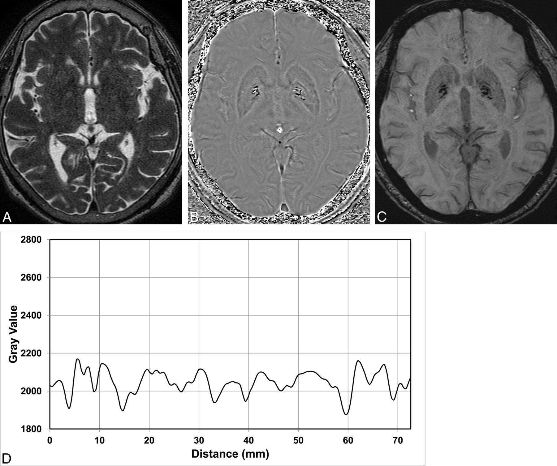
  • Fig 3.
    • Download figure
    • Open in new tab
    • Download powerpoint
    Fig 3.

    A 71-year-old woman who initially presented with right-leg dragging 3 years ago was clinically diagnosed as having probable MSA-p. A, On the T2-weighed axial image, the bilateral posterolateral putamen shows subtle hypointensity. The phase image (B) and final SWI (C) show a marked phase shift in the left posterolateral putamen with loss of lateral convexity of the posterolateral aspect of the putamen, suggesting atrophic change. D, Phase values along the far posterior of both putamina show asymmetric phase-shift values, and mean phase-shift values of the left putamina are measured as 2686.5 Siemens Phase Units.

  • Fig 4.
    • Download figure
    • Open in new tab
    • Download powerpoint
    Fig 4.

    A 69-year-old woman who initially presented with gait disturbance and bradykinesia 8 years ago was clinically diagnosed with IPD. The T2-weighed axial image (A), phase image (B), and final SWI (C) show no substantial signal alteration or atrophy in the bilateral putamina, while excessive iron deposition in the bilateral globus pallidus is observed. D, Phase values along the far posterior portion of both putamina show symmetric phase-shift values, and mean phase-shift values of the right and left putamina are measured as 2114.5 and 2111.7 Siemens Phase Units, respectively.

  • Fig 5.
    • Download figure
    • Open in new tab
    • Download powerpoint
    Fig 5.

    Receiver operating characteristic curves of values measured at the far posterior putamen to distinguish MSA-p from IPD.

Tables

  • Figures
    • View popup
    Table 1:

    Demographic and clinical characteristics of the study groupsa

    MSA-pIPDHealthy ControlP Value
    No.275027
    Mean age (yr)64.8 ± 8.6 (47–77)66.5 ± 6.7 (54–81)64.7 ± 8.5 (47–77).521b
    Male/female ratio13:1423:2711:16.850c
    Mean disease duration (yr)4.7 ± 2.97.8 ± 5.0NA.010d
    H&Y scale2.85 ± 0.721.95 ± 0.57NA<.001d
    No. of patients with asymmetric presentation1710NA<.001c
    • Note:—H&Y scale indicates modified Hoehn and Yahr scale; NA, not applicable.

    • ↵a Data are presented as mean ± SD, with ranges in the parentheses.

    • ↵b One-way analysis of variance test.

    • ↵c χ2 test.

    • ↵d Mann-Whitney U test.

    • View popup
    Table 2:

    Prevalence of each image finding by visual interpretation of SWI in each group

    Imaging FindingMSA-pIPDHealthy Control
    No.275027
    Putaminal atrophy
        Suspicious3 (11.1%)00
        Definite11 (40.7%)00
    Putaminal signal intensity
        Hyperintense10 (37.0%)24 (48.0%)15 (55.6%)
        Isointense3 (11.1%)22 (44.0%)10 (37.0%)
        Hypointense2 (7.4%)4 (8.0%)2 (7.4%)
        Markedly hypointense12 (44.4%)00
    Hot-cross bun sign2 (7.4%)00
    High signal intensity of middle cerebellar peduncle
        Suspicious3 (11.1%)00
        Definite1 (3.7%)00
    Cerebellar atrophy
        Suspicious11 (40.7%)10 (20.0%)0
        Definite3 (11.1%)00
    • View popup
    Table 3:

    Correlation between the clinically symptomatic side and asymmetry of imaging findings

    Clinical SymptomPutaminal Atrophy (n = 14)Marked Signal Hypointensity (n = 12)
    SymmetricRightLeftSymmetricRightLeft
    Symmetric012110
    Right-dominant104203
    Left-dominant051131
    • View popup
    Table 4:

    Quantitatively measured putaminal width and phase-shift values: the ratios of dominant-to-nondominant-side values in the far posterior portion of putamena

    MSA-pIPDHealthy ControlP Value
    Measured values: dominant side
        Putamen widthb3.17 ± 0.653.81 ± 0.533.87 ± 0.47<.001
        Phase-shift valueb2322.2 ± 236.92121.5 ± 44.42136.7 ± 45.8<.001
    Measured values: mean of both sides
        Putamen widthb3.42 ± 0.573.98 ± 0.524.05 ± 0.45<.001
        Phase-shift valueb2258.4 ± 180.12108.1 ± 39.82122.5 ± 37.6<.001
    Ratio of dominant/nondominant side
        Putamen width (shorter/longer side)0.866 ± 0.1230.921 ± 0.0700.912 ± 0.048.095
        Phase-shift valueb (higher/lower side)1.057 ± 0.0651.013 ± 0.0111.013 ± 0.011<.001
    • ↵a Data are presented as means.

    • ↵b MSA-p was significantly different (P < .05) from IPD and healthy controls by post hoc analysis.

PreviousNext
Back to top

In this issue

American Journal of Neuroradiology: 36 (12)
American Journal of Neuroradiology
Vol. 36, Issue 12
1 Dec 2015
  • Table of Contents
  • Index by author
  • Complete Issue (PDF)
Advertisement
Print
Download PDF
Email Article

Thank you for your interest in spreading the word on American Journal of Neuroradiology.

NOTE: We only request your email address so that the person you are recommending the page to knows that you wanted them to see it, and that it is not junk mail. We do not capture any email address.

Enter multiple addresses on separate lines or separate them with commas.
Differentiation of Parkinsonism-Predominant Multiple System Atrophy from Idiopathic Parkinson Disease Using 3T Susceptibility-Weighted MR Imaging, Focusing on Putaminal Change and Lesion Asymmetry
(Your Name) has sent you a message from American Journal of Neuroradiology
(Your Name) thought you would like to see the American Journal of Neuroradiology web site.
CAPTCHA
This question is for testing whether or not you are a human visitor and to prevent automated spam submissions.
Cite this article
I. Hwang, C.-H. Sohn, K.M. Kang, B.S. Jeon, H.-J. Kim, S.H. Choi, T.J. Yun, J.-h. Kim
Differentiation of Parkinsonism-Predominant Multiple System Atrophy from Idiopathic Parkinson Disease Using 3T Susceptibility-Weighted MR Imaging, Focusing on Putaminal Change and Lesion Asymmetry
American Journal of Neuroradiology Dec 2015, 36 (12) 2227-2234; DOI: 10.3174/ajnr.A4442

Citation Manager Formats

  • BibTeX
  • Bookends
  • EasyBib
  • EndNote (tagged)
  • EndNote 8 (xml)
  • Medlars
  • Mendeley
  • Papers
  • RefWorks Tagged
  • Ref Manager
  • RIS
  • Zotero
0 Responses
Respond to this article
Share
Bookmark this article
Differentiation of Parkinsonism-Predominant Multiple System Atrophy from Idiopathic Parkinson Disease Using 3T Susceptibility-Weighted MR Imaging, Focusing on Putaminal Change and Lesion Asymmetry
I. Hwang, C.-H. Sohn, K.M. Kang, B.S. Jeon, H.-J. Kim, S.H. Choi, T.J. Yun, J.-h. Kim
American Journal of Neuroradiology Dec 2015, 36 (12) 2227-2234; DOI: 10.3174/ajnr.A4442
del.icio.us logo Twitter logo Facebook logo Mendeley logo
  • Tweet Widget
  • Facebook Like
  • Google Plus One
Purchase

Jump to section

  • Article
    • Abstract
    • ABBREVIATIONS:
    • Materials and Methods
    • Results
    • Discussion
    • Conclusions
    • Footnotes
    • REFERENCES
  • Figures & Data
  • Supplemental
  • Info & Metrics
  • Responses
  • References
  • PDF

Related Articles

  • PubMed
  • Google Scholar

Cited By...

  • No citing articles found.
  • Crossref
  • Google Scholar

This article has not yet been cited by articles in journals that are participating in Crossref Cited-by Linking.

More in this TOC Section

  • Diagnostic Neuroradiology of Monoclonal Antibodies
  • NCCT vs. MRI for Brain Atrophy in Acute Stroke
  • Clinical Outcomes After Chiari I Decompression
Show more Adult Brain

Similar Articles

Advertisement

Indexed Content

  • Current Issue
  • Accepted Manuscripts
  • Article Preview
  • Past Issues
  • Editorials
  • Editor's Choice
  • Fellows' Journal Club
  • Letters to the Editor
  • Video Articles

Cases

  • Case Collection
  • Archive - Case of the Week
  • Archive - Case of the Month
  • Archive - Classic Case

More from AJNR

  • Trainee Corner
  • Imaging Protocols
  • MRI Safety Corner
  • Book Reviews

Multimedia

  • AJNR Podcasts
  • AJNR Scantastics

Resources

  • Turnaround Time
  • Submit a Manuscript
  • Submit a Video Article
  • Submit an eLetter to the Editor/Response
  • Manuscript Submission Guidelines
  • Statistical Tips
  • Fast Publishing of Accepted Manuscripts
  • Graphical Abstract Preparation
  • Imaging Protocol Submission
  • Evidence-Based Medicine Level Guide
  • Publishing Checklists
  • Author Policies
  • Become a Reviewer/Academy of Reviewers
  • News and Updates

About Us

  • About AJNR
  • Editorial Board
  • Editorial Board Alumni
  • Alerts
  • Permissions
  • Not an AJNR Subscriber? Join Now
  • Advertise with Us
  • Librarian Resources
  • Feedback
  • Terms and Conditions
  • AJNR Editorial Board Alumni

American Society of Neuroradiology

  • Not an ASNR Member? Join Now

© 2025 by the American Society of Neuroradiology All rights, including for text and data mining, AI training, and similar technologies, are reserved.
Print ISSN: 0195-6108 Online ISSN: 1936-959X

Powered by HighWire