
Article Figures & Data
Figures
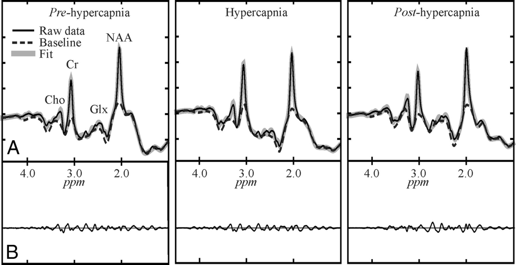
- Fig 1.
Automated spectral fitting of the pre- (left), during- (center), and posthypercapnia (right) WBNAA from 1 subject, all on the same intensity and chemical shift (parts per million [ppm]) scales. Top, A, Whole-head 1H-spectrum (thin black line), estimated baseline (dashed line), and fitted (metabolites + baseline) estimate (thick gray line). Bottom: B, Residual signals (raw-fitted data). Note: 1) The similarity of the pre-, during, and posthypercapnia spectra, suggesting a minimal effect of this physiologic challenge on the brain NAA; 2) the quality of the fit on A; and 3) the consequent vanishing residuals in B; and 4) although other metabolites are also visible in the spectrum, only NAA is implicitly localized by its biochemistry to just the brain.
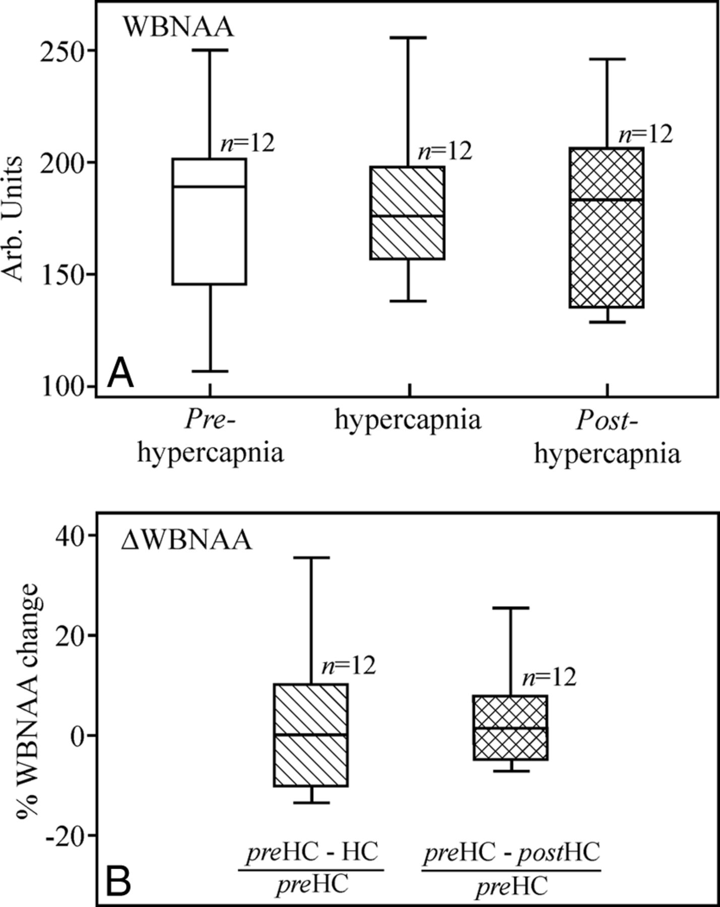
- Fig 2.
A, Boxplots showing the first, second (median), and third quartiles (box) and ±95% (whiskers) of the WBNAA distributions at normocapnia (white), hypercapnia (hatched), and posthypercapnia (cross-hatched). Note the insignificant WBNAA changes (P = .676). B, Boxplots show the percentage of NAA change from baseline normocapnia distribution of the 12 subjects. Note the ∼0% change from normocapnia (preHC) to hypercapnia (HC) (hatched) and from the former to the posthypercapnia (postHC) normocapnia (cross-hatched), underscoring the negligible NAA change as a response to the physiologic CO2 challenge. Arb indicates arbitrary.
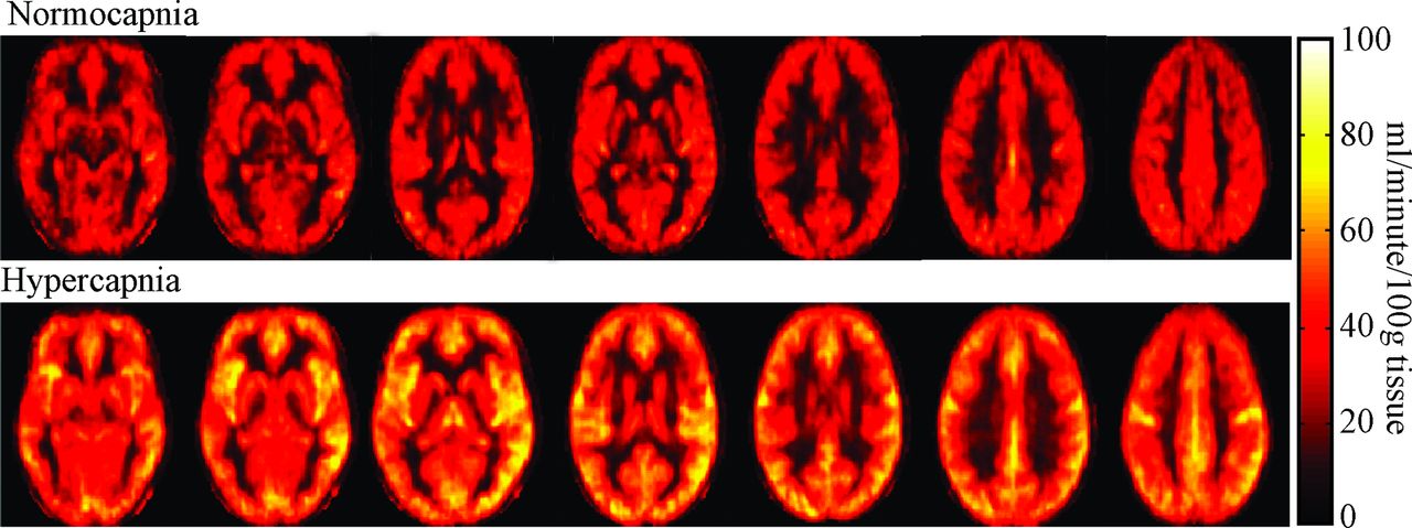
- Fig 3.
Average (n = 12) global CBF maps for 7 representative brain sections. Note the easily visible, ∼50% increase (P < 10−4) in CBF from normocapnia to hypercapnia.
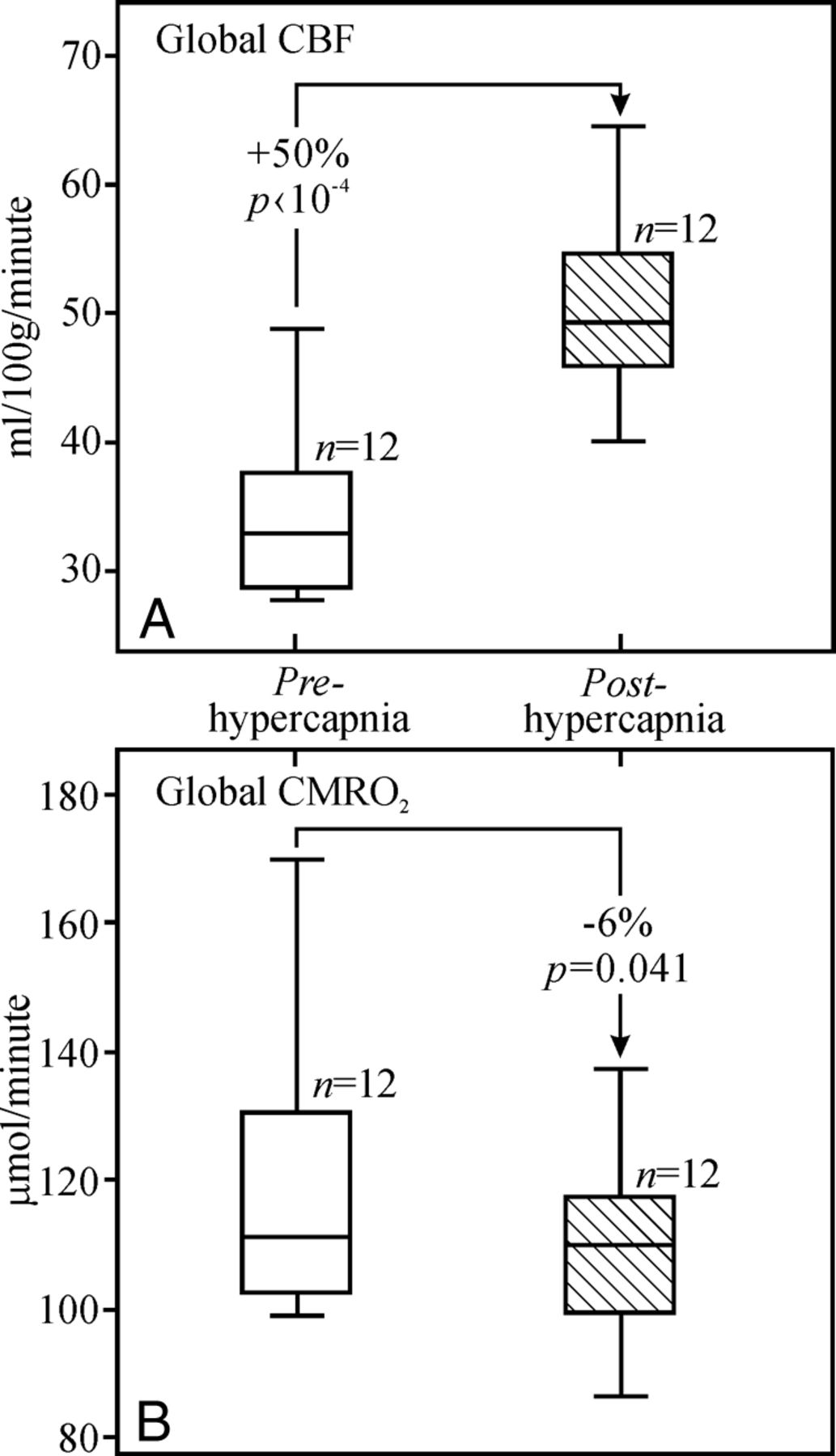
- Fig 4.
Boxplots of the distributions of global CBF (A) and CMRO2 (B) at normocapnia (white) and hypercapnia (hatched). Note the significant (P < 10−4) ∼47% increases in global CBF and the 6.4% (P = .04) decrease in global CMRO2 from normocapnia to hypercapnia.


Tables
Summary of ETCO2 and MRI metrics (mean) for normocapnia and hypercapnia
MRI/MRS and Measures Normocapnia Hypercapnia P Value 1H-MRSa ETCO2 (mm Hg) 44.40 ± 4.1 52.9 ± 2.4 <10−4 WBNAA (arb. unit) 177.9 ± 40.5 178.8 ± 32.3 .88 pCASL MRI ETCO2 (mm Hg) 40.6 ± 4.9 49.4 ± 3.9 <10−4 CBF (mL/100 g/min) 33.9 ± 6.3 50.2 ± 6.9 <10−4 TRUST MRI ETCO2 (mm Hg) 43.3 ± 4.7 52.5 ± 2.9 <10−4 Yv (%) 56.4 ± 5.6 71.9 ± 4.8 <10−4 TRUST and pCASL CMRO2 (μmol/100 g/min) 120.0 ± 23.9 111.2 ± 18.9 .04 Note:—Arb. indicates arbitrary; ETCO2, end-tidal carbon dioxide.
↵a WBNAA values measured with 1H-MRS of the posthypercapnic condition (the second normocapnia) are listed in the main text.


Jump to section
Related Articles
Cited By...
- No citing articles found.