Advertisement

Main menu

  • Home
  • Content
    • Current Issue
    • Accepted Manuscripts
    • Article Preview
    • Past Issue Archive
    • Video Articles
    • AJNR Case Collection
    • Case of the Week Archive
    • Case of the Month Archive
    • Classic Case Archive
  • Special Collections
    • AJNR Awards
    • Low-Field MRI
    • Alzheimer Disease
    • ASNR Foundation Special Collection
    • Photon-Counting CT
    • View All
  • Multimedia
    • AJNR Podcasts
    • AJNR SCANtastic
    • Trainee Corner
    • MRI Safety Corner
    • Imaging Protocols
  • For Authors
    • Submit a Manuscript
    • Submit a Video Article
    • Submit an eLetter to the Editor/Response
    • Manuscript Submission Guidelines
    • Statistical Tips
    • Fast Publishing of Accepted Manuscripts
    • Graphical Abstract Preparation
    • Imaging Protocol Submission
    • Author Policies
  • About Us
    • About AJNR
    • Editorial Board
    • Editorial Board Alumni
  • More
    • Become a Reviewer/Academy of Reviewers
    • Subscribers
    • Permissions
    • Alerts
    • Feedback
    • Advertisers
    • ASNR Home

User menu

  • Alerts
  • Log in

Search

  • Advanced search
American Journal of Neuroradiology

American Journal of Neuroradiology

ASHNR American Society of Functional Neuroradiology ASHNR American Society of Pediatric Neuroradiology ASSR
  • Alerts
  • Log in

Advanced Search

  • Home
  • Content
    • Current Issue
    • Accepted Manuscripts
    • Article Preview
    • Past Issue Archive
    • Video Articles
    • AJNR Case Collection
    • Case of the Week Archive
    • Case of the Month Archive
    • Classic Case Archive
  • Special Collections
    • AJNR Awards
    • Low-Field MRI
    • Alzheimer Disease
    • ASNR Foundation Special Collection
    • Photon-Counting CT
    • View All
  • Multimedia
    • AJNR Podcasts
    • AJNR SCANtastic
    • Trainee Corner
    • MRI Safety Corner
    • Imaging Protocols
  • For Authors
    • Submit a Manuscript
    • Submit a Video Article
    • Submit an eLetter to the Editor/Response
    • Manuscript Submission Guidelines
    • Statistical Tips
    • Fast Publishing of Accepted Manuscripts
    • Graphical Abstract Preparation
    • Imaging Protocol Submission
    • Author Policies
  • About Us
    • About AJNR
    • Editorial Board
    • Editorial Board Alumni
  • More
    • Become a Reviewer/Academy of Reviewers
    • Subscribers
    • Permissions
    • Alerts
    • Feedback
    • Advertisers
    • ASNR Home
  • Follow AJNR on Twitter
  • Visit AJNR on Facebook
  • Follow AJNR on Instagram
  • Join AJNR on LinkedIn
  • RSS Feeds

AJNR Awards, New Junior Editors, and more. Read the latest AJNR updates

Research ArticleAdult Brain
Open Access

Nigrosome 1 Detection at 3T MRI for the Diagnosis of Early-Stage Idiopathic Parkinson Disease: Assessment of Diagnostic Accuracy and Agreement on Imaging Asymmetry and Clinical Laterality

Y. Noh, Y.H. Sung, J. Lee and E.Y. Kim
American Journal of Neuroradiology November 2015, 36 (11) 2010-2016; DOI: https://doi.org/10.3174/ajnr.A4412
Y. Noh
aFrom the Departments of Neurology (Y.N., Y.H.S.)
  • Find this author on Google Scholar
  • Find this author on PubMed
  • Search for this author on this site
Y.H. Sung
aFrom the Departments of Neurology (Y.N., Y.H.S.)
  • Find this author on Google Scholar
  • Find this author on PubMed
  • Search for this author on this site
J. Lee
cDepartment of Electrical and Computer Engineering (Jongho Lee), Seoul National University, Seoul, Korea.
  • Find this author on Google Scholar
  • Find this author on PubMed
  • Search for this author on this site
E.Y. Kim
bRadiology (E.Y.K.), Gachon University Gil Medical Center, Incheon, Korea
  • Find this author on Google Scholar
  • Find this author on PubMed
  • Search for this author on this site
  • ORCID record for E.Y. Kim
  • Article
  • Figures & Data
  • Info & Metrics
  • Responses
  • References
  • PDF
Loading

Article Figures & Data

Figures

  • Tables
  • Fig 1.
    • Download figure
    • Open in new tab
    • Download powerpoint
    Fig 1.

    Visual assessment of nigrosome 1 in MR imaging. On the oblique axial MEDIC imaging, the evaluation of nigrosome 1 (arrows) was performed on 3 sections: an upper section at the lower tip of the red nucleus and the 2 successive sections (A). The oblique coronal MEDIC imaging was also assessed on 3 sections: an anterior section at the anterior tip of the red nucleus and the 2 consecutive sections (B). In this particular 71-year-old woman, the nigrosome 1 appears normal on both sides.
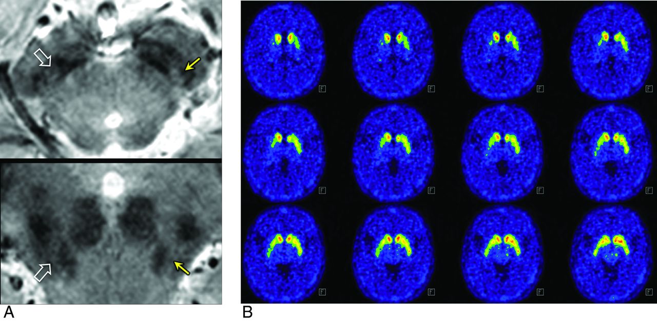
  • Fig 2.
    • Download figure
    • Open in new tab
    • Download powerpoint
    Fig 2.

    Degree of abnormality on MR imaging. Possibly abnormal and definitely abnormal were rated in the left (arrows) and right (open arrows) nigrosome 1, respectively (A). Similar asymmetry was observed on 18F-FP-CIT PET. This 53-year-old woman has H&Y stage 2 and UPDRS Part III (motor score) of 4 and 1 on the left and right, respectively, which is in concordance with asymmetry on both MR imaging and 18F-FP-CIT PET.
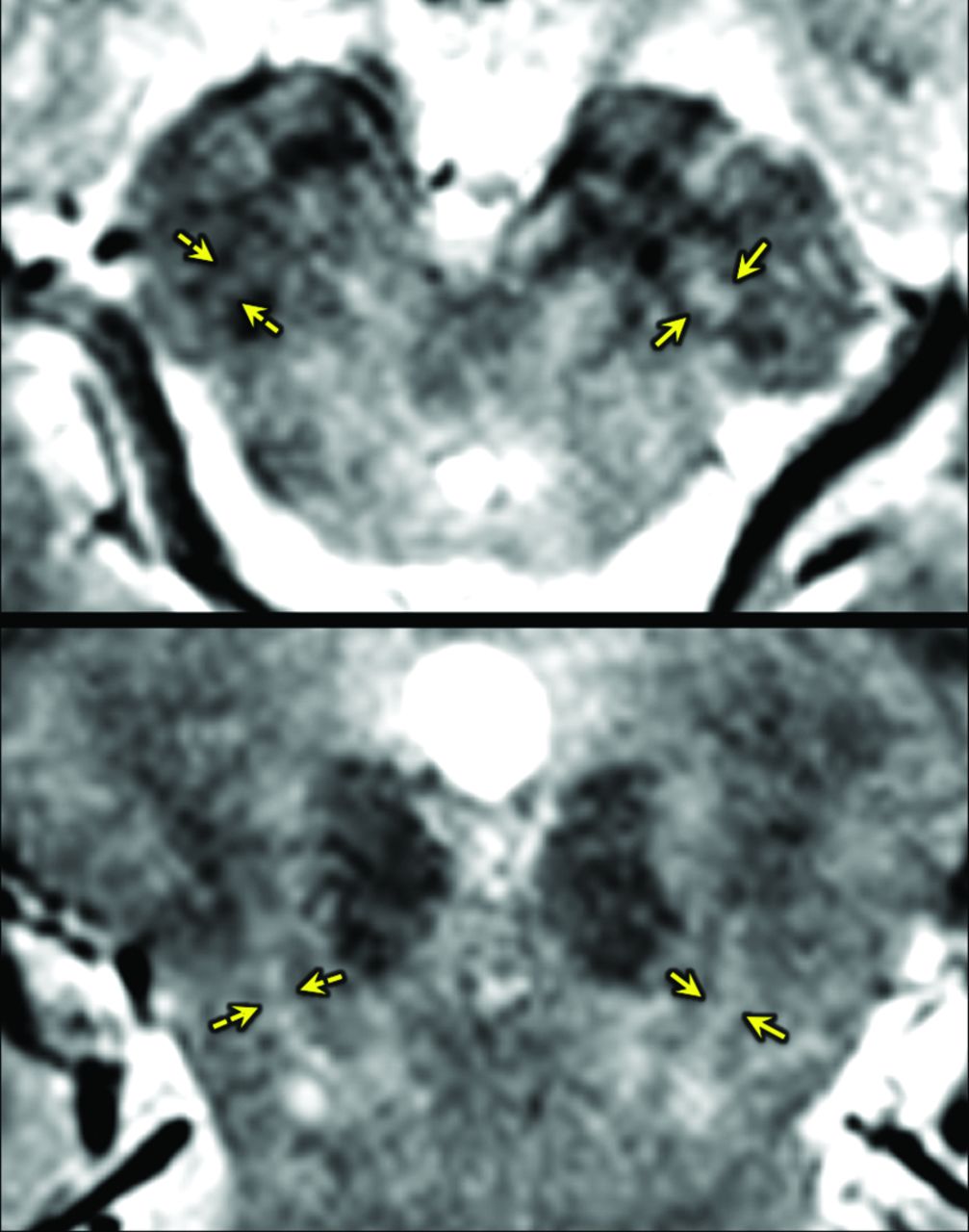
  • Fig 3.
    • Download figure
    • Open in new tab
    • Download powerpoint
    Fig 3.

    False-positive on MR imaging. In this 64-year-old healthy female subject, both reviewers interpreted the left nigrosome (arrows) was as normal, whereas they called the right one (broken arrows) abnormal.

  • Fig 4.
    • Download figure
    • Open in new tab
    • Download powerpoint
    Fig 4.

    Asymmetry on MR imaging but not on 18F-FP-CIT in a 78-year-old woman with H&Y stage 2 and UPDRS of 4 and 2 on the right and left shows that the left nigrosome 1 is more affected (arrows) than the right one (open arrows) (A), whereas 18F-FP-CIT PET shows relatively symmetric findings (B).

Tables

  • Figures
    • View popup
    Table 1:

    Demographic characteristics and imaging findings in study populationa

    IPD (n = 24)Healthy Control (n = 13)P Value
    Age (yr)63.6 ± 10.9761.6 ± 12.29.620
    Female sex (%)10 (41.7%)9 (69.2%).170
    MMSE27.5 (25.0–29.0)29.0 (28.0–29.5).036
    Onset age (yr)62.4 ± 11.59–
    Disease duration (mo)9.0 (3.0–12.0)–
    H&Y scale (1/1.5/2)2/2/20–
    UPDRS I1.0 (0–2.0)–
    UPDRS II6.75 ± 2.85–
    UPDRS III13.5 ± 6.19–
    • Note:—MMSE indicates Mini-Mental State Examination; UPDRS, Unified Parkinson Disease Rating Scale, –, not applicable.

    • ↵a Data are presented as mean ± SD for normally distributed variables and median (interquartile ranges) for non-normally distributed variables. The χ2 test with Yates continuity correction was used.

    • View popup
    Table 2:

    Interpretation of nigrosome 1 by 2 reviewers

    Reviewer 1Reviewer 2
    RightLeftRightLeft
    Normal (No.)10131211
    Possibly abnormal (No.)7523
    Definitely abnormal (No.)20192323
PreviousNext
Back to top

In this issue

American Journal of Neuroradiology: 36 (11)
American Journal of Neuroradiology
Vol. 36, Issue 11
1 Nov 2015
  • Table of Contents
  • Index by author
  • Complete Issue (PDF)
Advertisement
Print
Download PDF
Email Article

Thank you for your interest in spreading the word on American Journal of Neuroradiology.

NOTE: We only request your email address so that the person you are recommending the page to knows that you wanted them to see it, and that it is not junk mail. We do not capture any email address.

Enter multiple addresses on separate lines or separate them with commas.
Nigrosome 1 Detection at 3T MRI for the Diagnosis of Early-Stage Idiopathic Parkinson Disease: Assessment of Diagnostic Accuracy and Agreement on Imaging Asymmetry and Clinical Laterality
(Your Name) has sent you a message from American Journal of Neuroradiology
(Your Name) thought you would like to see the American Journal of Neuroradiology web site.
CAPTCHA
This question is for testing whether or not you are a human visitor and to prevent automated spam submissions.
Cite this article
Y. Noh, Y.H. Sung, J. Lee, E.Y. Kim
Nigrosome 1 Detection at 3T MRI for the Diagnosis of Early-Stage Idiopathic Parkinson Disease: Assessment of Diagnostic Accuracy and Agreement on Imaging Asymmetry and Clinical Laterality
American Journal of Neuroradiology Nov 2015, 36 (11) 2010-2016; DOI: 10.3174/ajnr.A4412

Citation Manager Formats

  • BibTeX
  • Bookends
  • EasyBib
  • EndNote (tagged)
  • EndNote 8 (xml)
  • Medlars
  • Mendeley
  • Papers
  • RefWorks Tagged
  • Ref Manager
  • RIS
  • Zotero
0 Responses
Respond to this article
Share
Bookmark this article
Nigrosome 1 Detection at 3T MRI for the Diagnosis of Early-Stage Idiopathic Parkinson Disease: Assessment of Diagnostic Accuracy and Agreement on Imaging Asymmetry and Clinical Laterality
Y. Noh, Y.H. Sung, J. Lee, E.Y. Kim
American Journal of Neuroradiology Nov 2015, 36 (11) 2010-2016; DOI: 10.3174/ajnr.A4412
del.icio.us logo Twitter logo Facebook logo Mendeley logo
  • Tweet Widget
  • Facebook Like
  • Google Plus One
Purchase

Jump to section

  • Article
    • Abstract
    • ABBREVIATIONS:
    • Materials and Methods
    • Results
    • Discussion
    • Conclusions
    • Footnotes
    • REFERENCES
  • Figures & Data
  • Info & Metrics
  • Responses
  • References
  • PDF

Related Articles

  • No related articles found.
  • PubMed
  • Google Scholar

Cited By...

  • Determining the Degree of Dopaminergic Denervation Based on the Loss of Nigral Hyperintensity on SMWI in Parkinsonism
  • Visualization of Nigrosome 1 from the Viewpoint of Anatomic Structure
  • Protocol of a single group prospective observational study on the diagnostic value of 3T susceptibility weighted MRI of nigrosome-1 in patients with parkinsonian symptoms: the N3iPD study (nigrosomal iron imaging in Parkinsons disease)
  • Crossref
  • Google Scholar

This article has not yet been cited by articles in journals that are participating in Crossref Cited-by Linking.

More in this TOC Section

  • Diagnostic Neuroradiology of Monoclonal Antibodies
  • Clinical Outcomes After Chiari I Decompression
  • Segmentation of Brain Metastases with BLAST
Show more Adult Brain

Similar Articles

Advertisement

Indexed Content

  • Current Issue
  • Accepted Manuscripts
  • Article Preview
  • Past Issues
  • Editorials
  • Editor's Choice
  • Fellows' Journal Club
  • Letters to the Editor
  • Video Articles

Cases

  • Case Collection
  • Archive - Case of the Week
  • Archive - Case of the Month
  • Archive - Classic Case

More from AJNR

  • Trainee Corner
  • Imaging Protocols
  • MRI Safety Corner
  • Book Reviews

Multimedia

  • AJNR Podcasts
  • AJNR Scantastics

Resources

  • Turnaround Time
  • Submit a Manuscript
  • Submit a Video Article
  • Submit an eLetter to the Editor/Response
  • Manuscript Submission Guidelines
  • Statistical Tips
  • Fast Publishing of Accepted Manuscripts
  • Graphical Abstract Preparation
  • Imaging Protocol Submission
  • Evidence-Based Medicine Level Guide
  • Publishing Checklists
  • Author Policies
  • Become a Reviewer/Academy of Reviewers
  • News and Updates

About Us

  • About AJNR
  • Editorial Board
  • Editorial Board Alumni
  • Alerts
  • Permissions
  • Not an AJNR Subscriber? Join Now
  • Advertise with Us
  • Librarian Resources
  • Feedback
  • Terms and Conditions
  • AJNR Editorial Board Alumni

American Society of Neuroradiology

  • Not an ASNR Member? Join Now

© 2025 by the American Society of Neuroradiology All rights, including for text and data mining, AI training, and similar technologies, are reserved.
Print ISSN: 0195-6108 Online ISSN: 1936-959X

Powered by HighWire