Advertisement

Main menu

  • Home
  • Content
    • Current Issue
    • Accepted Manuscripts
    • Article Preview
    • Past Issue Archive
    • Video Articles
    • AJNR Case Collection
    • Case of the Week Archive
    • Case of the Month Archive
    • Classic Case Archive
  • Special Collections
    • AJNR Awards
    • Low-Field MRI
    • Alzheimer Disease
    • ASNR Foundation Special Collection
    • Photon-Counting CT
    • View All
  • Multimedia
    • AJNR Podcasts
    • AJNR SCANtastic
    • Trainee Corner
    • MRI Safety Corner
    • Imaging Protocols
  • For Authors
    • Submit a Manuscript
    • Submit a Video Article
    • Submit an eLetter to the Editor/Response
    • Manuscript Submission Guidelines
    • Statistical Tips
    • Fast Publishing of Accepted Manuscripts
    • Graphical Abstract Preparation
    • Imaging Protocol Submission
    • Author Policies
  • About Us
    • About AJNR
    • Editorial Board
    • Editorial Board Alumni
  • More
    • Become a Reviewer/Academy of Reviewers
    • Subscribers
    • Permissions
    • Alerts
    • Feedback
    • Advertisers
    • ASNR Home

User menu

  • Alerts
  • Log in

Search

  • Advanced search
American Journal of Neuroradiology

American Journal of Neuroradiology

ASHNR American Society of Functional Neuroradiology ASHNR American Society of Pediatric Neuroradiology ASSR
  • Alerts
  • Log in

Advanced Search

  • Home
  • Content
    • Current Issue
    • Accepted Manuscripts
    • Article Preview
    • Past Issue Archive
    • Video Articles
    • AJNR Case Collection
    • Case of the Week Archive
    • Case of the Month Archive
    • Classic Case Archive
  • Special Collections
    • AJNR Awards
    • Low-Field MRI
    • Alzheimer Disease
    • ASNR Foundation Special Collection
    • Photon-Counting CT
    • View All
  • Multimedia
    • AJNR Podcasts
    • AJNR SCANtastic
    • Trainee Corner
    • MRI Safety Corner
    • Imaging Protocols
  • For Authors
    • Submit a Manuscript
    • Submit a Video Article
    • Submit an eLetter to the Editor/Response
    • Manuscript Submission Guidelines
    • Statistical Tips
    • Fast Publishing of Accepted Manuscripts
    • Graphical Abstract Preparation
    • Imaging Protocol Submission
    • Author Policies
  • About Us
    • About AJNR
    • Editorial Board
    • Editorial Board Alumni
  • More
    • Become a Reviewer/Academy of Reviewers
    • Subscribers
    • Permissions
    • Alerts
    • Feedback
    • Advertisers
    • ASNR Home
  • Follow AJNR on Twitter
  • Visit AJNR on Facebook
  • Follow AJNR on Instagram
  • Join AJNR on LinkedIn
  • RSS Feeds

AJNR Awards, New Junior Editors, and more. Read the latest AJNR updates

Getting new auth cookie, if you see this message a lot, tell someone!
Review ArticleAdult Brain
Open Access

Current Applications of MRI-Guided Laser Interstitial Thermal Therapy in the Treatment of Brain Neoplasms and Epilepsy: A Radiologic and Neurosurgical Overview

R. Medvid, A. Ruiz, R.J. Komotar, J.R. Jagid, M.E. Ivan, R.M. Quencer and M.B. Desai
American Journal of Neuroradiology November 2015, 36 (11) 1998-2006; DOI: https://doi.org/10.3174/ajnr.A4362
R. Medvid
aFrom the Department of Radiology (R.M., A.R., R.M.Q., M.B.D.), Division of Neuroradiology
  • Find this author on Google Scholar
  • Find this author on PubMed
  • Search for this author on this site
A. Ruiz
aFrom the Department of Radiology (R.M., A.R., R.M.Q., M.B.D.), Division of Neuroradiology
  • Find this author on Google Scholar
  • Find this author on PubMed
  • Search for this author on this site
R.J. Komotar
bDepartment of Neurological Surgery (J.R.J., R.J.K., M.E.I.), Jackson Memorial Hospital/University of Miami Hospital, Miami, Florida.
  • Find this author on Google Scholar
  • Find this author on PubMed
  • Search for this author on this site
J.R. Jagid
bDepartment of Neurological Surgery (J.R.J., R.J.K., M.E.I.), Jackson Memorial Hospital/University of Miami Hospital, Miami, Florida.
  • Find this author on Google Scholar
  • Find this author on PubMed
  • Search for this author on this site
M.E. Ivan
bDepartment of Neurological Surgery (J.R.J., R.J.K., M.E.I.), Jackson Memorial Hospital/University of Miami Hospital, Miami, Florida.
  • Find this author on Google Scholar
  • Find this author on PubMed
  • Search for this author on this site
R.M. Quencer
aFrom the Department of Radiology (R.M., A.R., R.M.Q., M.B.D.), Division of Neuroradiology
  • Find this author on Google Scholar
  • Find this author on PubMed
  • Search for this author on this site
M.B. Desai
aFrom the Department of Radiology (R.M., A.R., R.M.Q., M.B.D.), Division of Neuroradiology
  • Find this author on Google Scholar
  • Find this author on PubMed
  • Search for this author on this site
  • Article
  • Figures & Data
  • Info & Metrics
  • Responses
  • References
  • PDF
Loading

Article Figures & Data

Figures

  • Fig 1.
    • Download figure
    • Open in new tab
    • Download powerpoint
    Fig 1.

    Once the location of the laser probe is confirmed, ablation can proceed. A, T2 image demonstrates a properly positioned laser probe within a metastatic melanoma lesion. B, Heat map: during ablation, temperatures surrounding the laser tip are continuously updated and depicted with various colors. C, Damage zone images: orange color depicts the area of tissue that has been successfully ablated (damage zone) on the basis of the Arrhenius model of thermal tissue ablation.

  • Fig 2.
    • Download figure
    • Open in new tab
    • Download powerpoint
    Fig 2.

    Concentric zones: T1 contrast-enhanced (T1C) and T2WI 24 hours after laser ablation of a recurrent right cerebellar metastatic lesion in a 71-year-old female patient with history of breast carcinoma: 1) probe track, 2) central zone, 3) peripheral zone, 4) peripherally enhancing rim, 5) marginal zone. Note that the concentric zones appear as inverse images on T1C and T2 images.

  • Fig 3.
    • Download figure
    • Open in new tab
    • Download powerpoint
    Fig 3.

    T1 contrast-enhanced images demonstrating the normal evolution of a LITT-treated metastatic left cerebellar lesion, which recurred after SRS in a 70-year-old female patient with history of ovarian adenocarcinoma. Note the expected increase in the size of the treated lesion at 2-month follow-up and a steady decrease in size on subsequent follow-up.

  • Fig 4.
    • Download figure
    • Open in new tab
    • Download powerpoint
    Fig 4.

    T2WI demonstrating the normal evolution of an LITT-treated left splenial low-grade astrocytoma in a 30-year-old male patient with a history of type 1 neurofibromatosis. Note the expected increase in the size of the lesion and marginal zone at 1-month follow-up and subsequent decrease at 3 months.

  • Fig 5.
    • Download figure
    • Open in new tab
    • Download powerpoint
    Fig 5.

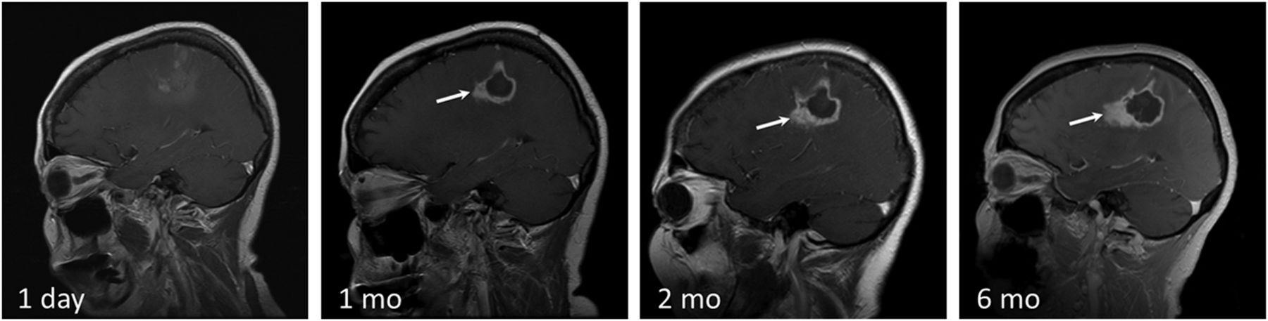
    Disease recurrence after treatment with LITT. T1 contrast-enhanced images in a 58-year-old man with GBM status post surgical excision, chemoradiation, and SRS for a recurrent left parietal lobe lesion. The second recurrence was treated with LITT. Note irregular peripheral nodular enhancement at 1-month follow-up (white arrow), which progressively increases in size at 2 and 6 months. Findings are consistent with disease recurrence.

  • Fig 6.
    • Download figure
    • Open in new tab
    • Download powerpoint
    Fig 6.

    LITT of radiation necrosis with subsequent disease recurrence in a 68-year-old female patient with lung squamous cell carcinoma status post surgical excision and SRS of a metastatic brain lesion in the left parietal lobe, which subsequently resulted in radiation necrosis. Medically intractable radiation necrosis was treated with LITT. Pre-LITT imaging demonstrated an enhancing lesion in the left parietal lobe on T1 contrast-enhanced (not shown) with significant vasogenic edema on T2 images (dashed white arrow on T2 image labeled “pre”). Dynamic imaging pre-LITT (not shown) did not demonstrate a significant increase in CBF or CBV. The patient was not treated with bevacizumab, and RN was favored over recurrence. Note a significant decrease in vasogenic edema 1 month after treatment (dashed white arrow), coinciding with symptomatic improvement. T2 images obtained at 4-month follow-up demonstrate a significant increase in peritumoral vasogenic edema. There is significant thickening of the peripheral zone of enhancement (white arrow) on T1 contrast-enhanced images at 4 months compared with 1 month. Findings are concerning for tumor recurrence within a treated RN lesion, which was corroborated on PET CT (not shown).

  • Fig 7.
    • Download figure
    • Open in new tab
    • Download powerpoint
    Fig 7.

    Mesial temporal sclerosis confirmed on preprocedural FDG-PET (black arrow), which showed decreased FDG uptake in the left hippocampal/parahippocampal region. The lesion was not noted on prior contrast-enhanced MR imaging and did not demonstrate enhancement (not shown). Intraprocedural T2 ablation map demonstrates the laser probe tip within the left medial temporal lobe (white arrow). T1 and T1 contrast-enhanced images 24 hours after LITT demonstrate an oval, rather than round, postablation lesion with signal characteristics similar to those of contrast-enhancing lesions (see Fig 2). The elongated shape is due to sequential probe retraction during ablation to cover the entire left hippocampo-amygdalar area.

PreviousNext
Back to top

In this issue

American Journal of Neuroradiology: 36 (11)
American Journal of Neuroradiology
Vol. 36, Issue 11
1 Nov 2015
  • Table of Contents
  • Index by author
  • Complete Issue (PDF)
Advertisement
Print
Download PDF
Email Article

Thank you for your interest in spreading the word on American Journal of Neuroradiology.

NOTE: We only request your email address so that the person you are recommending the page to knows that you wanted them to see it, and that it is not junk mail. We do not capture any email address.

Enter multiple addresses on separate lines or separate them with commas.
Current Applications of MRI-Guided Laser Interstitial Thermal Therapy in the Treatment of Brain Neoplasms and Epilepsy: A Radiologic and Neurosurgical Overview
(Your Name) has sent you a message from American Journal of Neuroradiology
(Your Name) thought you would like to see the American Journal of Neuroradiology web site.
CAPTCHA
This question is for testing whether or not you are a human visitor and to prevent automated spam submissions.
Cite this article
R. Medvid, A. Ruiz, R.J. Komotar, J.R. Jagid, M.E. Ivan, R.M. Quencer, M.B. Desai
Current Applications of MRI-Guided Laser Interstitial Thermal Therapy in the Treatment of Brain Neoplasms and Epilepsy: A Radiologic and Neurosurgical Overview
American Journal of Neuroradiology Nov 2015, 36 (11) 1998-2006; DOI: 10.3174/ajnr.A4362

Citation Manager Formats

  • BibTeX
  • Bookends
  • EasyBib
  • EndNote (tagged)
  • EndNote 8 (xml)
  • Medlars
  • Mendeley
  • Papers
  • RefWorks Tagged
  • Ref Manager
  • RIS
  • Zotero
0 Responses
Respond to this article
Share
Bookmark this article
Current Applications of MRI-Guided Laser Interstitial Thermal Therapy in the Treatment of Brain Neoplasms and Epilepsy: A Radiologic and Neurosurgical Overview
R. Medvid, A. Ruiz, R.J. Komotar, J.R. Jagid, M.E. Ivan, R.M. Quencer, M.B. Desai
American Journal of Neuroradiology Nov 2015, 36 (11) 1998-2006; DOI: 10.3174/ajnr.A4362
del.icio.us logo Twitter logo Facebook logo Mendeley logo
  • Tweet Widget
  • Facebook Like
  • Google Plus One
Purchase

Jump to section

  • Article
    • Abstract
    • ABBREVIATIONS:
    • Conclusions
    • REFERENCES
  • Figures & Data
  • Info & Metrics
  • Responses
  • References
  • PDF

Related Articles

  • No related articles found.
  • PubMed
  • Google Scholar

Cited By...

  • Long-term brain network reorganization predicts responsive neurostimulation outcomes for focal epilepsy
  • Theranostic OCT microneedle for fast ultrahigh-resolution deep-brain imaging and efficient laser ablation in vivo
  • Dynamic Contrast-Enhanced MRI in Patients with Brain Metastases Undergoing Laser Interstitial Thermal Therapy: A Pilot Study
  • MR Thermography-Guided Head and Neck Lesion Laser Ablation
  • Curative and palliative MRI-guided laser ablation for drug-resistant epilepsy
  • Epilepsy: Five new things
  • Crossref
  • Google Scholar

This article has not yet been cited by articles in journals that are participating in Crossref Cited-by Linking.

More in this TOC Section

  • Diagnostic Neuroradiology of Monoclonal Antibodies
  • Clinical Outcomes After Chiari I Decompression
  • Segmentation of Brain Metastases with BLAST
Show more Adult Brain

Similar Articles

Advertisement

Indexed Content

  • Current Issue
  • Accepted Manuscripts
  • Article Preview
  • Past Issues
  • Editorials
  • Editor's Choice
  • Fellows' Journal Club
  • Letters to the Editor
  • Video Articles

Cases

  • Case Collection
  • Archive - Case of the Week
  • Archive - Case of the Month
  • Archive - Classic Case

More from AJNR

  • Trainee Corner
  • Imaging Protocols
  • MRI Safety Corner
  • Book Reviews

Multimedia

  • AJNR Podcasts
  • AJNR Scantastics

Resources

  • Turnaround Time
  • Submit a Manuscript
  • Submit a Video Article
  • Submit an eLetter to the Editor/Response
  • Manuscript Submission Guidelines
  • Statistical Tips
  • Fast Publishing of Accepted Manuscripts
  • Graphical Abstract Preparation
  • Imaging Protocol Submission
  • Evidence-Based Medicine Level Guide
  • Publishing Checklists
  • Author Policies
  • Become a Reviewer/Academy of Reviewers
  • News and Updates

About Us

  • About AJNR
  • Editorial Board
  • Editorial Board Alumni
  • Alerts
  • Permissions
  • Not an AJNR Subscriber? Join Now
  • Advertise with Us
  • Librarian Resources
  • Feedback
  • Terms and Conditions
  • AJNR Editorial Board Alumni

American Society of Neuroradiology

  • Not an ASNR Member? Join Now

© 2025 by the American Society of Neuroradiology All rights, including for text and data mining, AI training, and similar technologies, are reserved.
Print ISSN: 0195-6108 Online ISSN: 1936-959X

Powered by HighWire