
Article Figures & Data
Figures
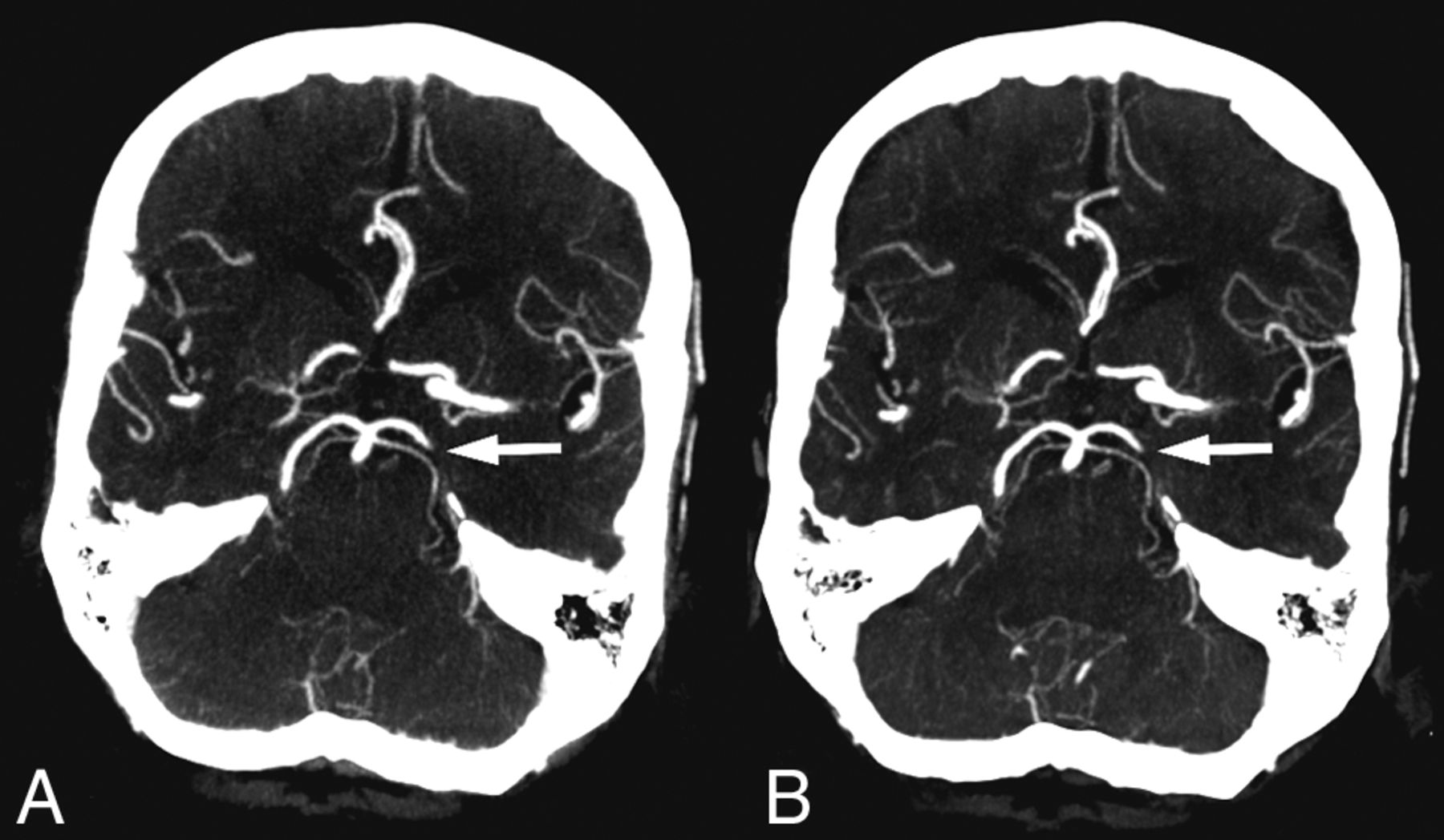
- Fig 1.
Standard CT angiography (A) and timing-invariant CT angiography (B) images in a patient with a left-sided middle cerebral artery occlusion in the M2 segment. The occlusion was detected on both CT angiography techniques.
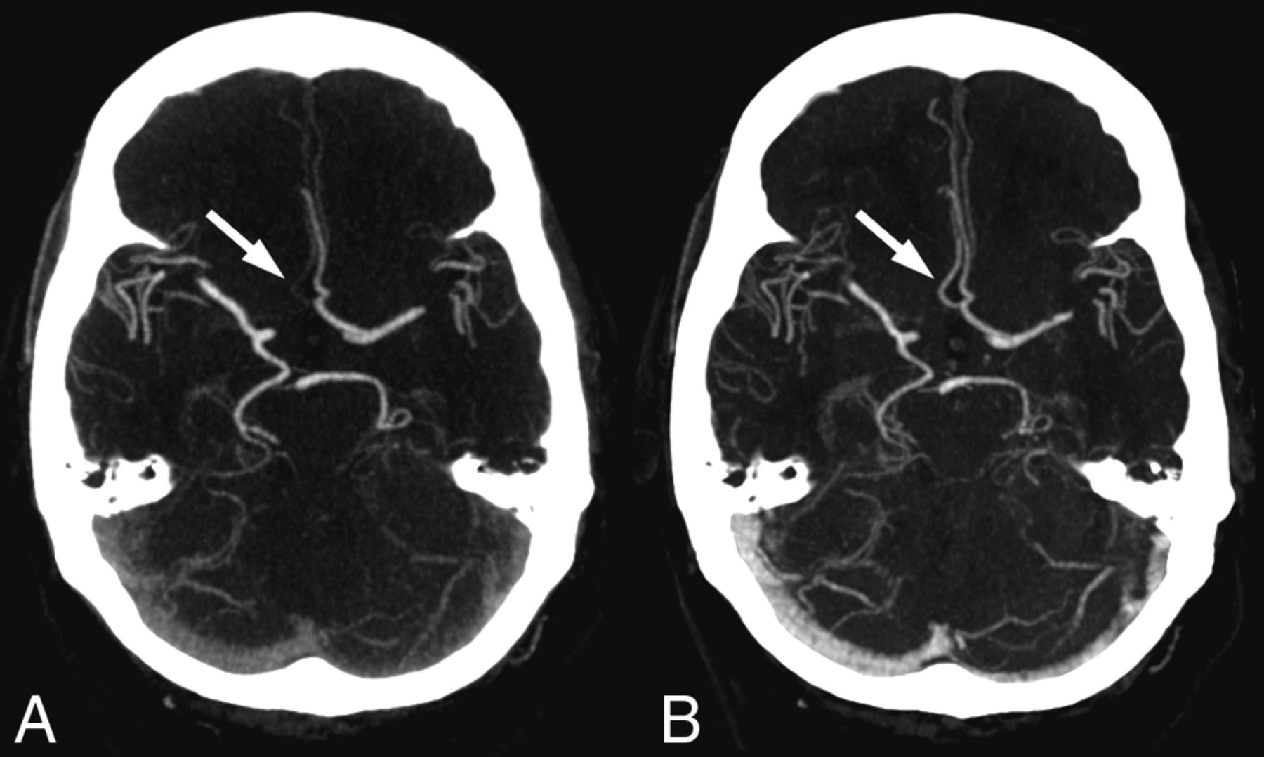
- Fig 2.
Standard CT angiography (A) and timing-invariant CT angiography (B) images in a patient with a left-sided posterior cerebral artery occlusion. This was the only occlusion missed on timing-invariant CTA and was considered a result of observer variation because it was scored as a false-negative finding by 3 observers on timing-invariant CTA and by 2 observers on standard CTA.
- Fig 3.
Images show the effect of delayed contrast material arrival in a patient with right-sided hemiparesis. A, On standard CT angiography, the right-sided anterior cerebral artery was considered occluded in both segments A1 and A2. B, On timing-invariant CTA, the right-sided anterior cerebral artery was considered hypoplastic in segment A1 and patent in segment A2. Standard CTA shows only faint enhancement in the right-sided A2 segment (arrow) because the bulk of contrast material arrived after the standard CTA acquisition. Timing-invariant CT shows strong A2 enhancement (arrow) because it is delay-insensitive and displays maximal contrast enhancement with time.


Tables
Diagnostic performance of standard CTA and timing-invariant CTA for assessment of artery occlusion in the various territoriesa
Territory Occlusions Standard CTA Timing-Invariant CTA Sensitivity Specificity Sensitivity Specificity Overall 47 96% (90–100) 100% (99–100) 98% (94–100) 100% (100–100) Internal carotid artery 9 100% (100–100) 100% (100–100) 100% (100–100) 100% (100–100) Anterior cerebral artery 2 100% (100–100) 99% (98–100) 100% (100–100) 99% (98–100) Middle cerebral artery 33 94% (86–100) 100% (100–100) 100% (100–100) 99% (98–100) Segment 1 (M1) 25 100% (100–100) 100% (100–100) 100% (100–100) 100% (100–100) Segment 2+ (M2+) 8 75% (45–100) 100% (100–100) 100% (100–100) 100% (100–100) Basilar artery 1 100% (100–100) 100% (100–100) 100% (100–100) 100% (100–100) Posterior cerebral artery 2 100% (100–100) 99% (98–100) 50% (0–100) 100% (100–100) ↵a Data in parentheses are 95% confidence intervals.


