Advertisement

Main menu

  • Home
  • Content
    • Current Issue
    • Accepted Manuscripts
    • Article Preview
    • Past Issue Archive
    • Video Articles
    • AJNR Case Collection
    • Case of the Week Archive
    • Case of the Month Archive
    • Classic Case Archive
  • Special Collections
    • AJNR Awards
    • Low-Field MRI
    • Alzheimer Disease
    • ASNR Foundation Special Collection
    • Photon-Counting CT
    • View All
  • Multimedia
    • AJNR Podcasts
    • AJNR SCANtastic
    • Trainee Corner
    • MRI Safety Corner
    • Imaging Protocols
  • For Authors
    • Submit a Manuscript
    • Submit a Video Article
    • Submit an eLetter to the Editor/Response
    • Manuscript Submission Guidelines
    • Statistical Tips
    • Fast Publishing of Accepted Manuscripts
    • Graphical Abstract Preparation
    • Imaging Protocol Submission
    • Author Policies
  • About Us
    • About AJNR
    • Editorial Board
    • Editorial Board Alumni
  • More
    • Become a Reviewer/Academy of Reviewers
    • Subscribers
    • Permissions
    • Alerts
    • Feedback
    • Advertisers
    • ASNR Home

User menu

  • Alerts
  • Log in

Search

  • Advanced search
American Journal of Neuroradiology

American Journal of Neuroradiology

ASHNR American Society of Functional Neuroradiology ASHNR American Society of Pediatric Neuroradiology ASSR
  • Alerts
  • Log in

Advanced Search

  • Home
  • Content
    • Current Issue
    • Accepted Manuscripts
    • Article Preview
    • Past Issue Archive
    • Video Articles
    • AJNR Case Collection
    • Case of the Week Archive
    • Case of the Month Archive
    • Classic Case Archive
  • Special Collections
    • AJNR Awards
    • Low-Field MRI
    • Alzheimer Disease
    • ASNR Foundation Special Collection
    • Photon-Counting CT
    • View All
  • Multimedia
    • AJNR Podcasts
    • AJNR SCANtastic
    • Trainee Corner
    • MRI Safety Corner
    • Imaging Protocols
  • For Authors
    • Submit a Manuscript
    • Submit a Video Article
    • Submit an eLetter to the Editor/Response
    • Manuscript Submission Guidelines
    • Statistical Tips
    • Fast Publishing of Accepted Manuscripts
    • Graphical Abstract Preparation
    • Imaging Protocol Submission
    • Author Policies
  • About Us
    • About AJNR
    • Editorial Board
    • Editorial Board Alumni
  • More
    • Become a Reviewer/Academy of Reviewers
    • Subscribers
    • Permissions
    • Alerts
    • Feedback
    • Advertisers
    • ASNR Home
  • Follow AJNR on Twitter
  • Visit AJNR on Facebook
  • Follow AJNR on Instagram
  • Join AJNR on LinkedIn
  • RSS Feeds

AJNR Awards, New Junior Editors, and more. Read the latest AJNR updates

Research ArticleAdult Brain
Open Access

Gray Matter Volume Reduction Is Associated with Cognitive Impairment in Neuromyelitis Optica

Q. Wang, N. Zhang, W. Qin, Y. Li, Y. Fu, T. Li, J. Shao, L. Yang, F.-D. Shi and C. Yu
American Journal of Neuroradiology October 2015, 36 (10) 1822-1829; DOI: https://doi.org/10.3174/ajnr.A4403
Q. Wang
aFrom the Department of Radiology and Tianjin Key Laboratory of Functional Imaging (Q.W., N.Z., W.Q., C.Y.)
  • Find this author on Google Scholar
  • Find this author on PubMed
  • Search for this author on this site
N. Zhang
aFrom the Department of Radiology and Tianjin Key Laboratory of Functional Imaging (Q.W., N.Z., W.Q., C.Y.)
  • Find this author on Google Scholar
  • Find this author on PubMed
  • Search for this author on this site
W. Qin
aFrom the Department of Radiology and Tianjin Key Laboratory of Functional Imaging (Q.W., N.Z., W.Q., C.Y.)
  • Find this author on Google Scholar
  • Find this author on PubMed
  • Search for this author on this site
Y. Li
bDepartment of Neurology and Tianjin Neurological Institute (Y.L., Y.F., T.L., J.S., L.Y., F.-D.S.), Tianjin Medical University General Hospital, Tianjin, China
  • Find this author on Google Scholar
  • Find this author on PubMed
  • Search for this author on this site
Y. Fu
bDepartment of Neurology and Tianjin Neurological Institute (Y.L., Y.F., T.L., J.S., L.Y., F.-D.S.), Tianjin Medical University General Hospital, Tianjin, China
  • Find this author on Google Scholar
  • Find this author on PubMed
  • Search for this author on this site
T. Li
bDepartment of Neurology and Tianjin Neurological Institute (Y.L., Y.F., T.L., J.S., L.Y., F.-D.S.), Tianjin Medical University General Hospital, Tianjin, China
  • Find this author on Google Scholar
  • Find this author on PubMed
  • Search for this author on this site
J. Shao
bDepartment of Neurology and Tianjin Neurological Institute (Y.L., Y.F., T.L., J.S., L.Y., F.-D.S.), Tianjin Medical University General Hospital, Tianjin, China
  • Find this author on Google Scholar
  • Find this author on PubMed
  • Search for this author on this site
L. Yang
bDepartment of Neurology and Tianjin Neurological Institute (Y.L., Y.F., T.L., J.S., L.Y., F.-D.S.), Tianjin Medical University General Hospital, Tianjin, China
  • Find this author on Google Scholar
  • Find this author on PubMed
  • Search for this author on this site
F.-D. Shi
bDepartment of Neurology and Tianjin Neurological Institute (Y.L., Y.F., T.L., J.S., L.Y., F.-D.S.), Tianjin Medical University General Hospital, Tianjin, China
cDepartment of Neurology (F.-D.S.), Barrow Neurological Institute, St. Joseph's Hospital and Medical Center, Phoenix, Arizona.
  • Find this author on Google Scholar
  • Find this author on PubMed
  • Search for this author on this site
C. Yu
aFrom the Department of Radiology and Tianjin Key Laboratory of Functional Imaging (Q.W., N.Z., W.Q., C.Y.)
  • Find this author on Google Scholar
  • Find this author on PubMed
  • Search for this author on this site
  • Article
  • Figures & Data
  • Supplemental
  • Info & Metrics
  • Responses
  • References
  • PDF
Loading

Article Figures & Data

Figures

  • Tables
  • Fig 1.
    • Download figure
    • Open in new tab
    • Download powerpoint
    Fig 1.

    Brain regions with GMV differences between patients with NMO and healthy controls (P < .05, false discovery rate correction). Blue represents significant GMV reductions in the patients with NMO. L indicates left; R, right.
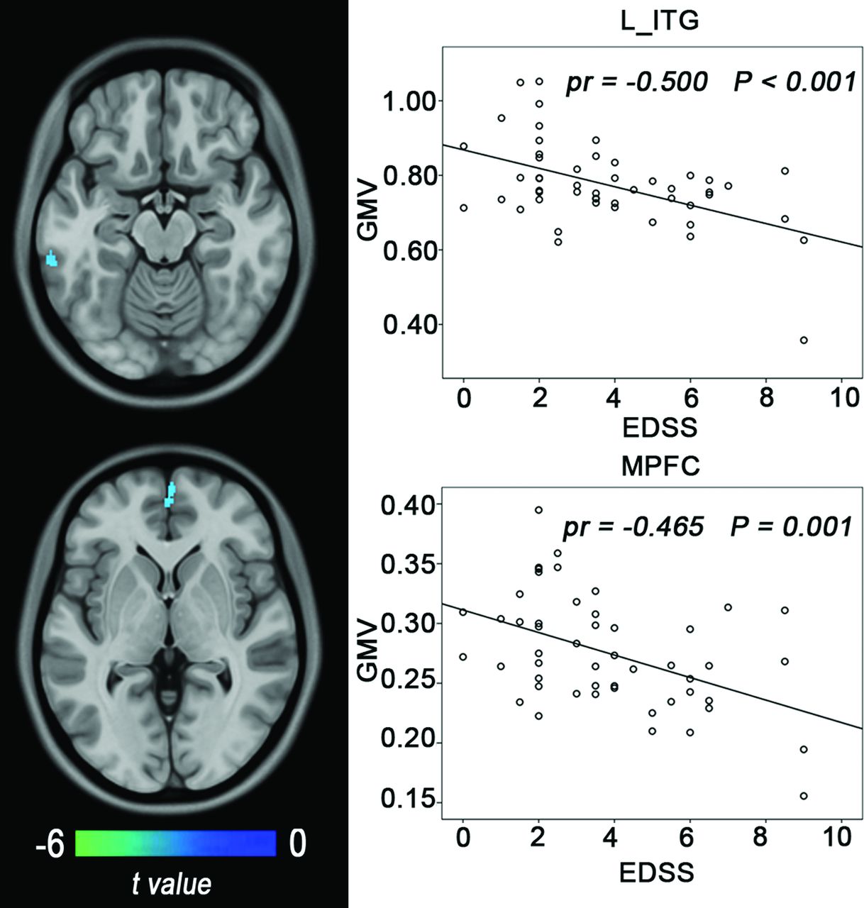
  • Fig 2.
    • Download figure
    • Open in new tab
    • Download powerpoint
    Fig 2.

    Correlations between the GMV and clinical variables in patients with NMO. The left column shows brain regions where the GMV is correlated with EDSS scores (Alphasim correction, P < .05). The right column shows scatter diagrams of these correlations. ITG indicates inferior temporal gyrus; MPFC, medial prefrontal cortex.

  • Fig 3.
    • Download figure
    • Open in new tab
    • Download powerpoint
    Fig 3.

    Correlations between the GMV and cognitive scores in patients with NMO. The upper row shows brain regions where the GMV is correlated with cognitive scores (Alphasim correction, P < .05). The middle row shows scatter diagrams of these correlations. The lower row shows brain regions where the GMV is correlated with cognitive scores after further controlling for the brain lesion volume. Immediate recall of Trial 2 (A), long-delay free recall (B), and short-delay free recall (C). MPFC indicates medial prefrontal cortex; R_Th, right thalamus.

  • Fig 4.
    • Download figure
    • Open in new tab
    • Download powerpoint
    Fig 4.

    Correlations between the GMV and BLV in patients with NMO (Alphasim correction, P < .05). A, Before removing 2 patients with thalamic and inferior temporal gyrus (ITG) lesions. B, After removing 2 patients with thalamic and ITG lesions.

Tables

  • Figures
    • View popup
    Table 1:

    Demographic and clinical characteristics of the subjectsa

    SubjectsPatients with NMOHCsP Value
    No. of subjects5050NA
    Sex (female/male)42:843:7.779
    Age (yr)47.4 (13.4)49.8 (8.9).294
    Education (yr)11.2 (3.4)12.1 (3.1).192
    NMO-IgG (±)32/18NANA
    Brain lesion volumes (cm3)b3.3 (5.1)NANA
    Onset age (yr)41.6 (14.1)NANA
    Relapsing frequency (times/yr)0.8 (0.7)NANA
    No. of attacks (times)4.5 (5.1)NANA
    Disease duration (yr)6.6 (6.7)NANA
    EDSS score3.8 (2.3)NANA
    • Note:—NA indicates not applicable.

    • ↵a All values are expressed as mean (SD).

    • ↵b Thirty-seven patients had visible brain lesions.

    • View popup
    Table 2:

    Neuropsychological tests for patients with NMO and healthy controlsa

    Neuropsychological TestsPatients with NMOHCsP Value
    CVLT-II
        IR28.7 (2.2) (n = 44)9.6 (2.5) (n = 45).075
        SDFR11.1 (2.6) (n = 44)11.8 (2.3) (n = 45).176
        SDCR11.0 (2.2) (n = 44)12.0 (2.2) (n = 45).030b
        LDFR10.9 (2.9) (n = 44)12.2 (2.2) (n = 45).017b
        LDCR10.5 (3.1) (n = 44)12.1 (2.2) (n = 45).007b
    PASAT34.4 (12.3) (n = 43)37.6 (7.7) (n = 45).152
    SDMT42.1 (12.2) (n = 39)49.5 (15.1) (n = 45).018b
    WCST8.8 (3.9) (n = 34)10.0 (5.2) (n = 44).245
    COWAT (semantic)17.5 (4.7) (n = 43)19.5 (4.0) (n = 44).035b
    • Note:—CVLT-II indicates California Verbal Learning Test–Second Edition; COWAT, Controlled Oral Word Association Test; IR2, Immediate Recall of Trail 2; LDCR, Long-Delay Cued Recall; LDFR, Long-Delay Free Recall; PASAT, Paced Auditory Serial Addition Test; SDCR, Short-Delay Cued Recall; SDFR, Short-Delay Free Recall; SDMT, Symbol Digit Modalities Test; WCST, Wisconsin Card Sorting Test.

    • ↵a Data are shown as mean (SD).

    • ↵b Significant.

PreviousNext
Back to top

In this issue

American Journal of Neuroradiology: 36 (10)
American Journal of Neuroradiology
Vol. 36, Issue 10
1 Oct 2015
  • Table of Contents
  • Index by author
  • Complete Issue (PDF)
Advertisement
Print
Download PDF
Email Article

Thank you for your interest in spreading the word on American Journal of Neuroradiology.

NOTE: We only request your email address so that the person you are recommending the page to knows that you wanted them to see it, and that it is not junk mail. We do not capture any email address.

Enter multiple addresses on separate lines or separate them with commas.
Gray Matter Volume Reduction Is Associated with Cognitive Impairment in Neuromyelitis Optica
(Your Name) has sent you a message from American Journal of Neuroradiology
(Your Name) thought you would like to see the American Journal of Neuroradiology web site.
CAPTCHA
This question is for testing whether or not you are a human visitor and to prevent automated spam submissions.
Cite this article
Q. Wang, N. Zhang, W. Qin, Y. Li, Y. Fu, T. Li, J. Shao, L. Yang, F.-D. Shi, C. Yu
Gray Matter Volume Reduction Is Associated with Cognitive Impairment in Neuromyelitis Optica
American Journal of Neuroradiology Oct 2015, 36 (10) 1822-1829; DOI: 10.3174/ajnr.A4403

Citation Manager Formats

  • BibTeX
  • Bookends
  • EasyBib
  • EndNote (tagged)
  • EndNote 8 (xml)
  • Medlars
  • Mendeley
  • Papers
  • RefWorks Tagged
  • Ref Manager
  • RIS
  • Zotero
0 Responses
Respond to this article
Share
Bookmark this article
Gray Matter Volume Reduction Is Associated with Cognitive Impairment in Neuromyelitis Optica
Q. Wang, N. Zhang, W. Qin, Y. Li, Y. Fu, T. Li, J. Shao, L. Yang, F.-D. Shi, C. Yu
American Journal of Neuroradiology Oct 2015, 36 (10) 1822-1829; DOI: 10.3174/ajnr.A4403
del.icio.us logo Twitter logo Facebook logo Mendeley logo
  • Tweet Widget
  • Facebook Like
  • Google Plus One
Purchase

Jump to section

  • Article
    • Abstract
    • ABBREVIATIONS:
    • Materials and Methods
    • Results
    • Discussion
    • Conclusions
    • Acknowledgments
    • Footnotes
    • REFERENCES
  • Figures & Data
  • Supplemental
  • Info & Metrics
  • Responses
  • References
  • PDF

Related Articles

  • PubMed
  • Google Scholar

Cited By...

  • Silent burden: recognising and managing invisible symptoms in neuromyelitis optica
  • Crossref
  • Google Scholar

This article has not yet been cited by articles in journals that are participating in Crossref Cited-by Linking.

More in this TOC Section

  • Diagnostic Neuroradiology of Monoclonal Antibodies
  • Clinical Outcomes After Chiari I Decompression
  • Segmentation of Brain Metastases with BLAST
Show more Adult Brain

Similar Articles

Advertisement

Indexed Content

  • Current Issue
  • Accepted Manuscripts
  • Article Preview
  • Past Issues
  • Editorials
  • Editor's Choice
  • Fellows' Journal Club
  • Letters to the Editor
  • Video Articles

Cases

  • Case Collection
  • Archive - Case of the Week
  • Archive - Case of the Month
  • Archive - Classic Case

More from AJNR

  • Trainee Corner
  • Imaging Protocols
  • MRI Safety Corner
  • Book Reviews

Multimedia

  • AJNR Podcasts
  • AJNR Scantastics

Resources

  • Turnaround Time
  • Submit a Manuscript
  • Submit a Video Article
  • Submit an eLetter to the Editor/Response
  • Manuscript Submission Guidelines
  • Statistical Tips
  • Fast Publishing of Accepted Manuscripts
  • Graphical Abstract Preparation
  • Imaging Protocol Submission
  • Evidence-Based Medicine Level Guide
  • Publishing Checklists
  • Author Policies
  • Become a Reviewer/Academy of Reviewers
  • News and Updates

About Us

  • About AJNR
  • Editorial Board
  • Editorial Board Alumni
  • Alerts
  • Permissions
  • Not an AJNR Subscriber? Join Now
  • Advertise with Us
  • Librarian Resources
  • Feedback
  • Terms and Conditions
  • AJNR Editorial Board Alumni

American Society of Neuroradiology

  • Not an ASNR Member? Join Now

© 2025 by the American Society of Neuroradiology All rights, including for text and data mining, AI training, and similar technologies, are reserved.
Print ISSN: 0195-6108 Online ISSN: 1936-959X

Powered by HighWire