Advertisement

Main menu

  • Home
  • Content
    • Current Issue
    • Accepted Manuscripts
    • Article Preview
    • Past Issue Archive
    • Video Articles
    • AJNR Case Collection
    • Case of the Week Archive
    • Case of the Month Archive
    • Classic Case Archive
  • Special Collections
    • AJNR Awards
    • Low-Field MRI
    • Alzheimer Disease
    • ASNR Foundation Special Collection
    • Photon-Counting CT
    • View All
  • Multimedia
    • AJNR Podcasts
    • AJNR SCANtastic
    • Trainee Corner
    • MRI Safety Corner
    • Imaging Protocols
  • For Authors
    • Submit a Manuscript
    • Submit a Video Article
    • Submit an eLetter to the Editor/Response
    • Manuscript Submission Guidelines
    • Statistical Tips
    • Fast Publishing of Accepted Manuscripts
    • Graphical Abstract Preparation
    • Imaging Protocol Submission
    • Author Policies
  • About Us
    • About AJNR
    • Editorial Board
    • Editorial Board Alumni
  • More
    • Become a Reviewer/Academy of Reviewers
    • Subscribers
    • Permissions
    • Alerts
    • Feedback
    • Advertisers
    • ASNR Home

User menu

  • Alerts
  • Log in

Search

  • Advanced search
American Journal of Neuroradiology

American Journal of Neuroradiology

ASHNR American Society of Functional Neuroradiology ASHNR American Society of Pediatric Neuroradiology ASSR
  • Alerts
  • Log in

Advanced Search

  • Home
  • Content
    • Current Issue
    • Accepted Manuscripts
    • Article Preview
    • Past Issue Archive
    • Video Articles
    • AJNR Case Collection
    • Case of the Week Archive
    • Case of the Month Archive
    • Classic Case Archive
  • Special Collections
    • AJNR Awards
    • Low-Field MRI
    • Alzheimer Disease
    • ASNR Foundation Special Collection
    • Photon-Counting CT
    • View All
  • Multimedia
    • AJNR Podcasts
    • AJNR SCANtastic
    • Trainee Corner
    • MRI Safety Corner
    • Imaging Protocols
  • For Authors
    • Submit a Manuscript
    • Submit a Video Article
    • Submit an eLetter to the Editor/Response
    • Manuscript Submission Guidelines
    • Statistical Tips
    • Fast Publishing of Accepted Manuscripts
    • Graphical Abstract Preparation
    • Imaging Protocol Submission
    • Author Policies
  • About Us
    • About AJNR
    • Editorial Board
    • Editorial Board Alumni
  • More
    • Become a Reviewer/Academy of Reviewers
    • Subscribers
    • Permissions
    • Alerts
    • Feedback
    • Advertisers
    • ASNR Home
  • Follow AJNR on Twitter
  • Visit AJNR on Facebook
  • Follow AJNR on Instagram
  • Join AJNR on LinkedIn
  • RSS Feeds

AJNR Awards, New Junior Editors, and more. Read the latest AJNR updates

Research ArticleSpine Imaging and Spine Image-Guided Interventions

Acute Myelopathy or Cauda Equina Syndrome in HIV-Positive Adults in a Tuberculosis Endemic Setting: MRI, Clinical, and Pathologic Findings

S. Candy, G. Chang and S. Andronikou
American Journal of Neuroradiology August 2014, 35 (8) 1634-1641; DOI: https://doi.org/10.3174/ajnr.A3958
S. Candy
aFrom the Department of Radiology (S.C., G.C.), Groote Schuur Hospital, Faculty of Health Sciences, University of Cape Town, Cape Town, South Africa
  • Find this author on Google Scholar
  • Find this author on PubMed
  • Search for this author on this site
G. Chang
aFrom the Department of Radiology (S.C., G.C.), Groote Schuur Hospital, Faculty of Health Sciences, University of Cape Town, Cape Town, South Africa
  • Find this author on Google Scholar
  • Find this author on PubMed
  • Search for this author on this site
S. Andronikou
bDepartment of Radiology (S.A.), Faculty of Health Sciences, University of the Witwatersrand, Johannesburg, South Africa.
  • Find this author on Google Scholar
  • Find this author on PubMed
  • Search for this author on this site
  • Article
  • Figures & Data
  • Info & Metrics
  • Responses
  • References
  • PDF
Loading

Article Figures & Data

Figures

  • Tables
  • Fig 1.
    • Download figure
    • Open in new tab
    • Download powerpoint
    Fig 1.

    Summary of causation of myelopathy. Abbreviations: CC indicates cryptococcus; Zoster, Varicella zoster; MA, Mycobacterium avium; EC, enterococcus; and MEAS, measles; CMV, cytomegalovirus.
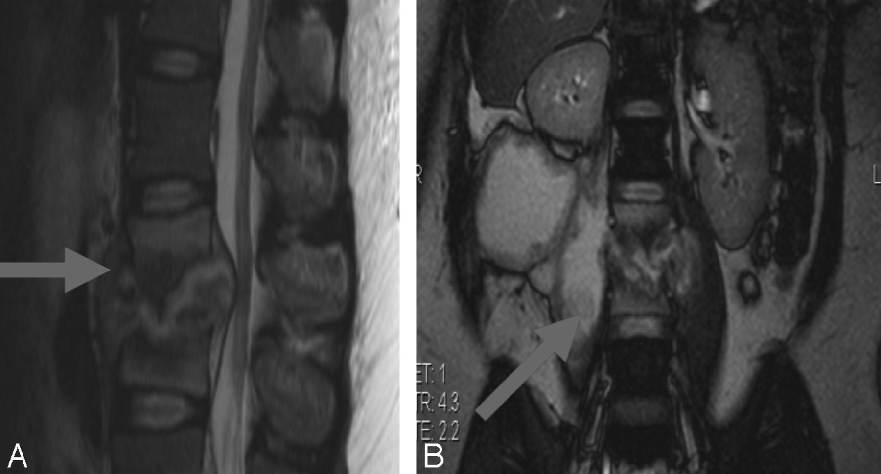
  • Fig 2.
    • Download figure
    • Open in new tab
    • Download powerpoint
    Fig 2.

    Tuberculous spondylo-diskitis. A, Sagittal T2-weighted MR imaging demonstrates caseous destruction of the L2–3 disk with epidural collection causing compression of the conus. B, Coronal FISP demonstrates large psoas abscesses communicating with the L2–3 disk. A thick walled collection displaces the right kidney superiorly.

  • Fig 3.
    • Download figure
    • Open in new tab
    • Download powerpoint
    Fig 3.

    Nonspondylitic spinal tuberculosis. A, T1 sagittal pregadolinium image with increased signal of the CSF. B, T2 sagittal shows root thickening. C and D, T1 sagittal postgadolinium demonstrates circumferential cord and root enhancement. E, T2 sagittal shows loculated CSF with extensive cord signal abnormality.

  • Fig 4.
    • Download figure
    • Open in new tab
    • Download powerpoint
    Fig 4.

    Burkitt lymphoma. A, Coronal FISP showing nonfluid-containing right L2/3 paraspinal and right pelvic masses infiltrating the right iliac bone and multiple low-signal lesions in both renal cortices. B, Sagittal T1 shows diffusely low marrow signal with isointense epidural masses posterior to the bodies of L5, S1, and S2.

  • Fig 5.
    • Download figure
    • Open in new tab
    • Download powerpoint
    Fig 5.

    Plasmablastic lymphoma. A, Axial T2-weighted MR imaging shows a large destructive lobulated mass in the left hemithorax infiltrating the thoracic spinal canal via the neural foramen. B, T2-weighted sagittal MR imaging showing posterior epidural cord compression.

  • Fig 6.
    • Download figure
    • Open in new tab
    • Download powerpoint
    Fig 6.

    EB-associated myopericytoma. A, Sagittal T2-weighted MR imaging shows predominantly low-signal epidural mass causing significant cord compression at T6/7 with abnormal marrow in the adjacent vertebral body. B, Coronal FISP demonstrates improved conspicuity of marrow signal abnormality. C, Axial T2-weighted image shows tumor displacing and compressing the cord.

Tables

  • Figures
    • View popup
    Table 1:

    MRI-based pathologic categories in 216 patients with HIV and acute onset myelopathy/cauda equina

    MRI NormalMRI Abnormal
    Normal GroupSpondylitis GroupNonspondylitis Groups
    SpondylitisArachnoiditis-Myelitis Intramedullary TuberculomasNeoplasm NonboneNeoplasm BoneOther
    HIV AssociatedLymphomaLymphomaPlasmacytomaMetsDiskSyrinx?
    55656421034823
    • Note:—? indicates no final diagnosis.

    • View popup
    Table 2:

    Mycobacterium tuberculosis as a cause of myelopathy/cauda equina syndrome in subset of patients with a confirmed aetiology on laboratory testing

    MRI SpondylitisMRI NonspondylitisTotal
    TB
        Yes572683
        No04040
    Total5766123
    • View popup
    Table 3:

    Comparison of myelitis group and normal MRI groups with regard to CSF parameters using Mann-Whitney U test

    MRI GroupLymphocyte Count Median (per mm3) and RangePolymorphonuclear Median (per mm3) and RangeProtein Median (g/l) Normal 0.15–0.45 and Range
    Myelitis38.5 (0–265)0 (0–300)2 (0.14–2)
    Normal1 (0–160)0 (0–5)0.49 (0.11–2.6)
    P value.0053a.2082.0017a
    • ↵a Indicates significance.

PreviousNext
Back to top

In this issue

American Journal of Neuroradiology: 35 (8)
American Journal of Neuroradiology
Vol. 35, Issue 8
1 Aug 2014
  • Table of Contents
  • Index by author
  • Complete Issue (PDF)
Advertisement
Print
Download PDF
Email Article

Thank you for your interest in spreading the word on American Journal of Neuroradiology.

NOTE: We only request your email address so that the person you are recommending the page to knows that you wanted them to see it, and that it is not junk mail. We do not capture any email address.

Enter multiple addresses on separate lines or separate them with commas.
Acute Myelopathy or Cauda Equina Syndrome in HIV-Positive Adults in a Tuberculosis Endemic Setting: MRI, Clinical, and Pathologic Findings
(Your Name) has sent you a message from American Journal of Neuroradiology
(Your Name) thought you would like to see the American Journal of Neuroradiology web site.
CAPTCHA
This question is for testing whether or not you are a human visitor and to prevent automated spam submissions.
Cite this article
S. Candy, G. Chang, S. Andronikou
Acute Myelopathy or Cauda Equina Syndrome in HIV-Positive Adults in a Tuberculosis Endemic Setting: MRI, Clinical, and Pathologic Findings
American Journal of Neuroradiology Aug 2014, 35 (8) 1634-1641; DOI: 10.3174/ajnr.A3958

Citation Manager Formats

  • BibTeX
  • Bookends
  • EasyBib
  • EndNote (tagged)
  • EndNote 8 (xml)
  • Medlars
  • Mendeley
  • Papers
  • RefWorks Tagged
  • Ref Manager
  • RIS
  • Zotero
0 Responses
Respond to this article
Share
Bookmark this article
Acute Myelopathy or Cauda Equina Syndrome in HIV-Positive Adults in a Tuberculosis Endemic Setting: MRI, Clinical, and Pathologic Findings
S. Candy, G. Chang, S. Andronikou
American Journal of Neuroradiology Aug 2014, 35 (8) 1634-1641; DOI: 10.3174/ajnr.A3958
del.icio.us logo Twitter logo Facebook logo Mendeley logo
  • Tweet Widget
  • Facebook Like
  • Google Plus One
Purchase

Jump to section

  • Article
    • Abstract
    • ABBREVIATIONS:
    • Method
    • Results
    • Discussion
    • Conclusions
    • Acknowledgments
    • Appendix 1
    • Appendix 2
    • REFERENCES
  • Figures & Data
  • Info & Metrics
  • Responses
  • References
  • PDF

Related Articles

  • No related articles found.
  • PubMed
  • Google Scholar

Cited By...

  • Is water carriage associated with the water carriers health? A systematic review of quantitative and qualitative evidence
  • Crossref
  • Google Scholar

This article has not yet been cited by articles in journals that are participating in Crossref Cited-by Linking.

More in this TOC Section

  • Optimization of Photon Counting CT Myelography
  • Characteristics of SIH Type I Culprit Lesions
  • Management Outcomes For VO Spine Biopsy
Show more Spine Imaging and Spine Image-Guided Interventions

Similar Articles

Advertisement

Indexed Content

  • Current Issue
  • Accepted Manuscripts
  • Article Preview
  • Past Issues
  • Editorials
  • Editor's Choice
  • Fellows' Journal Club
  • Letters to the Editor
  • Video Articles

Cases

  • Case Collection
  • Archive - Case of the Week
  • Archive - Case of the Month
  • Archive - Classic Case

More from AJNR

  • Trainee Corner
  • Imaging Protocols
  • MRI Safety Corner
  • Book Reviews

Multimedia

  • AJNR Podcasts
  • AJNR Scantastics

Resources

  • Turnaround Time
  • Submit a Manuscript
  • Submit a Video Article
  • Submit an eLetter to the Editor/Response
  • Manuscript Submission Guidelines
  • Statistical Tips
  • Fast Publishing of Accepted Manuscripts
  • Graphical Abstract Preparation
  • Imaging Protocol Submission
  • Evidence-Based Medicine Level Guide
  • Publishing Checklists
  • Author Policies
  • Become a Reviewer/Academy of Reviewers
  • News and Updates

About Us

  • About AJNR
  • Editorial Board
  • Editorial Board Alumni
  • Alerts
  • Permissions
  • Not an AJNR Subscriber? Join Now
  • Advertise with Us
  • Librarian Resources
  • Feedback
  • Terms and Conditions
  • AJNR Editorial Board Alumni

American Society of Neuroradiology

  • Not an ASNR Member? Join Now

© 2025 by the American Society of Neuroradiology All rights, including for text and data mining, AI training, and similar technologies, are reserved.
Print ISSN: 0195-6108 Online ISSN: 1936-959X

Powered by HighWire