
Article Figures & Data
Figures
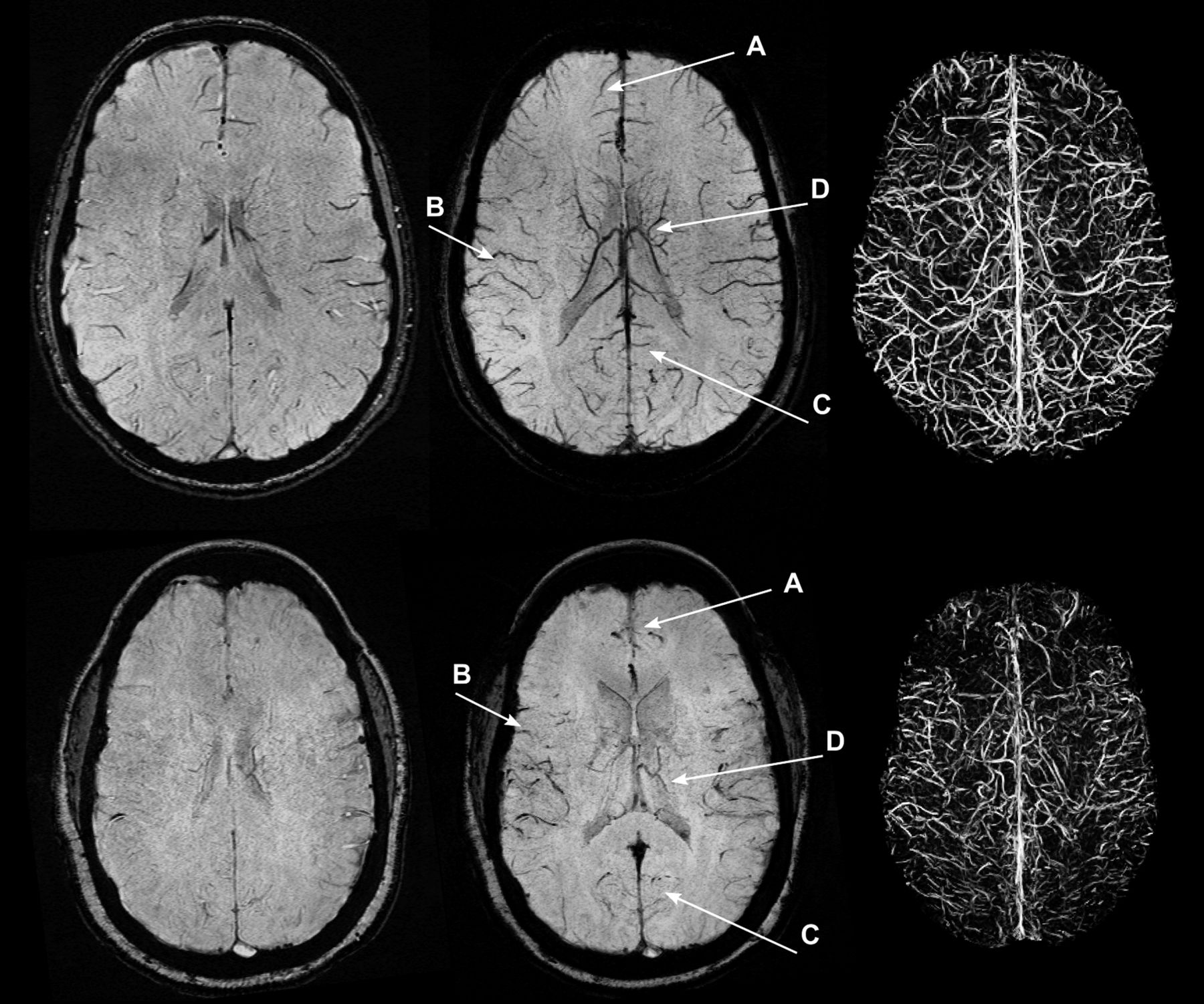
- Fig 1.
SWI, mIP SWI, and segmented vein maps for a representative control (top row, NVVV = 0.032) and patient with SCD (bottom row, NVVV = 0.013). In the mIP SWI and segmented vein maps, there is visually less venous contrast in the examination from the patient with SCD. The arrows point to loss of cortical venous conspicuity in the frontal (A), temporal (B), occipital (C), and deep medullary (D) areas, indicating a global decrease in signal.
- Fig 2.
An SCD mIP SWI (A) with several hypointense-appearing vessels that could be interpreted as veins (black arrowheads). The corresponding MIP TOF (B) indicated that many of the hypointense-appearing vessels on the mIP SWI correlate with a hyperintense arterial signal from the TOF (white arrowheads). The arrows indicated arterial contribution of more distal branches of the left and right (anterior to posterior) bilateral anterior, middle, and posterior cerebral arteries. This example received a grade of II because of a hypointense arterial signal on the mIP SWI in the anterior Sylvian fissure through to the posterior temporal lobe.
- Fig 3.
Representative mIP SWI from the same volunteer who underwent scanning at 1.5T (A) and 3T (B) depicts similar venous anatomy. The combination of higher SNR and venous contrast at 3T results in an improved venous conspicuity and a higher venous vessel likelihood from the Frangi filter. Several veins (eg, deep medullary veins) appear larger at 3T. The combination of larger-appearing veins and a higher vessel likelihood we believe were attributed to the increased conspicuity of NVVV at 3T.
- Fig 4.
Boxplot of the NVVV in the SCD group (1.5T = 0.013 ± 0.004; 3T = 0.011 ± 0.006), control group (3T = 0.031 ± 0.009), and volunteers (1.5T = 0.024 ± 0.006; 3T = 0.028 ± 0.009). The difference in NVVV between the 3T SCD and control groups was significant (P < .001) when assessed by the Wilcoxon rank sum test. Volunteer measurements at 1.5T and 3T indicate increased conspicuity of NVVV at 3T (P = .03) when assessed by the Wilcoxon signed rank test.


Tables
Correlation of NVVV with hematologic parameters
Parameter N Interval between SWI and Laboratory Measurements (d) Value R (P Value)* Hemoglobin (g/dL) 21 −0.05 ± 2.4 8.51 ± 1.10 0.25 (.28) Absolute reticulocyte count (×106/L) 21 −0.05 ± 2.4 0.23 ± 0.10 0.10 (.66) White blood cell count (×106/L) 21 −0.05 ± 2.4 10.10 ± 4.50 0.19 (.40) Hemoglobin F (%) 18 −3.22 ± 9.38 12.84 ± 9.40 −0.09 (.71) Hemoglobin S (%) 18 −3.22 ± 9.38 68.06 ± 18.22 −0.25 (.31) Note:—Results presented as mean ± standard deviation.
↵* R(P Value) value for the correlation of each hematologic parameter and NVVV.


Jump to section
Related Articles
Cited By...
- No citing articles found.