Advertisement

Main menu

  • Home
  • Content
    • Current Issue
    • Accepted Manuscripts
    • Article Preview
    • Past Issue Archive
    • Video Articles
    • AJNR Case Collection
    • Case of the Week Archive
    • Case of the Month Archive
    • Classic Case Archive
  • Special Collections
    • AJNR Awards
    • Low-Field MRI
    • Alzheimer Disease
    • ASNR Foundation Special Collection
    • Photon-Counting CT
    • View All
  • Multimedia
    • AJNR Podcasts
    • AJNR SCANtastic
    • Trainee Corner
    • MRI Safety Corner
    • Imaging Protocols
  • For Authors
    • Submit a Manuscript
    • Submit a Video Article
    • Submit an eLetter to the Editor/Response
    • Manuscript Submission Guidelines
    • Statistical Tips
    • Fast Publishing of Accepted Manuscripts
    • Graphical Abstract Preparation
    • Imaging Protocol Submission
    • Author Policies
  • About Us
    • About AJNR
    • Editorial Board
    • Editorial Board Alumni
  • More
    • Become a Reviewer/Academy of Reviewers
    • Subscribers
    • Permissions
    • Alerts
    • Feedback
    • Advertisers
    • ASNR Home

User menu

  • Alerts
  • Log in

Search

  • Advanced search
American Journal of Neuroradiology

American Journal of Neuroradiology

ASHNR American Society of Functional Neuroradiology ASHNR American Society of Pediatric Neuroradiology ASSR
  • Alerts
  • Log in

Advanced Search

  • Home
  • Content
    • Current Issue
    • Accepted Manuscripts
    • Article Preview
    • Past Issue Archive
    • Video Articles
    • AJNR Case Collection
    • Case of the Week Archive
    • Case of the Month Archive
    • Classic Case Archive
  • Special Collections
    • AJNR Awards
    • Low-Field MRI
    • Alzheimer Disease
    • ASNR Foundation Special Collection
    • Photon-Counting CT
    • View All
  • Multimedia
    • AJNR Podcasts
    • AJNR SCANtastic
    • Trainee Corner
    • MRI Safety Corner
    • Imaging Protocols
  • For Authors
    • Submit a Manuscript
    • Submit a Video Article
    • Submit an eLetter to the Editor/Response
    • Manuscript Submission Guidelines
    • Statistical Tips
    • Fast Publishing of Accepted Manuscripts
    • Graphical Abstract Preparation
    • Imaging Protocol Submission
    • Author Policies
  • About Us
    • About AJNR
    • Editorial Board
    • Editorial Board Alumni
  • More
    • Become a Reviewer/Academy of Reviewers
    • Subscribers
    • Permissions
    • Alerts
    • Feedback
    • Advertisers
    • ASNR Home
  • Follow AJNR on Twitter
  • Visit AJNR on Facebook
  • Follow AJNR on Instagram
  • Join AJNR on LinkedIn
  • RSS Feeds

AJNR Awards, New Junior Editors, and more. Read the latest AJNR updates

Research ArticlePediatric Neuroimaging
Open Access

Regional Cerebral Blood Flow in Children From 3 to 5 Months of Age

A.F. Duncan, A. Caprihan, E.Q. Montague, J. Lowe, R. Schrader and J.P. Phillips
American Journal of Neuroradiology March 2014, 35 (3) 593-598; DOI: https://doi.org/10.3174/ajnr.A3728
A.F. Duncan
aFrom the Department of Pediatrics, Division of Neonatology (A.F.D., J.L.)
  • Find this author on Google Scholar
  • Find this author on PubMed
  • Search for this author on this site
A. Caprihan
dThe MIND Research Network (A.C.), Albuquerque, New Mexico
  • Find this author on Google Scholar
  • Find this author on PubMed
  • Search for this author on this site
E.Q. Montague
eNeuroDevelopmental Science Center (E.Q.M.), Akron Children's Hospital, Akron, Ohio.
  • Find this author on Google Scholar
  • Find this author on PubMed
  • Search for this author on this site
J. Lowe
aFrom the Department of Pediatrics, Division of Neonatology (A.F.D., J.L.)
  • Find this author on Google Scholar
  • Find this author on PubMed
  • Search for this author on this site
R. Schrader
bClinical and Translational Science Center (R.S.)
  • Find this author on Google Scholar
  • Find this author on PubMed
  • Search for this author on this site
J.P. Phillips
cDepartment of Neurology (J.P.P.), University of New Mexico, Albuquerque, New Mexico
  • Find this author on Google Scholar
  • Find this author on PubMed
  • Search for this author on this site
  • Article
  • Figures & Data
  • Info & Metrics
  • Responses
  • References
  • PDF
Loading

Article Figures & Data

Figures

  • Tables
  • Fig 1.
    • Download figure
    • Open in new tab
    • Download powerpoint
    Fig 1.

    A and B, Brain segmentation process of the 9-month template is shown; C, 4-month T1 image; D, corresponding atlas after mathematica transformation. E, CBF image; F, CBF image with the atlas overlaid to demonstrate how well they are registered; G, the 8 segments projected onto the cortical surface.
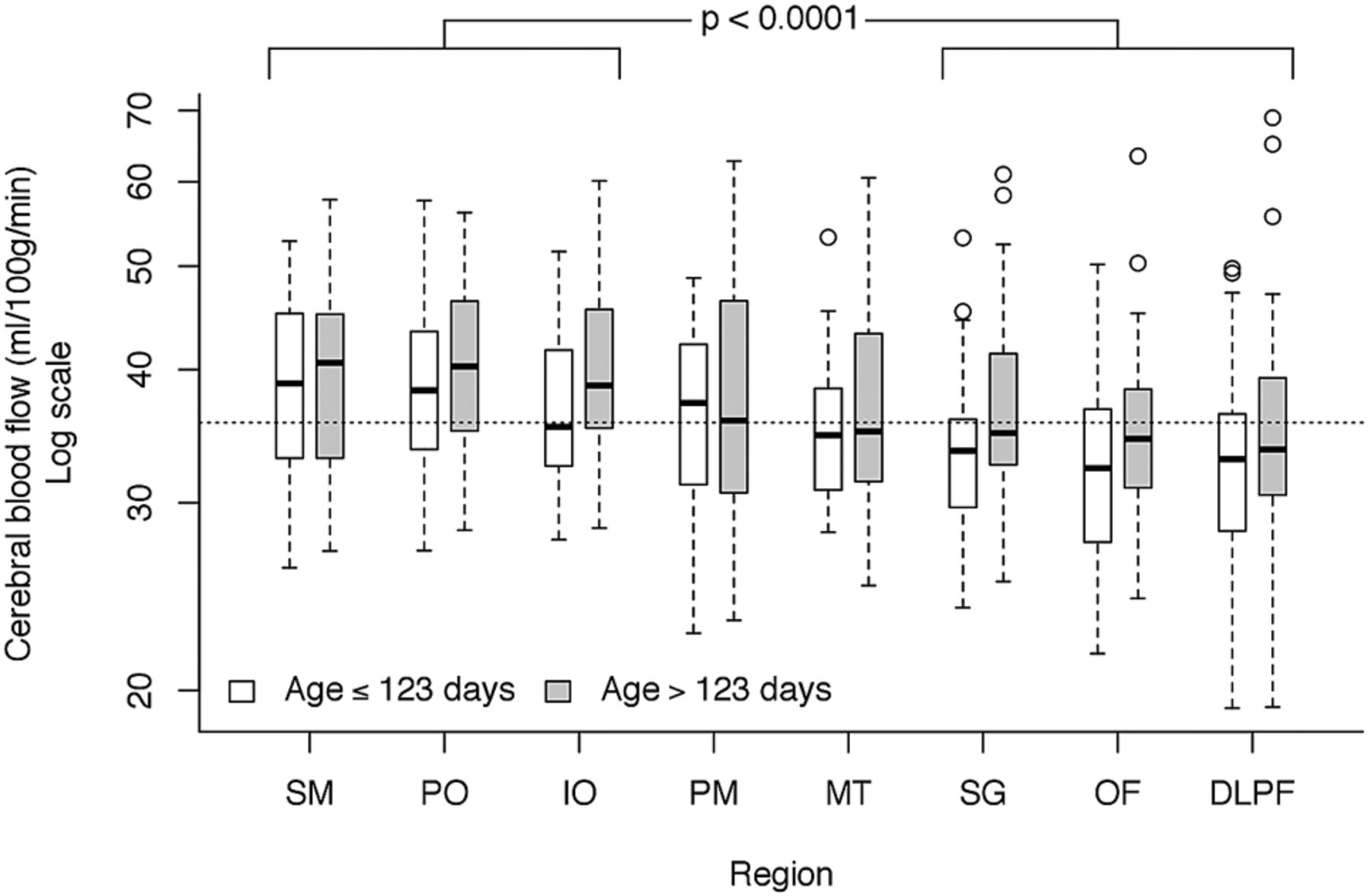
  • Fig 2.
    • Download figure
    • Open in new tab
    • Download powerpoint
    Fig 2.

    Regional differences in CBF. Although there were no significant differences within the 3 higher flow regions (sensorimotor, parieto-occipital, and inferior-occipital; P = .43) or within the 3 lower flow regions (subgenual, orbito-frontal, and dorsolateral prefrontal; P = .15), mean CBF between the high and low groups was highly significant. The mixed model fit age in a continuous manner, but, for illustrative purposes, age is split at the median. Horizontal dotted line is the median CBF value across all regions and ages.

  • Fig 3.
    • Download figure
    • Open in new tab
    • Download powerpoint
    Fig 3.

    Regions with CBF above study population median are shown in shades of red and those regions with CBF below the median are shown in shades of blue. Darker color indicates greater deviation from the median flow. Thus, the sensorimotor (SM) region had the highest flow above the median and the dorsolateral prefrontal (DLPF) region had the lowest flow below the median.

  • Fig 4.
    • Download figure
    • Open in new tab
    • Download powerpoint
    Fig 4.

    Cerebral blood flow versus age within each region; ρ is Spearman rank correlation coefficient.

Tables

  • Figures
  • Demographic information for study subjects

    Total (n = 61)Male (n = 37) (61%)Female (n = 24) (39%)
    Birth weight, g3,294.95 ± 432.23,363.6 ± 408.83,190.5 ± 554.6
    Gestational age, wk39.2 ± 1.437.3 ± 9.138.9 ± 1.5
    Age at scanning, d123.6 ± 13122.3 ± 12.6125.5 ± 13.4a
    Sex, % girlsa39%
    Ethnicitya
    White15 (24.59%)8 (21.62%)7 (29.16%)
    Black1 (1.64%)1 (2.70%)0 (0%)
    Hispanic34 (55.73%)21 (56.75%)13 (54.16%)
    Native American3 (4.92%)2 (5.40%)1 (4.16%)
    Other4 (6.55%)3 (8.10%)1 (4.16%)
    Unknown4 (6.55%)2 (5.40%)2 (8.33%)
    • Note:—Values are depicted as means ± standard deviations unless otherwise indicated.

    • ↵a P < .05.

PreviousNext
Back to top

In this issue

American Journal of Neuroradiology: 35 (3)
American Journal of Neuroradiology
Vol. 35, Issue 3
1 Mar 2014
  • Table of Contents
  • Index by author
  • Complete Issue (PDF)
Advertisement
Print
Download PDF
Email Article

Thank you for your interest in spreading the word on American Journal of Neuroradiology.

NOTE: We only request your email address so that the person you are recommending the page to knows that you wanted them to see it, and that it is not junk mail. We do not capture any email address.

Enter multiple addresses on separate lines or separate them with commas.
Regional Cerebral Blood Flow in Children From 3 to 5 Months of Age
(Your Name) has sent you a message from American Journal of Neuroradiology
(Your Name) thought you would like to see the American Journal of Neuroradiology web site.
CAPTCHA
This question is for testing whether or not you are a human visitor and to prevent automated spam submissions.
Cite this article
A.F. Duncan, A. Caprihan, E.Q. Montague, J. Lowe, R. Schrader, J.P. Phillips
Regional Cerebral Blood Flow in Children From 3 to 5 Months of Age
American Journal of Neuroradiology Mar 2014, 35 (3) 593-598; DOI: 10.3174/ajnr.A3728

Citation Manager Formats

  • BibTeX
  • Bookends
  • EasyBib
  • EndNote (tagged)
  • EndNote 8 (xml)
  • Medlars
  • Mendeley
  • Papers
  • RefWorks Tagged
  • Ref Manager
  • RIS
  • Zotero
0 Responses
Respond to this article
Share
Bookmark this article
Regional Cerebral Blood Flow in Children From 3 to 5 Months of Age
A.F. Duncan, A. Caprihan, E.Q. Montague, J. Lowe, R. Schrader, J.P. Phillips
American Journal of Neuroradiology Mar 2014, 35 (3) 593-598; DOI: 10.3174/ajnr.A3728
del.icio.us logo Twitter logo Facebook logo Mendeley logo
  • Tweet Widget
  • Facebook Like
  • Google Plus One
Purchase

Jump to section

  • Article
    • Abstract
    • ABBREVIATION:
    • Materials and Methods
    • Results
    • Discussion
    • Conclusions
    • Acknowledgments
    • Footnotes
    • REFERENCES
  • Figures & Data
  • Info & Metrics
  • Responses
  • References
  • PDF

Related Articles

  • No related articles found.
  • PubMed
  • Google Scholar

Cited By...

  • Cerebral blood perfusion across biological systems and the human lifespan
  • Rest functional brain maturation during the first year of life
  • Cerebral Perfusion After Repair of Congenital Diaphragmatic Hernia with Common Carotid Artery Occlusion After ECMO Therapy
  • Brain Perfusion Imaging in Neonates: An Overview
  • Gray Matter Growth Is Accompanied by Increasing Blood Flow and Decreasing Apparent Diffusion Coefficient during Childhood
  • Crossref
  • Google Scholar

This article has not yet been cited by articles in journals that are participating in Crossref Cited-by Linking.

More in this TOC Section

  • Frontal Paraventricular Cysts
  • Sodium MRI in Pediatric Brain Tumors
  • FRACTURE MR in Congenital Vertebral Anomalies
Show more Pediatric Neuroimaging

Similar Articles

Advertisement

Indexed Content

  • Current Issue
  • Accepted Manuscripts
  • Article Preview
  • Past Issues
  • Editorials
  • Editor's Choice
  • Fellows' Journal Club
  • Letters to the Editor
  • Video Articles

Cases

  • Case Collection
  • Archive - Case of the Week
  • Archive - Case of the Month
  • Archive - Classic Case

More from AJNR

  • Trainee Corner
  • Imaging Protocols
  • MRI Safety Corner
  • Book Reviews

Multimedia

  • AJNR Podcasts
  • AJNR Scantastics

Resources

  • Turnaround Time
  • Submit a Manuscript
  • Submit a Video Article
  • Submit an eLetter to the Editor/Response
  • Manuscript Submission Guidelines
  • Statistical Tips
  • Fast Publishing of Accepted Manuscripts
  • Graphical Abstract Preparation
  • Imaging Protocol Submission
  • Evidence-Based Medicine Level Guide
  • Publishing Checklists
  • Author Policies
  • Become a Reviewer/Academy of Reviewers
  • News and Updates

About Us

  • About AJNR
  • Editorial Board
  • Editorial Board Alumni
  • Alerts
  • Permissions
  • Not an AJNR Subscriber? Join Now
  • Advertise with Us
  • Librarian Resources
  • Feedback
  • Terms and Conditions
  • AJNR Editorial Board Alumni

American Society of Neuroradiology

  • Not an ASNR Member? Join Now

© 2025 by the American Society of Neuroradiology All rights, including for text and data mining, AI training, and similar technologies, are reserved.
Print ISSN: 0195-6108 Online ISSN: 1936-959X

Powered by HighWire