Advertisement

Main menu

  • Home
  • Content
    • Current Issue
    • Accepted Manuscripts
    • Article Preview
    • Past Issue Archive
    • Video Articles
    • AJNR Case Collection
    • Case of the Week Archive
    • Case of the Month Archive
    • Classic Case Archive
  • Special Collections
    • AJNR Awards
    • Low-Field MRI
    • Alzheimer Disease
    • ASNR Foundation Special Collection
    • Photon-Counting CT
    • View All
  • Multimedia
    • AJNR Podcasts
    • AJNR SCANtastic
    • Trainee Corner
    • MRI Safety Corner
    • Imaging Protocols
  • For Authors
    • Submit a Manuscript
    • Submit a Video Article
    • Submit an eLetter to the Editor/Response
    • Manuscript Submission Guidelines
    • Statistical Tips
    • Fast Publishing of Accepted Manuscripts
    • Graphical Abstract Preparation
    • Imaging Protocol Submission
    • Author Policies
  • About Us
    • About AJNR
    • Editorial Board
    • Editorial Board Alumni
  • More
    • Become a Reviewer/Academy of Reviewers
    • Subscribers
    • Permissions
    • Alerts
    • Feedback
    • Advertisers
    • ASNR Home

User menu

  • Alerts
  • Log in

Search

  • Advanced search
American Journal of Neuroradiology

American Journal of Neuroradiology

ASHNR American Society of Functional Neuroradiology ASHNR American Society of Pediatric Neuroradiology ASSR
  • Alerts
  • Log in

Advanced Search

  • Home
  • Content
    • Current Issue
    • Accepted Manuscripts
    • Article Preview
    • Past Issue Archive
    • Video Articles
    • AJNR Case Collection
    • Case of the Week Archive
    • Case of the Month Archive
    • Classic Case Archive
  • Special Collections
    • AJNR Awards
    • Low-Field MRI
    • Alzheimer Disease
    • ASNR Foundation Special Collection
    • Photon-Counting CT
    • View All
  • Multimedia
    • AJNR Podcasts
    • AJNR SCANtastic
    • Trainee Corner
    • MRI Safety Corner
    • Imaging Protocols
  • For Authors
    • Submit a Manuscript
    • Submit a Video Article
    • Submit an eLetter to the Editor/Response
    • Manuscript Submission Guidelines
    • Statistical Tips
    • Fast Publishing of Accepted Manuscripts
    • Graphical Abstract Preparation
    • Imaging Protocol Submission
    • Author Policies
  • About Us
    • About AJNR
    • Editorial Board
    • Editorial Board Alumni
  • More
    • Become a Reviewer/Academy of Reviewers
    • Subscribers
    • Permissions
    • Alerts
    • Feedback
    • Advertisers
    • ASNR Home
  • Follow AJNR on Twitter
  • Visit AJNR on Facebook
  • Follow AJNR on Instagram
  • Join AJNR on LinkedIn
  • RSS Feeds

AJNR Awards, New Junior Editors, and more. Read the latest AJNR updates

Research ArticleNeurointervention

Acute Bleeding in the Head and Neck: Angiographic Findings and Endovascular Management

L.-B. Zhao, H.B. Shi, S. Park, D.g. Lee, J.H. Shim, D.H. Lee and D.C. Suh
American Journal of Neuroradiology February 2014, 35 (2) 360-366; DOI: https://doi.org/10.3174/ajnr.A3761
L.-B. Zhao
aFrom the Departments of Radiology and Research Institute of Radiology (L.-B.Z., H.B.S., S.P., D.-g.L., J.H.S., D.H.L., D.C.S.), University of Ulsan, College of Medicine, Asan Medical Center, Seoul, Korea
bDepartment of Radiology (L.-B.Z., H.B.S.), First Affiliated Hospital of Nanjing Medical University, Nanjing, Nanjing, China.
  • Find this author on Google Scholar
  • Find this author on PubMed
  • Search for this author on this site
H.B. Shi
aFrom the Departments of Radiology and Research Institute of Radiology (L.-B.Z., H.B.S., S.P., D.-g.L., J.H.S., D.H.L., D.C.S.), University of Ulsan, College of Medicine, Asan Medical Center, Seoul, Korea
bDepartment of Radiology (L.-B.Z., H.B.S.), First Affiliated Hospital of Nanjing Medical University, Nanjing, Nanjing, China.
  • Find this author on Google Scholar
  • Find this author on PubMed
  • Search for this author on this site
S. Park
aFrom the Departments of Radiology and Research Institute of Radiology (L.-B.Z., H.B.S., S.P., D.-g.L., J.H.S., D.H.L., D.C.S.), University of Ulsan, College of Medicine, Asan Medical Center, Seoul, Korea
  • Find this author on Google Scholar
  • Find this author on PubMed
  • Search for this author on this site
D.g. Lee
aFrom the Departments of Radiology and Research Institute of Radiology (L.-B.Z., H.B.S., S.P., D.-g.L., J.H.S., D.H.L., D.C.S.), University of Ulsan, College of Medicine, Asan Medical Center, Seoul, Korea
  • Find this author on Google Scholar
  • Find this author on PubMed
  • Search for this author on this site
J.H. Shim
aFrom the Departments of Radiology and Research Institute of Radiology (L.-B.Z., H.B.S., S.P., D.-g.L., J.H.S., D.H.L., D.C.S.), University of Ulsan, College of Medicine, Asan Medical Center, Seoul, Korea
  • Find this author on Google Scholar
  • Find this author on PubMed
  • Search for this author on this site
D.H. Lee
aFrom the Departments of Radiology and Research Institute of Radiology (L.-B.Z., H.B.S., S.P., D.-g.L., J.H.S., D.H.L., D.C.S.), University of Ulsan, College of Medicine, Asan Medical Center, Seoul, Korea
  • Find this author on Google Scholar
  • Find this author on PubMed
  • Search for this author on this site
D.C. Suh
aFrom the Departments of Radiology and Research Institute of Radiology (L.-B.Z., H.B.S., S.P., D.-g.L., J.H.S., D.H.L., D.C.S.), University of Ulsan, College of Medicine, Asan Medical Center, Seoul, Korea
  • Find this author on Google Scholar
  • Find this author on PubMed
  • Search for this author on this site
  • Article
  • Figures & Data
  • Supplemental
  • Info & Metrics
  • Responses
  • References
  • PDF
Loading

Article Figures & Data

Figures

  • Fig 1.
    • Download figure
    • Open in new tab
    • Download powerpoint
    Fig 1.

    A 24-year-old male patient who presented with intractable epistaxis after sagittal split ramus osteotomy. A, Lateral left ECA arteriogram shows a pseudoaneurysm at the internal maxillary artery segment at the pterygopalatine fossa. B, A microcatheter was advanced to the distal maxillary artery beyond the pseudoaneurysm that was embolized by using a coil. C, Selective injection of the left facial artery shows collaterals from the ascending artery to the distal maxillary artery via the infraorbital artery. Note the absence of filling of the pseudoaneurysm from retrograde flow of the distal maxillary artery.
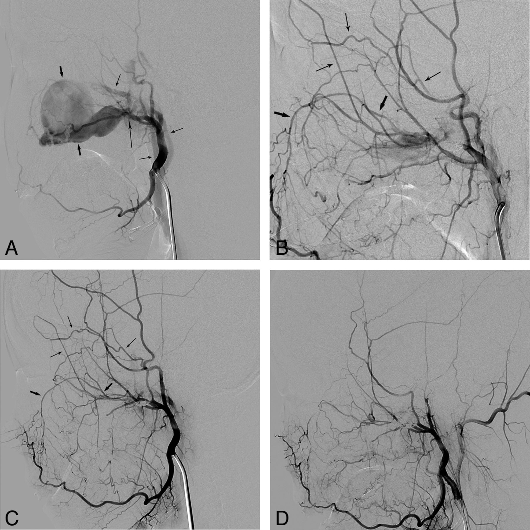
  • Fig 2.
    • Download figure
    • Open in new tab
    • Download powerpoint
    Fig 2.

    A 19-year-old female patient who presented with intractable epistaxis after maxillofacial plastic surgery. A, An external carotid angiogram shows a large pseudoaneurysm (short thick arrows) and a fistula (long thin arrow) with an arteriovenous shunt. Note the early filling veins (short thin arrows). B, An angiogram obtained following coil embolization of the maxillary artery at the aneurysm site shows retrograde filling of the remaining shunt through the infraorbital artery (short thick arrows) via the facial artery and the zygomatico-orbital branch (short thin arrows) of the superficial temporal artery. C, An angiogram obtained the following day revealed a slightly smaller but still remaining fistula with persistent bleeding. Note the retrograde filling as indicated on B by the same arrows. D, The bleeding was finally controlled after additional coil embolization via the zygomatico-orbital branch (short thin arrows, B) into the fistula.
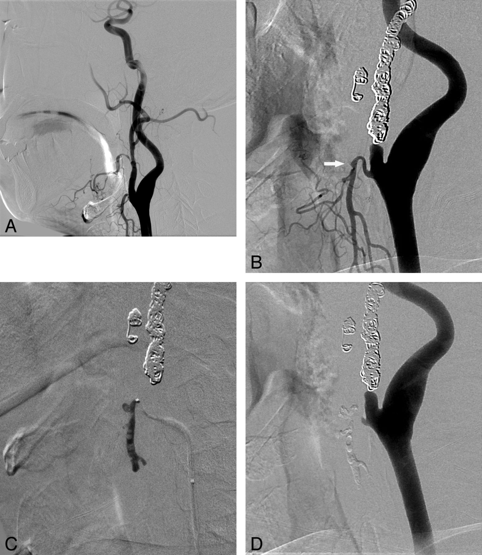
  • Fig 3.
    • Download figure
    • Open in new tab
    • Download powerpoint
    Fig 3.

    Endovascular treatment for recurrent bleeding approximately 2 months and 15 days after the initial embolization of the left ECA in a 45-year-old male patient with tonsil cancer. A, At the time of the first bleeding, the right facial and nearby maxillary arteries showing irregularities were regarded as the origin of the initial bleeding site and were embolized with coils as shown in B. B, Left common carotid arteriogram obtained at the time of the second bleeding reveals a tiny pseudoaneurysm (white arrow) in the proximal segment of the left superior thyroidal artery. Note the coils used in the previous embolization. C, Glue was used for embolization of the left superior thyroidal artery, and the final angiogram shows no residual pseudoaneurysm filling. The patient's bleeding was controlled thereafter.

  • Fig 4.
    • Download figure
    • Open in new tab
    • Download powerpoint
    Fig 4.

    A 51-year-old male patient with a history of supraglottic cancer who presented with massive bleeding at the ulcerative wound site in the right neck. After tracheostomy, he became drowsy and developed left-sided weakness in both the upper and lower extremities. A, A common carotid arteriogram shows extensive contrast leakage due to massive bleeding. No filling of the internal carotid artery from the common carotid artery (arrow) was seen. B, Bleeding was stopped following deployment of a covered stent. C, There is an occlusion of the right middle cerebral artery at the proximal M2 segment (arrow). D, Diffusion-weighted image obtained the next day shows a territorial infarct in the right MCA territory. E, CT scan obtained 17 months later shows the old infarction in the right middle cerebral artery territory, though the patient's weakness had improved and he walked without assistance.

PreviousNext
Back to top

In this issue

American Journal of Neuroradiology: 35 (2)
American Journal of Neuroradiology
Vol. 35, Issue 2
1 Feb 2014
  • Table of Contents
  • Index by author
  • Complete Issue (PDF)
Advertisement
Print
Download PDF
Email Article

Thank you for your interest in spreading the word on American Journal of Neuroradiology.

NOTE: We only request your email address so that the person you are recommending the page to knows that you wanted them to see it, and that it is not junk mail. We do not capture any email address.

Enter multiple addresses on separate lines or separate them with commas.
Acute Bleeding in the Head and Neck: Angiographic Findings and Endovascular Management
(Your Name) has sent you a message from American Journal of Neuroradiology
(Your Name) thought you would like to see the American Journal of Neuroradiology web site.
CAPTCHA
This question is for testing whether or not you are a human visitor and to prevent automated spam submissions.
Cite this article
L.-B. Zhao, H.B. Shi, S. Park, D.g. Lee, J.H. Shim, D.H. Lee, D.C. Suh
Acute Bleeding in the Head and Neck: Angiographic Findings and Endovascular Management
American Journal of Neuroradiology Feb 2014, 35 (2) 360-366; DOI: 10.3174/ajnr.A3761

Citation Manager Formats

  • BibTeX
  • Bookends
  • EasyBib
  • EndNote (tagged)
  • EndNote 8 (xml)
  • Medlars
  • Mendeley
  • Papers
  • RefWorks Tagged
  • Ref Manager
  • RIS
  • Zotero
0 Responses
Respond to this article
Share
Bookmark this article
Acute Bleeding in the Head and Neck: Angiographic Findings and Endovascular Management
L.-B. Zhao, H.B. Shi, S. Park, D.g. Lee, J.H. Shim, D.H. Lee, D.C. Suh
American Journal of Neuroradiology Feb 2014, 35 (2) 360-366; DOI: 10.3174/ajnr.A3761
del.icio.us logo Twitter logo Facebook logo Mendeley logo
  • Tweet Widget
  • Facebook Like
  • Google Plus One
Purchase

Jump to section

  • Article
    • Abstract
    • ABBREVIATIONS:
    • Materials and Methods
    • Results
    • Discussion
    • Conclusions
    • Footnotes
    • References
  • Figures & Data
  • Supplemental
  • Info & Metrics
  • Responses
  • References
  • PDF

Related Articles

  • PubMed
  • Google Scholar

Cited By...

  • Comparison of Prophylactic Endovascular Treatments for Threatened or Impending Internal/Common Carotid Artery Blowout Syndrome
  • Crossref
  • Google Scholar

This article has not yet been cited by articles in journals that are participating in Crossref Cited-by Linking.

More in this TOC Section

  • Rescue Reentry in Carotid Near-Occlusion
  • Contour Neurovascular System: Five Year Follow Up
  • Effect of SARS-CoV2 on Endovascular Thrombectomy
Show more Neurointervention

Similar Articles

Advertisement

Indexed Content

  • Current Issue
  • Accepted Manuscripts
  • Article Preview
  • Past Issues
  • Editorials
  • Editor's Choice
  • Fellows' Journal Club
  • Letters to the Editor
  • Video Articles

Cases

  • Case Collection
  • Archive - Case of the Week
  • Archive - Case of the Month
  • Archive - Classic Case

More from AJNR

  • Trainee Corner
  • Imaging Protocols
  • MRI Safety Corner
  • Book Reviews

Multimedia

  • AJNR Podcasts
  • AJNR Scantastics

Resources

  • Turnaround Time
  • Submit a Manuscript
  • Submit a Video Article
  • Submit an eLetter to the Editor/Response
  • Manuscript Submission Guidelines
  • Statistical Tips
  • Fast Publishing of Accepted Manuscripts
  • Graphical Abstract Preparation
  • Imaging Protocol Submission
  • Evidence-Based Medicine Level Guide
  • Publishing Checklists
  • Author Policies
  • Become a Reviewer/Academy of Reviewers
  • News and Updates

About Us

  • About AJNR
  • Editorial Board
  • Editorial Board Alumni
  • Alerts
  • Permissions
  • Not an AJNR Subscriber? Join Now
  • Advertise with Us
  • Librarian Resources
  • Feedback
  • Terms and Conditions
  • AJNR Editorial Board Alumni

American Society of Neuroradiology

  • Not an ASNR Member? Join Now

© 2025 by the American Society of Neuroradiology All rights, including for text and data mining, AI training, and similar technologies, are reserved.
Print ISSN: 0195-6108 Online ISSN: 1936-959X

Powered by HighWire