
Article Figures & Data
Figures
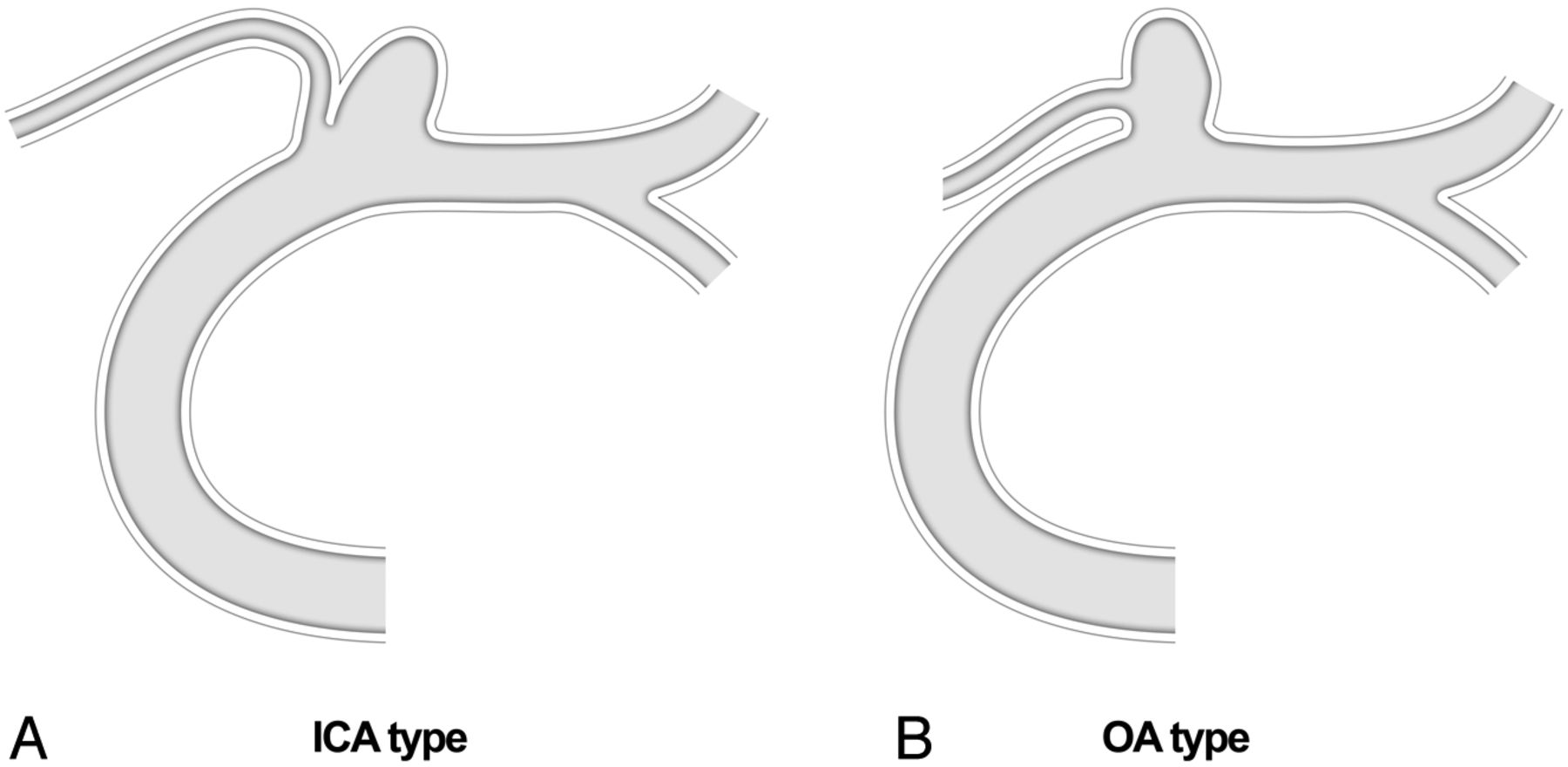
- Fig 1.
Classification of OA aneurysms (ICA type versus OA type).
- Fig 2.
A and B, Conventional angiography and 3D imaging of a medially directed OA aneurysm (OA originating from the aneurysmal neck and an aneurysm incorporating most of the OA entry). C, Balloon test occlusion performed after planned sacrifice of the OA orifice during coil embolization; the balloon catheter was placed at the ophthalmic segment (arrow) and inflated. D, With the balloon inflated, angiographic image of the common carotid artery reveals choroidal blush (arrow) and contrast filling (arrowhead) of the OA through the middle meningeal artery. E, Final angiographic view of the occluded aneurysm and OA orifice. F, Angiography of the external carotid artery confirms a patent choroidal blush (arrow) and contrast filling of the OA (arrowhead) after coiling.
- Fig 3.
A, 3D imaging of a superiorly directed OA aneurysm; balloon test occlusion was not performed due to the aneurysm type (ICA) and a relatively well-demarcated margin between the OA orifice and the aneurysmal neck in the working projection. B, Simple technique via a steam-shaped S-configured microcatheter results in persistent herniation of coils into the parent artery. C, A Neuroform stent (arrows, stent markers) stabilizes the coil mass and secures the OA orifice. D, Completion angiography of successfully occluded aneurysm, with the OA preserved.
- Fig 4.
Schematic depicting the treatment algorithm in the study population.
- Fig 5.
A, 3D imaging of a superiorly directed OA aneurysm; balloon test occlusion was not performed due to the aneurysm type (ICA) and relatively well-demarcated margin, B, Completion angiography of a successfully occluded aneurysm with OA compromise (arrow). C, Angiography of the external carotid artery confirms no patent contrast filling of the OA. D, Restoration of OA flow after intra-arterial tirofiban infusion (0.75 mg).


Tables
Characteristics No. of aneurysms and patients 43 aneurysms, 43 patients Age (yr) (mean) 53.3 ± 10.4 Female/male 33:10 Presentation Incidental 42 Visual disturbance 0 Ruptured 1 Aneurysm direction Superior 28 (65.1%) Medial 14 (32.6%) Inferior 1 (2.3%) Aneurysm type ICA type 19 (44.2%) OA type 24 (55.8%) Aneurysm size ≤5 mm 23 (53.5%) 5∼10 mm 18 (41.8%) ≥10 mm 2 (4.7%) No. of aneurysms of other locations OA aneurysm only 25 (58.1%) Multiple aneurysm 18 (41.9%) Depth-to-neck ratio ≤1 22 (51.2%) 1.0∼1.5 16 (37.2%) ≥1.5 5 (11.6%) Direction of aneurysm Superior 27 (62.8%) Medial 14 (32.5) Other 2 (4.7%) Initial occlusion result Complete occlusion 14 (32.6%) Residual neck 23 (53.5%) Residual aneurysm 6 (13.9%) Follow-up result Stable occlusion 30 (85.7%) Minor recanalization 4 (11.4%) Major recanalization 1 (2.9%) Superior (n = 28) Medial (n = 14) Inferior (n = 1) Technique Single microcatheter 5 1 0 Multiple microcatheter 4 2 0 Balloon remodeling 15 7 0 Stent protection 4 4 1 Microcatheter shape Steam-shaped S 19 2 0 Straight 5 0 0 Steam-shaped pigtail 3 10 1 Preshaped 45° or 90° 1 2 0


