
Article Figures & Data
Figures
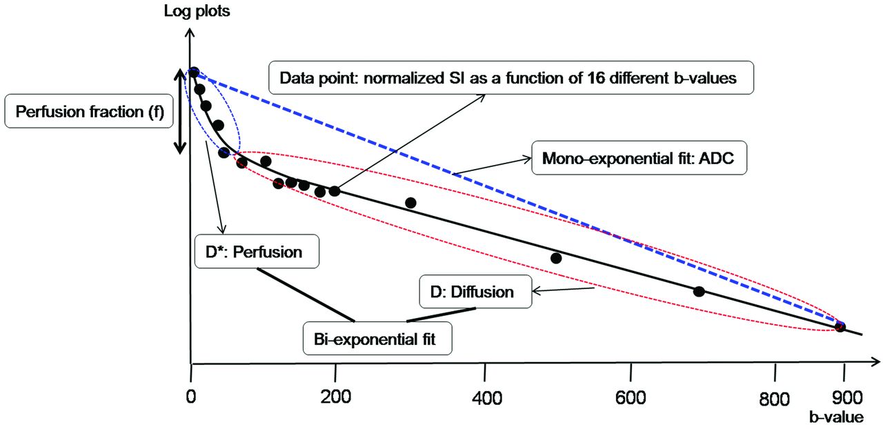
- Fig 1.
Illustration for biexponential signal decay as a function of the 16 different diffusion b-values in a given voxel of a recurrent tumor. The bold, solid line is the IVIM nonlinear regression fit providing D, D*, and f. The biexponential fit provides the fast decay associated with perfusion (blue dotted circle), and the red dotted circle represents the slow decay of the biexponential fit, thus indicating true diffusion. The blue dotted line shows the monoexponential fit providing the ADC.
- Fig 2.
A 61-year-old woman with treatment effect following GKRS. Axial contrast-enhanced T1-weighted images, obtained 3 (A) and 6 months (B) after GKRS, show a progressively enlarging necrotic contrast-enhancing lesion in the left parietal lobe. C, The necrotic contrast-enhancing lesion is stabilized on a subsequent follow-up image obtained 9 months after GKRS, thus indicating treatment effect. The ADC (D) and nCBV (E) maps show no visual decrease of the ADC and no visual increase of the nCBV in the corresponding area of the contrast-enhancing lesion in B, respectively. The D (F) and f (G) maps show no visual decrease of the D value and no visual increase of the f value in the corresponding area of the contrast-enhancing lesion in B, respectively. H, The signal decay curve, plotted as a function of the diffusion b-values, is monoexponential.
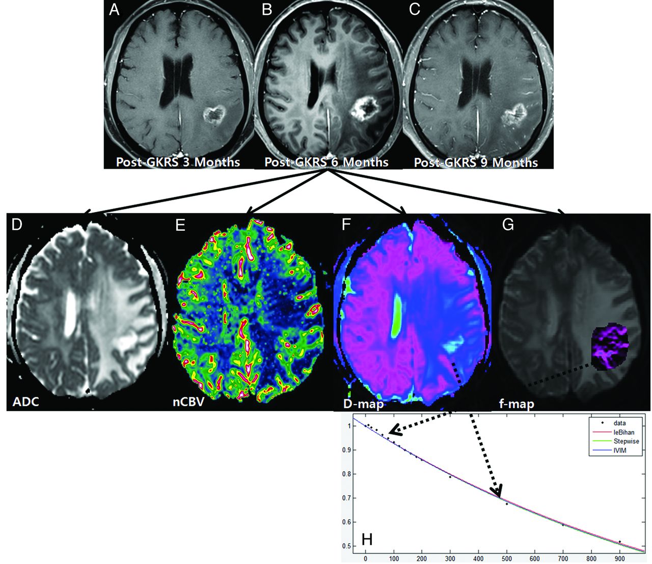
- Fig 3.
A 54-year-old man with recurrent tumor following GKRS. Axial contrast-enhanced T1-weighted images, obtained 3 (A), 6 (B), and 9 months (C) after GKRS, show a progressively enlarging necrotic contrast-enhancing lesion in the right parietal lobe. The ADC (D) and nCBV (E) maps show a visual decrease of the ADC and a visual increase of the nCBV in the corresponding area of the contrast-enhancing lesion in B, respectively. The D (F) and f (G) maps show a visual decrease of the D value and a visual increase of the f value in the corresponding area of the contrast-enhancing lesion in B, respectively. H, The signal-decay curve, plotted as a function of the diffusion b-values, is biexponential.
- Fig 4.
Box-and-whisker plots for the IVIM-derived f and D values between recurrent tumor and treatment effect for both readers.
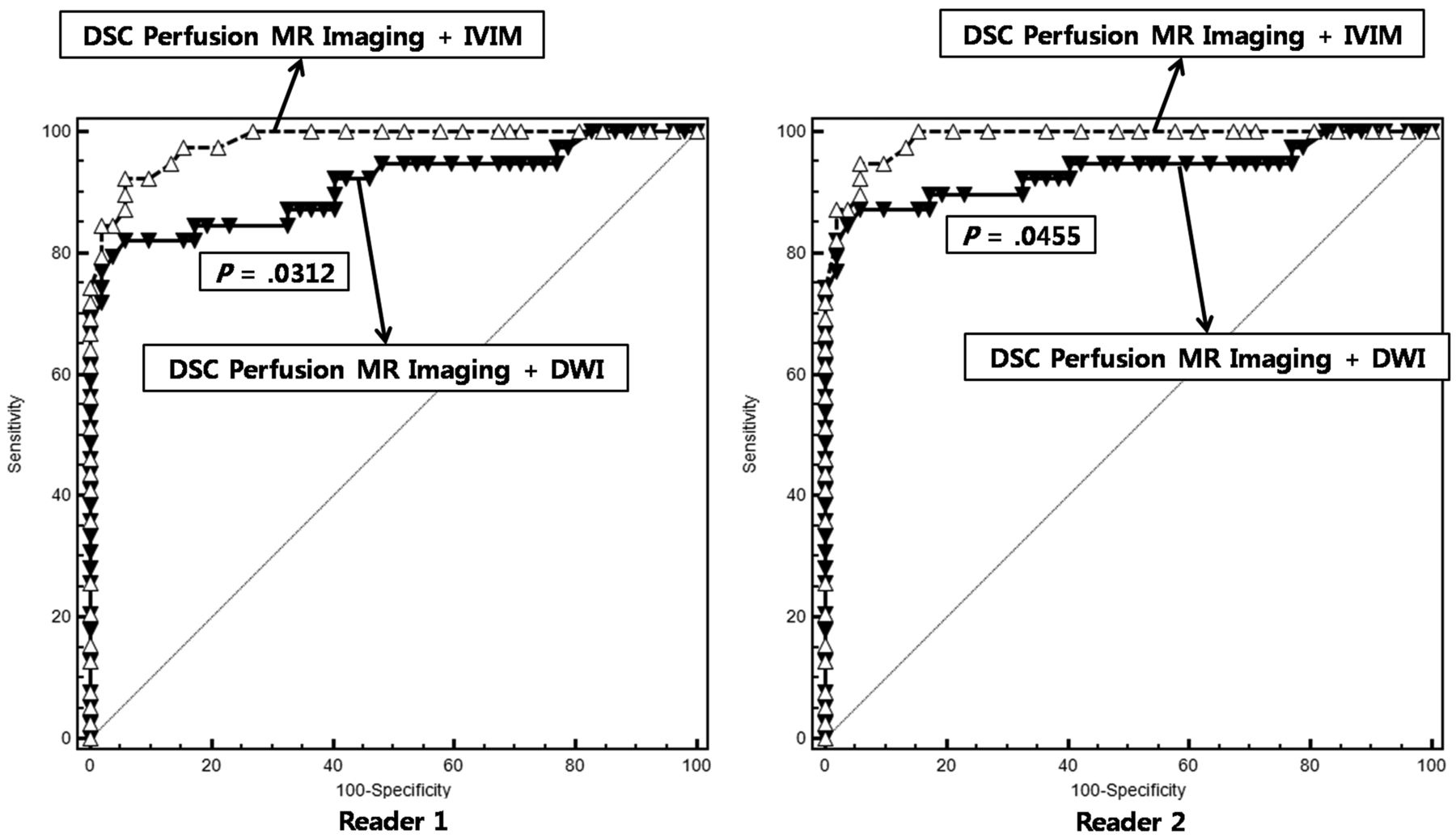
- Fig 5.
The graph shows the comparison between the receiver operating characteristic curve for the combination of DSC MR imaging and IVIM and for the combination of DSC MR imaging and DWI. The combination of DSC MR imaging and IVIM resulted in a significantly higher area under the receiver operating characteristic curve than the combination of DSC MR imaging and DWI for both readers, thus indicating improved diagnostic performance.


Tables
Variables Recurrent Tumor Treatment Effect P Value No. of male patients 19 (53.3%) 27 (61.9%) .272 No. of female patients 20 (46.7%) 25 (38.1%) Age (yr)a 47.4 ± 6.7 51.5 ± 8.5 .395 Mean gamma knife dose (Gy)a 17.5 ± 0.7 17.3 ± 0.5 .872 Target volume (mL)a 6.52 ± 7.01 5.97 ± 5.19 .312 Time interval between GKRS and detection of a new or enlarging, contrast-enhancing lesion (wk)a 34.7 ± 15.4 37.9 ± 17.3 .159 Time interval between GKRS and the last follow-up (wk)a 85.1 ± 22.1 89.2 ± 27.2 .576 ↵a Data are means.
- Table 2:
Differences in the imaging parameters in patients with recurrent tumor and those with treatment effect
Parameters Reader 1 Reader 2 Recurrent Tumor Treatment Effect P Value Recurrent Tumor Treatment Effect P Value f90a 0.079 ± 0.019 0.048 ± 0.009 <.001 0.081 ± 0.017 0.046 ± 0.012 <.001 D*90a (10−3mm2s−1) 39.1 ± 21.2 16.4 ± 12.6 .009 32..4 ± 22.5 19.5 ± 11.6 .024 D10a (10−3mm2s−1) 0.970 ± 0.082 1.043 ± 0.062 <.001 0.967 ± 0.071 1.045 ± 0.055 <.001 nCBV90a 4.457 ± 1.301 2.674 ± 0.348 <.001 4.782 ± 1.122 2.551 ± 0.416 <.001 ADC10a (10−3mm2s−1) 0.986 ± 0.079 1.052 ± 0.059 <.001 0.991 ± 0.092 1.055 ± 0.072 <.001 Note:—D*90 indicates the 90th percentile histogram cutoff of D*.
↵a Data are means.
MR Imaging Method and Comparison Reader 1 P Value Reader 2 P Value AUC 95% CI AUC 95% CI MR imaging method IVIM 0.939 0.868–0.978 0.947 0.879–0.983 DSC + DWI 0.911 0.832–0.960 0.933 0.861–0.975 DSC + IVIM 0.982 0.928–0.998 0.987 0.938–1.000 Comparison IVIM vs DSC + DWI .3762 .5897 IVIM vs DSC + IVIM .0471 .0951 DSC + DWI vs DSC + IVIM .0312 .0455 Note:—ROC indicates receiver operating characteristic analysis; AUC, area under the ROC curve.
Reader and MR Imaging Method Sensitivity Specificity Accuracy Reader 1 IVIM 79.5% 92.3% 86.8% DSC + DWI 69.2% 100.0% 86.8% DSC + IVIM 89.7% 94.2% 92.3% Reader 2 IVIM 84.6% 94.2% 90.1% DSC + DWI 74.3% 100.0% 89.0% DSC + IVIM 92.3% 94.2% 93.4%


