Advertisement

Main menu

  • Home
  • Content
    • Current Issue
    • Accepted Manuscripts
    • Article Preview
    • Past Issue Archive
    • Video Articles
    • AJNR Case Collection
    • Case of the Week Archive
    • Case of the Month Archive
    • Classic Case Archive
  • Special Collections
    • AJNR Awards
    • Low-Field MRI
    • Alzheimer Disease
    • ASNR Foundation Special Collection
    • Photon-Counting CT
    • View All
  • Multimedia
    • AJNR Podcasts
    • AJNR SCANtastic
    • Trainee Corner
    • MRI Safety Corner
    • Imaging Protocols
  • For Authors
    • Submit a Manuscript
    • Submit a Video Article
    • Submit an eLetter to the Editor/Response
    • Manuscript Submission Guidelines
    • Statistical Tips
    • Fast Publishing of Accepted Manuscripts
    • Graphical Abstract Preparation
    • Imaging Protocol Submission
    • Author Policies
  • About Us
    • About AJNR
    • Editorial Board
    • Editorial Board Alumni
  • More
    • Become a Reviewer/Academy of Reviewers
    • Subscribers
    • Permissions
    • Alerts
    • Feedback
    • Advertisers
    • ASNR Home

User menu

  • Alerts
  • Log in

Search

  • Advanced search
American Journal of Neuroradiology

American Journal of Neuroradiology

ASHNR American Society of Functional Neuroradiology ASHNR American Society of Pediatric Neuroradiology ASSR
  • Alerts
  • Log in

Advanced Search

  • Home
  • Content
    • Current Issue
    • Accepted Manuscripts
    • Article Preview
    • Past Issue Archive
    • Video Articles
    • AJNR Case Collection
    • Case of the Week Archive
    • Case of the Month Archive
    • Classic Case Archive
  • Special Collections
    • AJNR Awards
    • Low-Field MRI
    • Alzheimer Disease
    • ASNR Foundation Special Collection
    • Photon-Counting CT
    • View All
  • Multimedia
    • AJNR Podcasts
    • AJNR SCANtastic
    • Trainee Corner
    • MRI Safety Corner
    • Imaging Protocols
  • For Authors
    • Submit a Manuscript
    • Submit a Video Article
    • Submit an eLetter to the Editor/Response
    • Manuscript Submission Guidelines
    • Statistical Tips
    • Fast Publishing of Accepted Manuscripts
    • Graphical Abstract Preparation
    • Imaging Protocol Submission
    • Author Policies
  • About Us
    • About AJNR
    • Editorial Board
    • Editorial Board Alumni
  • More
    • Become a Reviewer/Academy of Reviewers
    • Subscribers
    • Permissions
    • Alerts
    • Feedback
    • Advertisers
    • ASNR Home
  • Follow AJNR on Twitter
  • Visit AJNR on Facebook
  • Follow AJNR on Instagram
  • Join AJNR on LinkedIn
  • RSS Feeds

AJNR Awards, New Junior Editors, and more. Read the latest AJNR updates

Research ArticleSpine Imaging and Spine Image-Guided Interventions

Distinguishing Imaging Features between Spinal Hyperplastic Hematopoietic Bone Marrow and Bone Metastasis

Y. Shigematsu, T. Hirai, K. Kawanaka, S. Shiraishi, M. Yoshida, M. Kitajima, H. Uetani, M. Azuma, Y. Iryo and Y. Yamashita
American Journal of Neuroradiology October 2014, 35 (10) 2013-2020; DOI: https://doi.org/10.3174/ajnr.A4012
Y. Shigematsu
aFrom the Department of Diagnostic Radiology, Graduate School of Medical Sciences, Kumamoto University, Kumamoto, Japan.
  • Find this author on Google Scholar
  • Find this author on PubMed
  • Search for this author on this site
T. Hirai
aFrom the Department of Diagnostic Radiology, Graduate School of Medical Sciences, Kumamoto University, Kumamoto, Japan.
  • Find this author on Google Scholar
  • Find this author on PubMed
  • Search for this author on this site
K. Kawanaka
aFrom the Department of Diagnostic Radiology, Graduate School of Medical Sciences, Kumamoto University, Kumamoto, Japan.
  • Find this author on Google Scholar
  • Find this author on PubMed
  • Search for this author on this site
S. Shiraishi
aFrom the Department of Diagnostic Radiology, Graduate School of Medical Sciences, Kumamoto University, Kumamoto, Japan.
  • Find this author on Google Scholar
  • Find this author on PubMed
  • Search for this author on this site
M. Yoshida
aFrom the Department of Diagnostic Radiology, Graduate School of Medical Sciences, Kumamoto University, Kumamoto, Japan.
  • Find this author on Google Scholar
  • Find this author on PubMed
  • Search for this author on this site
M. Kitajima
aFrom the Department of Diagnostic Radiology, Graduate School of Medical Sciences, Kumamoto University, Kumamoto, Japan.
  • Find this author on Google Scholar
  • Find this author on PubMed
  • Search for this author on this site
H. Uetani
aFrom the Department of Diagnostic Radiology, Graduate School of Medical Sciences, Kumamoto University, Kumamoto, Japan.
  • Find this author on Google Scholar
  • Find this author on PubMed
  • Search for this author on this site
M. Azuma
aFrom the Department of Diagnostic Radiology, Graduate School of Medical Sciences, Kumamoto University, Kumamoto, Japan.
  • Find this author on Google Scholar
  • Find this author on PubMed
  • Search for this author on this site
Y. Iryo
aFrom the Department of Diagnostic Radiology, Graduate School of Medical Sciences, Kumamoto University, Kumamoto, Japan.
  • Find this author on Google Scholar
  • Find this author on PubMed
  • Search for this author on this site
Y. Yamashita
aFrom the Department of Diagnostic Radiology, Graduate School of Medical Sciences, Kumamoto University, Kumamoto, Japan.
  • Find this author on Google Scholar
  • Find this author on PubMed
  • Search for this author on this site
  • Article
  • Figures & Data
  • Info & Metrics
  • Responses
  • References
  • PDF
Loading

Article Figures & Data

Figures

  • Tables
  • Fig 1.
    • Download figure
    • Open in new tab
    • Download powerpoint
    Fig 1.

    A, Schema of the distribution pattern of a lesion in a single vertebral body. “Diffuse pattern” indicates involvement of the entire region in a single vertebral body; “patchy pattern,” involvement of the partial region in a single vertebral body; and “nodular pattern,” involvement of the nodular area in a single vertebral body. B, Schema of the appearance of homogeneity of a lesion in a single vertebral body. Lesions were considered homogeneous if no evidence of necrosis, cyst, hemorrhage, or fracture was seen on T1- and T2-weighted images. If ≥1 of these features or mixed signal intensity (ie, high and low signal intensity on the MR images) was present, the vertebral lesion was considered heterogeneous.
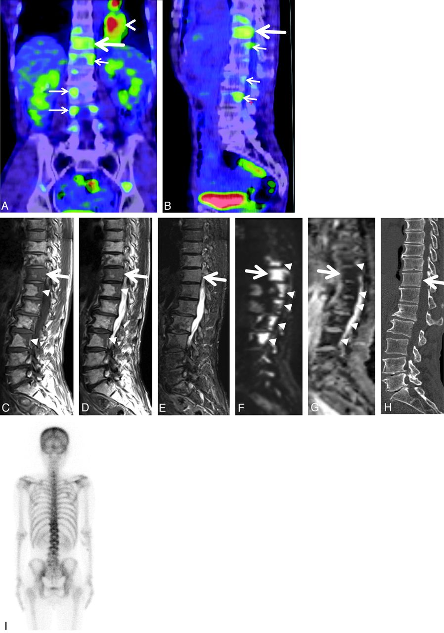
  • Fig 2.
    • Download figure
    • Open in new tab
    • Download powerpoint
    Fig 2.

    A 54-year-old man with biopsy-proved hyperplastic hematopoietic bone marrow in the T11 vertebral body. Coronal (A) and sagittal (B) reconstructed FDG-PET/CT images reveal the highest uptake (large arrow) in the T11 vertebral body. The SUVmax of the vertebra was 2.4. Subtle uptake is also seen in some thoracic and lumbar vertebral bodies (small arrows). Primary lung cancer is demonstrated in the left lower lung (arrowhead in A). Sagittal T1- (C) and T2-weighted (D) spin-echo MR images of the thoracolumbar spine show diffuse hypointensity (large arrow) in the T11 vertebral body. Patchy hypointense areas are seen in some thoracic and lumbar vertebral bodies (arrowheads). E, Sagittal STIR image of the thoracolumbar spine depicts slight hyperintensity (arrow) in the vertebrae from T10 to L3. F, Sagittal diffusion-weighted image of the thoracolumbar spine reveals marked hyperintense areas (arrowheads) in the vertebrae from T10 to L3. G, The ADC map shows restricted diffusion (arrowheads), corresponding to the hyperintense areas in the vertebrae on diffusion-weighted images. The ADC value in the T11 vertebral body was 0.56 × 10⁻³mm2/s. H, On the sagittal reconstructed CT image of the thoracolumbar spine, there is diffuse, slightly high attenuation in the T11 vertebral body (large arrow). I, Posterior bone scintigraphy shows no apparent uptake in the thoracolumbar spine.

  • Fig 3.
    • Download figure
    • Open in new tab
    • Download powerpoint
    Fig 3.

    An 84-year-old man with lung cancer and biopsy-proved metastatic bone tumor in the L3 vertebral body. Sagittal T1- (A) and T2-weighted (B) spin-echo MR images of the lumbar spine show diffuse hypointensity (arrow) in the L3 vertebra. There are no vertebral lesions adjacent to the L3 lesion. C, Sagittal STIR image of the lumbar spine depicts hyperintensity (arrow) in the same vertebra. D, On the axial CT image at the level of the L3 lumbar vertebra, there is an osteolytic area in the vertebral body (arrow). E, Axial FDG-PET/CT image at the level of the L3 lumbar vertebra reveals a high uptake area in the vertebral body (arrow). SUVmax of the vertebral lesion was 12.4.

  • Fig 4.
    • Download figure
    • Open in new tab
    • Download powerpoint
    Fig 4.

    Comparison of SUVmax between HHBM and bone metastasis. The SUVmax of the biopsy-proved vertebral lesions is shown. A significant difference is seen between HHBM and bone metastasis (Mann-Whitney test, P = .037). The horizontal short lines indicate 95% CI for the median.
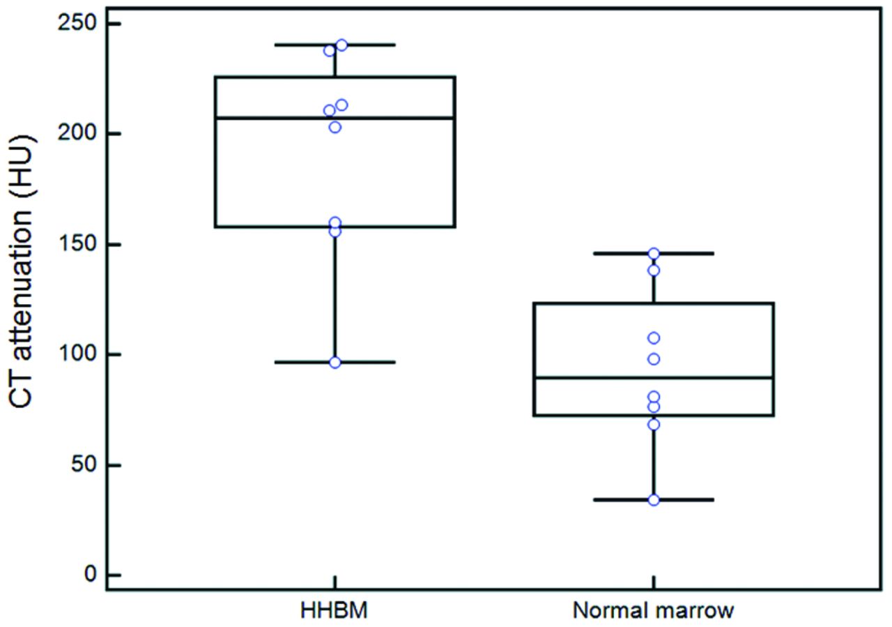
  • Fig 5.
    • Download figure
    • Open in new tab
    • Download powerpoint
    Fig 5.

    Comparison of CT attenuation between HHBM and normal-appearing bone marrow. The mean CT attenuation of HHBM was significantly higher than that of adjacent normal-appearing vertebrae (Wilcoxon signed rank test, P = .0078). The horizontal short lines indicate 95% CI for the median.

  • Fig 6.
    • Download figure
    • Open in new tab
    • Download powerpoint
    Fig 6.

    A proposed diagnostic scheme to be followed by practitioners/radiologists when faced with the problem of differentiating HHBM from bone metastasis. Asterisk indicates that bone biopsy might be still needed in indeterminate cases.

Tables

  • Figures
    • View popup
    Table 1:

    Summary of characteristics of patients with spinal HHBM

    Case No.AgeSexClinical Diagnosis (TNM stage)SymptomLocation of Spinal LesionsBiopsy SiteBrinkman IndexHemoglobin (g/dL)
    157MaleLung ca. (T1N0)NoneT2–L3T568412.4↓
    254MaleLung ca. (T2N1)NoneT11–L3T1175011.4↓
    359MaleEsop. ca. (T3N2)NoneC2–T12, S1T1178010.9↓
    473MaleEsop. ca. (T1N0)NoneT9–11T11106012.6↓
    578MaleEsop. ca. (T3N2)NoneC2–T9, L1,3L1011.9↓
    670MaleEsop. ca. (T2N0)NoneL3L390011.6↓
    770MaleLumbar spondylosisLBPC6–T12T12107514.1
    852MaleLumbar spondylosisLBPC9–L5L364013.7↓
    • Note:—Lung ca. indicates lung cancer; Esop. ca., esophageal cancer; LBP, low back pain; C, cervical spine; T, thoracic spine; L, lumbar spine; S, sacral spine; ↓, anemia.

    • View popup
    Table 2:

    Summary of visual assessments of vertebral lesions on MRIa

    T1WIT2WIDistribution PatternAppearance
    HyperIsoHypoHyperIsoHypoDiffusePatchyNodularHomoHetero
    HHBM (n = 8)00800852180
    Metastasis (n = 21)002136122001156
    • Note:—Hyper indicates hyperintensity; Iso, isointensity; Hypo, hypointensity; Homo, homogeneous; Hetero, heterogeneous.

    • ↵a When the signal intensity was divided into 2 categories (hypointensity vs iso- to hyperintensity), there was no significant difference in the T1 signal intensity of the lesions between patients with HHBM and those with bone metastasis. On the other hand, there was a significant difference in the T2 signal intensity of the lesions between the 2 groups (Fisher exact test, P = .033). There was no significant difference in the distribution pattern and appearance of the lesions between patients with HHBM and those with bone metastasis.

    • View popup
    Table 3:

    Summary of visual inspections of vertebral lesions on CTa

    CT Appearanceb
    Grade 1Grade 2Grade 3Grade 4
    HHBM (n = 8)8000
    Bone metastasis (n = 24)11751
    • ↵a When the CT appearance was divided into 2 categories (grade 1 vs grades 2–4), HHBM and metastasis differed significantly (Fisher exact test, P < .01).

    • ↵b CT appearance of each vertebral body lesion was classified using the 4-point grading system: grade 1, normal appearance with normal or subtle high attenuation; grade 2, osteolytic lesions; grade 3, osteoblastic lesions; and grade 4, mixed osteolytic and osteoblastic lesions.

    • View popup
    Table 4:

    Summary of visual inspections for abnormal uptake on bone scintigraphy

    Abnormal Uptakea
    NonePresent
    HHBM (n = 5)50
    Bone metastasis (n = 13)013
    • ↵a The uptake assessment between the HHBM and bone metastasis was significantly different (Fisher exact test, P < .01).

PreviousNext
Back to top

In this issue

American Journal of Neuroradiology: 35 (10)
American Journal of Neuroradiology
Vol. 35, Issue 10
1 Oct 2014
  • Table of Contents
  • Index by author
  • Complete Issue (PDF)
Advertisement
Print
Download PDF
Email Article

Thank you for your interest in spreading the word on American Journal of Neuroradiology.

NOTE: We only request your email address so that the person you are recommending the page to knows that you wanted them to see it, and that it is not junk mail. We do not capture any email address.

Enter multiple addresses on separate lines or separate them with commas.
Distinguishing Imaging Features between Spinal Hyperplastic Hematopoietic Bone Marrow and Bone Metastasis
(Your Name) has sent you a message from American Journal of Neuroradiology
(Your Name) thought you would like to see the American Journal of Neuroradiology web site.
CAPTCHA
This question is for testing whether or not you are a human visitor and to prevent automated spam submissions.
Cite this article
Y. Shigematsu, T. Hirai, K. Kawanaka, S. Shiraishi, M. Yoshida, M. Kitajima, H. Uetani, M. Azuma, Y. Iryo, Y. Yamashita
Distinguishing Imaging Features between Spinal Hyperplastic Hematopoietic Bone Marrow and Bone Metastasis
American Journal of Neuroradiology Oct 2014, 35 (10) 2013-2020; DOI: 10.3174/ajnr.A4012

Citation Manager Formats

  • BibTeX
  • Bookends
  • EasyBib
  • EndNote (tagged)
  • EndNote 8 (xml)
  • Medlars
  • Mendeley
  • Papers
  • RefWorks Tagged
  • Ref Manager
  • RIS
  • Zotero
0 Responses
Respond to this article
Share
Bookmark this article
Distinguishing Imaging Features between Spinal Hyperplastic Hematopoietic Bone Marrow and Bone Metastasis
Y. Shigematsu, T. Hirai, K. Kawanaka, S. Shiraishi, M. Yoshida, M. Kitajima, H. Uetani, M. Azuma, Y. Iryo, Y. Yamashita
American Journal of Neuroradiology Oct 2014, 35 (10) 2013-2020; DOI: 10.3174/ajnr.A4012
del.icio.us logo Twitter logo Facebook logo Mendeley logo
  • Tweet Widget
  • Facebook Like
  • Google Plus One
Purchase

Jump to section

  • Article
    • Abstract
    • ABBREVIATIONS:
    • Materials and Methods
    • Results
    • Discussion
    • Conclusions
    • REFERENCES
  • Figures & Data
  • Info & Metrics
  • Responses
  • References
  • PDF

Related Articles

  • No related articles found.
  • PubMed
  • Google Scholar

Cited By...

  • Spinal hyperplastic bone marrow with a lumbar vertebral compression fracture mimicking vertebral metastasis
  • Crossref
  • Google Scholar

This article has not yet been cited by articles in journals that are participating in Crossref Cited-by Linking.

More in this TOC Section

  • Optimization of Photon Counting CT Myelography
  • Characteristics of SIH Type I Culprit Lesions
  • Management Outcomes For VO Spine Biopsy
Show more Spine Imaging and Spine Image-Guided Interventions

Similar Articles

Advertisement

Indexed Content

  • Current Issue
  • Accepted Manuscripts
  • Article Preview
  • Past Issues
  • Editorials
  • Editor's Choice
  • Fellows' Journal Club
  • Letters to the Editor
  • Video Articles

Cases

  • Case Collection
  • Archive - Case of the Week
  • Archive - Case of the Month
  • Archive - Classic Case

More from AJNR

  • Trainee Corner
  • Imaging Protocols
  • MRI Safety Corner
  • Book Reviews

Multimedia

  • AJNR Podcasts
  • AJNR Scantastics

Resources

  • Turnaround Time
  • Submit a Manuscript
  • Submit a Video Article
  • Submit an eLetter to the Editor/Response
  • Manuscript Submission Guidelines
  • Statistical Tips
  • Fast Publishing of Accepted Manuscripts
  • Graphical Abstract Preparation
  • Imaging Protocol Submission
  • Evidence-Based Medicine Level Guide
  • Publishing Checklists
  • Author Policies
  • Become a Reviewer/Academy of Reviewers
  • News and Updates

About Us

  • About AJNR
  • Editorial Board
  • Editorial Board Alumni
  • Alerts
  • Permissions
  • Not an AJNR Subscriber? Join Now
  • Advertise with Us
  • Librarian Resources
  • Feedback
  • Terms and Conditions
  • AJNR Editorial Board Alumni

American Society of Neuroradiology

  • Not an ASNR Member? Join Now

© 2025 by the American Society of Neuroradiology All rights, including for text and data mining, AI training, and similar technologies, are reserved.
Print ISSN: 0195-6108 Online ISSN: 1936-959X

Powered by HighWire