Advertisement

Main menu

  • Home
  • Content
    • Current Issue
    • Accepted Manuscripts
    • Article Preview
    • Past Issue Archive
    • Video Articles
    • AJNR Case Collection
    • Case of the Week Archive
    • Case of the Month Archive
    • Classic Case Archive
  • Special Collections
    • AJNR Awards
    • Low-Field MRI
    • Alzheimer Disease
    • ASNR Foundation Special Collection
    • Photon-Counting CT
    • View All
  • Multimedia
    • AJNR Podcasts
    • AJNR SCANtastic
    • Trainee Corner
    • MRI Safety Corner
    • Imaging Protocols
  • For Authors
    • Submit a Manuscript
    • Submit a Video Article
    • Submit an eLetter to the Editor/Response
    • Manuscript Submission Guidelines
    • Statistical Tips
    • Fast Publishing of Accepted Manuscripts
    • Graphical Abstract Preparation
    • Imaging Protocol Submission
    • Author Policies
  • About Us
    • About AJNR
    • Editorial Board
    • Editorial Board Alumni
  • More
    • Become a Reviewer/Academy of Reviewers
    • Subscribers
    • Permissions
    • Alerts
    • Feedback
    • Advertisers
    • ASNR Home

User menu

  • Alerts
  • Log in

Search

  • Advanced search
American Journal of Neuroradiology

American Journal of Neuroradiology

ASHNR American Society of Functional Neuroradiology ASHNR American Society of Pediatric Neuroradiology ASSR
  • Alerts
  • Log in

Advanced Search

  • Home
  • Content
    • Current Issue
    • Accepted Manuscripts
    • Article Preview
    • Past Issue Archive
    • Video Articles
    • AJNR Case Collection
    • Case of the Week Archive
    • Case of the Month Archive
    • Classic Case Archive
  • Special Collections
    • AJNR Awards
    • Low-Field MRI
    • Alzheimer Disease
    • ASNR Foundation Special Collection
    • Photon-Counting CT
    • View All
  • Multimedia
    • AJNR Podcasts
    • AJNR SCANtastic
    • Trainee Corner
    • MRI Safety Corner
    • Imaging Protocols
  • For Authors
    • Submit a Manuscript
    • Submit a Video Article
    • Submit an eLetter to the Editor/Response
    • Manuscript Submission Guidelines
    • Statistical Tips
    • Fast Publishing of Accepted Manuscripts
    • Graphical Abstract Preparation
    • Imaging Protocol Submission
    • Author Policies
  • About Us
    • About AJNR
    • Editorial Board
    • Editorial Board Alumni
  • More
    • Become a Reviewer/Academy of Reviewers
    • Subscribers
    • Permissions
    • Alerts
    • Feedback
    • Advertisers
    • ASNR Home
  • Follow AJNR on Twitter
  • Visit AJNR on Facebook
  • Follow AJNR on Instagram
  • Join AJNR on LinkedIn
  • RSS Feeds

AJNR Awards, New Junior Editors, and more. Read the latest AJNR updates

Research ArticleHead and Neck Imaging
Open Access

Orbital Lymphoproliferative Disorders (OLPDs): Value of MR Imaging for Differentiating Orbital Lymphoma from Benign OPLDs

K. Haradome, H. Haradome, Y. Usui, S. Ueda, T.C. Kwee, K. Saito, K. Tokuuye, J. Matsubayashi, T. Nagao and H. Goto
American Journal of Neuroradiology October 2014, 35 (10) 1976-1982; DOI: https://doi.org/10.3174/ajnr.A3986
K. Haradome
aFrom the Departments of Ophthalmology (K.H., Y.U., S.U., H.G.)
  • Find this author on Google Scholar
  • Find this author on PubMed
  • Search for this author on this site
H. Haradome
dDepartment of Radiology (H.H.), Nihon University School of Medicine, Tokyo, Japan
  • Find this author on Google Scholar
  • Find this author on PubMed
  • Search for this author on this site
Y. Usui
aFrom the Departments of Ophthalmology (K.H., Y.U., S.U., H.G.)
  • Find this author on Google Scholar
  • Find this author on PubMed
  • Search for this author on this site
S. Ueda
aFrom the Departments of Ophthalmology (K.H., Y.U., S.U., H.G.)
  • Find this author on Google Scholar
  • Find this author on PubMed
  • Search for this author on this site
T.C. Kwee
eDepartment of Radiology (T.C.K.), University Medical Center Utrecht, Utrecht, the Netherlands.
  • Find this author on Google Scholar
  • Find this author on PubMed
  • Search for this author on this site
K. Saito
bRadiology (K.S., K.T.)
  • Find this author on Google Scholar
  • Find this author on PubMed
  • Search for this author on this site
K. Tokuuye
bRadiology (K.S., K.T.)
  • Find this author on Google Scholar
  • Find this author on PubMed
  • Search for this author on this site
J. Matsubayashi
cAnatomic Pathology (J.M., T.N.), Tokyo Medical University, Tokyo, Japan
  • Find this author on Google Scholar
  • Find this author on PubMed
  • Search for this author on this site
T. Nagao
cAnatomic Pathology (J.M., T.N.), Tokyo Medical University, Tokyo, Japan
  • Find this author on Google Scholar
  • Find this author on PubMed
  • Search for this author on this site
H. Goto
aFrom the Departments of Ophthalmology (K.H., Y.U., S.U., H.G.)
  • Find this author on Google Scholar
  • Find this author on PubMed
  • Search for this author on this site
  • Article
  • Figures & Data
  • Info & Metrics
  • Responses
  • References
  • PDF
Loading

Article Figures & Data

Figures

  • Tables
  • Fig 1.
    • Download figure
    • Open in new tab
    • Download powerpoint
    Fig 1.

    Orbital lymphoma (MALT lymphoma) in a 79-year-old woman. A, Transverse fat-saturated T2-weighted image; B, Transverse postcontrast fat-saturated T1-weighted image; C, Transverse DWI; D, ADC map; and E, Hematoxylin and eosin (HE)-stained biopsied specimen of the orbital tumor at low power field. An infiltrative mass involves the left lacrimal gland and extraocular muscles, appears isointense compared with the brain cortex on a fat-saturated T2-weighted MR image (A), and shows homogeneous contrast enhancement, similar to that of the right extraocular muscle (arrow) (B). The mass appears as strongly hyperintense on DWI (C) and hypointense on the ADC map (D). Notably, noninvolved extraocular muscles do not show hyperintensity similar to that of the mass on DWI (C). Lesion ADC and CER were 0.56 × 10⁻³ mm2/s and 1.69, respectively. HE-stained biopsied specimen (E) demonstrated numerous small-to-medium-sized atypical lymphocytes around reactive lymph follicles, with greater high cellular attenuation, however, interstitial fibrosis or edematous change was not prominent.

  • Fig 2.
    • Download figure
    • Open in new tab
    • Download powerpoint
    Fig 2.

    IgG4-related ophthalmic disease in a 67-year-old woman. A, Transverse fat-saturated T2-weighted image; B, transverse postcontrast fat-saturated T1-weighted image; C, transverse DWI; D, ADC map; and E, Hematoxylin and eosin (HE)-stained biopsied specimen of the lacrimal gland at low power field. The mass-like enlarged lacrimal glands are isointense compared with the brain cortex on the fat-saturated T2-weighted image (A) and show homogeneous contrast enhancement, similar to that of the extraocular muscles (B). The lesions appear mildly hyperintense on DWI (C) and slightly hypointense on the ADC map (D). Lesion ADC and CER were 0.94/0.75 (right/left) ×10−3 mm2/s and 2.05/2.07 (right/left), respectively. HE-stained biopsied specimen (E) showed a large germinal center with accompanying lymphoplasmacytic infiltration and abundant interstitial fibrosis with edematous changes. On immunochemical stained section analysis (not shown), many IgG4-positive plasma cell (>40%) were identified, and this is compatible with IgG4-related ophthalmic disease.
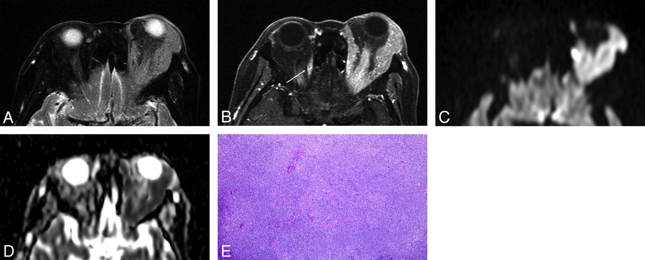
  • Fig 3.
    • Download figure
    • Open in new tab
    • Download powerpoint
    Fig 3.

    Orbital lymphoma (mantle cell lymphoma) in a 71-year-old man. A, Transverse fat-saturated T2-weighted image; B, transverse fat-saturated postcontrast T1-weighted image; C, transverse DWI; and D, ADC map. The lesions involve the bilateral lacrimal glands, appear isointense compared with the brain cortex on fat-saturated T2-weighted image (A), and show homogeneous contrast enhancement, similar that of the extraocular muscles (B). The lesions are strongly hyperintense on the DWI (C) and hypointense on the ADC map (D). Lesion ADC and CER were 0.47/0.48 (right/left) ×10−3 mm2/s and 1.49/1.46 (right/left), respectively. The low ADC and CER values could suggest orbital lymphoma though the imaging features are similar to that of IgG4-related ophthalmic disease (Fig 2).

  • Fig 4.
    • Download figure
    • Open in new tab
    • Download powerpoint
    Fig 4.

    Reactive lymphoid hyperplasia in a 65-year-old man. A, Coronal fat-saturated T2-weighted image. A number of vessel signal voids are observed in the bilaterally enlarged lacrimal glands on T2-weighted image (“flow void sign”) (arrows).

  • Fig 5.
    • Download figure
    • Open in new tab
    • Download powerpoint
    Fig 5.

    ADCs and CERs of orbital lymphoma and benign OPLDs. A and B, Scatterplot and box-and-whisker plot show the distribution of ADCs and CERs, and each mean value in the orbital lymphoma and benign OLPDs lesions. The mean ADC and CER of orbital lymphomas are significantly lower than those of benign OLPDs (P < .01). The standard deviation of the ADC of orbital lymphomas was lower (±0.051) compared with that of benign OLPDs (±0.246), and ADCs of only 2 benign OLPDs (encircled) overlapped with those of orbital lymphomas in the ADC range.

Tables

  • Figures
    • View popup
    Table 1:

    Patient characteristics and pathologic findings of OLPD cases

    Orbital Lymphoma (n = 29)Benign OLPDs (n = 18)P Value
    Age72.2 ± 11.3 (47–88)57.6 ± 14.7 (27–80)P = .001a
    SexMale 18, Female 11Male 10, Female 8P = .763
    Histologic subtypesMALT lymphoma 21 (72)IgG4-related ophthalmic disease 14 (78)
    DLBCL 4 (14)RLH 4 (22)
    Follicular lymphoma 3 (10)
    Mantle cell lymphoma 1 (3)
    LateralityBilateral 12 (41)Bilateral 15 (83)P = .006a
    Unilateral 17 (59)Unilateral 3 (17)
    • Note:—Data in parentheses are ranges for age and percentages for histologic subtypes and laterality. DLBCL indicates diffuse large B-cell lymphoma; RLH, reactive lymphoid hyperplasia.

    • ↵a There were significant differences in age and laterality between the 2 groups.

    • View popup
    Table 2:

    MR imaging characteristics

    Orbital Lymphoma (n = 29)Benign OLPDs (n = 18)P Value
    ShapeWell-defined 7 (24)Well-defined 10 (56)P = .0006a
    Ill-defined 21 (72)Ill-defined 3 (17)
    Lobulated 1 (3)Lobulated 5 (17)
    Signal intensity
        T1WIIso 28 (97)Iso 18 (100)P = .2548
    Low 1 (3)
        T2WIIso 27 (93)Iso 16 (89)P = .6147
    Low 2 (7)Low 2 (11)
        DWIHigh 17 (100)High 14 (93)P = .2794
    Iso 1 (7)
    Homogeneity and degree of contrast enhancementHomogeneous 23 (85)Homogeneous 15 (100)P = .2787
    Inhomogeneous 4 (15)
    High 2 (13)P = .0519
    Iso 27 (100)Iso 13 (87)
    Presence of “flow void sign”17 (59)17 (94)P = .0084a
    Findings suggestive of sinusitis8 (28)16 (89)P < .001a
    • Note:—Data in parentheses are percentages.

    • ↵a There were significant differences in shape of the lesions, presence of the “flow void sign,” and findings suggestive of sinusitis between the 2 groups.

    • View popup
    Table 3:

    ADCs of each group of OLPDs

    Mean ADC ± SDADC rangeP Value
    Orbital lymphoma (n = 17)0.54 ± 0.050.44–0.64P < .001a
    MALT lymphoma (n = 12)0.47–0.64
    DLBCL (n = 2)0.44–0.56
    Follicular lymphoma (n = 2)0.53–0.57
    Mantle cell lymphoma (n = 1)0.47
    Benign OLPDs (n = 15)0.81 ± 0.180.58–1.24
    IgG4-related ophthalmic disease (n = 12)0.58–1.24
    RLH (n = 3)0.73–0.85
    • Note:—There were significant differences in ADC between the 2 groups. SD indicates standard deviation; DLBCL diffuse large B-cell lymphoma; RLH, reactive lymphoid hyperplasia.

    • View popup
    Table 4:

    CERs of each group of OLPDs

    Mean CER ± SDCER RangeP Value
    Orbital lymphoma (n = 27)1.70 ± 0.251.27–2.24P = .0096a
    MALT lymphoma (n = 20)1.27–2.24
    DLBCL (n = 3)1.36–1.71
    Follicular lymphoma (n = 3)1.76–1.94
    Mantle cell lymphoma (n = 1)1.55
    Benign OLPDs (n = 15)2.07 ± 0.461.27–3.03
    IgG4-related ophthalmic disease (n = 11)1.27–3.03
    RLH (n = 4)1.66–2.14
    • ↵a There were significant differences in CER between the 2 groups. SD indicates standard deviation; DLBCL, diffuse large B-cell lymphoma; RLH, reactive lymphoid hyperplasia.

PreviousNext
Back to top

In this issue

American Journal of Neuroradiology: 35 (10)
American Journal of Neuroradiology
Vol. 35, Issue 10
1 Oct 2014
  • Table of Contents
  • Index by author
  • Complete Issue (PDF)
Advertisement
Print
Download PDF
Email Article

Thank you for your interest in spreading the word on American Journal of Neuroradiology.

NOTE: We only request your email address so that the person you are recommending the page to knows that you wanted them to see it, and that it is not junk mail. We do not capture any email address.

Enter multiple addresses on separate lines or separate them with commas.
Orbital Lymphoproliferative Disorders (OLPDs): Value of MR Imaging for Differentiating Orbital Lymphoma from Benign OPLDs
(Your Name) has sent you a message from American Journal of Neuroradiology
(Your Name) thought you would like to see the American Journal of Neuroradiology web site.
CAPTCHA
This question is for testing whether or not you are a human visitor and to prevent automated spam submissions.
Cite this article
K. Haradome, H. Haradome, Y. Usui, S. Ueda, T.C. Kwee, K. Saito, K. Tokuuye, J. Matsubayashi, T. Nagao, H. Goto
Orbital Lymphoproliferative Disorders (OLPDs): Value of MR Imaging for Differentiating Orbital Lymphoma from Benign OPLDs
American Journal of Neuroradiology Oct 2014, 35 (10) 1976-1982; DOI: 10.3174/ajnr.A3986

Citation Manager Formats

  • BibTeX
  • Bookends
  • EasyBib
  • EndNote (tagged)
  • EndNote 8 (xml)
  • Medlars
  • Mendeley
  • Papers
  • RefWorks Tagged
  • Ref Manager
  • RIS
  • Zotero
0 Responses
Respond to this article
Share
Bookmark this article
Orbital Lymphoproliferative Disorders (OLPDs): Value of MR Imaging for Differentiating Orbital Lymphoma from Benign OPLDs
K. Haradome, H. Haradome, Y. Usui, S. Ueda, T.C. Kwee, K. Saito, K. Tokuuye, J. Matsubayashi, T. Nagao, H. Goto
American Journal of Neuroradiology Oct 2014, 35 (10) 1976-1982; DOI: 10.3174/ajnr.A3986
del.icio.us logo Twitter logo Facebook logo Mendeley logo
  • Tweet Widget
  • Facebook Like
  • Google Plus One
Purchase

Jump to section

  • Article
    • Abstract
    • ABBREVIATIONS:
    • Materials and Methods
    • Results
    • Discussion
    • Conclusions
    • Acknowledgments
    • REFERENCES
  • Figures & Data
  • Info & Metrics
  • Responses
  • References
  • PDF

Related Articles

  • No related articles found.
  • PubMed
  • Google Scholar

Cited By...

  • Diagnostic Performance of Dynamic Contrast-Enhanced 3T MR Imaging for Characterization of Orbital Lesions: Validation in a Large Prospective Study
  • Droopy eyelid due to IgG4-related dacryoadenitis
  • Crossref
  • Google Scholar

This article has not yet been cited by articles in journals that are participating in Crossref Cited-by Linking.

More in this TOC Section

  • MRI of the Parasellar Ligaments
  • ASL Sensitivity for Head and Neck Paraganglioma
  • Post SRS Peritumoral Hyperintense Signal of VSs
Show more Head and Neck Imaging

Similar Articles

Advertisement

Indexed Content

  • Current Issue
  • Accepted Manuscripts
  • Article Preview
  • Past Issues
  • Editorials
  • Editor's Choice
  • Fellows' Journal Club
  • Letters to the Editor
  • Video Articles

Cases

  • Case Collection
  • Archive - Case of the Week
  • Archive - Case of the Month
  • Archive - Classic Case

More from AJNR

  • Trainee Corner
  • Imaging Protocols
  • MRI Safety Corner
  • Book Reviews

Multimedia

  • AJNR Podcasts
  • AJNR Scantastics

Resources

  • Turnaround Time
  • Submit a Manuscript
  • Submit a Video Article
  • Submit an eLetter to the Editor/Response
  • Manuscript Submission Guidelines
  • Statistical Tips
  • Fast Publishing of Accepted Manuscripts
  • Graphical Abstract Preparation
  • Imaging Protocol Submission
  • Evidence-Based Medicine Level Guide
  • Publishing Checklists
  • Author Policies
  • Become a Reviewer/Academy of Reviewers
  • News and Updates

About Us

  • About AJNR
  • Editorial Board
  • Editorial Board Alumni
  • Alerts
  • Permissions
  • Not an AJNR Subscriber? Join Now
  • Advertise with Us
  • Librarian Resources
  • Feedback
  • Terms and Conditions
  • AJNR Editorial Board Alumni

American Society of Neuroradiology

  • Not an ASNR Member? Join Now

© 2025 by the American Society of Neuroradiology All rights, including for text and data mining, AI training, and similar technologies, are reserved.
Print ISSN: 0195-6108 Online ISSN: 1936-959X

Powered by HighWire