Advertisement

Main menu

  • Home
  • Content
    • Current Issue
    • Accepted Manuscripts
    • Article Preview
    • Past Issue Archive
    • Video Articles
    • AJNR Case Collection
    • Case of the Week Archive
    • Case of the Month Archive
    • Classic Case Archive
  • Special Collections
    • AJNR Awards
    • Low-Field MRI
    • Alzheimer Disease
    • ASNR Foundation Special Collection
    • Photon-Counting CT
    • View All
  • Multimedia
    • AJNR Podcasts
    • AJNR SCANtastic
    • Trainee Corner
    • MRI Safety Corner
    • Imaging Protocols
  • For Authors
    • Submit a Manuscript
    • Submit a Video Article
    • Submit an eLetter to the Editor/Response
    • Manuscript Submission Guidelines
    • Statistical Tips
    • Fast Publishing of Accepted Manuscripts
    • Graphical Abstract Preparation
    • Imaging Protocol Submission
    • Author Policies
  • About Us
    • About AJNR
    • Editorial Board
    • Editorial Board Alumni
  • More
    • Become a Reviewer/Academy of Reviewers
    • Subscribers
    • Permissions
    • Alerts
    • Feedback
    • Advertisers
    • ASNR Home

User menu

  • Alerts
  • Log in

Search

  • Advanced search
American Journal of Neuroradiology

American Journal of Neuroradiology

ASHNR American Society of Functional Neuroradiology ASHNR American Society of Pediatric Neuroradiology ASSR
  • Alerts
  • Log in

Advanced Search

  • Home
  • Content
    • Current Issue
    • Accepted Manuscripts
    • Article Preview
    • Past Issue Archive
    • Video Articles
    • AJNR Case Collection
    • Case of the Week Archive
    • Case of the Month Archive
    • Classic Case Archive
  • Special Collections
    • AJNR Awards
    • Low-Field MRI
    • Alzheimer Disease
    • ASNR Foundation Special Collection
    • Photon-Counting CT
    • View All
  • Multimedia
    • AJNR Podcasts
    • AJNR SCANtastic
    • Trainee Corner
    • MRI Safety Corner
    • Imaging Protocols
  • For Authors
    • Submit a Manuscript
    • Submit a Video Article
    • Submit an eLetter to the Editor/Response
    • Manuscript Submission Guidelines
    • Statistical Tips
    • Fast Publishing of Accepted Manuscripts
    • Graphical Abstract Preparation
    • Imaging Protocol Submission
    • Author Policies
  • About Us
    • About AJNR
    • Editorial Board
    • Editorial Board Alumni
  • More
    • Become a Reviewer/Academy of Reviewers
    • Subscribers
    • Permissions
    • Alerts
    • Feedback
    • Advertisers
    • ASNR Home
  • Follow AJNR on Twitter
  • Visit AJNR on Facebook
  • Follow AJNR on Instagram
  • Join AJNR on LinkedIn
  • RSS Feeds

AJNR Awards, New Junior Editors, and more. Read the latest AJNR updates

Research ArticleBrain
Open Access

Computer-Aided Diagnosis Improves Detection of Small Intracranial Aneurysms on MRA in a Clinical Setting

I.L. Štep̌án-Buksakowska, J.M. Accurso, F.E. Diehn, J. Huston, T.J. Kaufmann, P.H. Luetmer, C.P. Wood, X. Yang, D.J. Blezek, R. Carter, C. Hagen, D. Hořínek, A. Hejčl, M. Roček and B.J. Erickson
American Journal of Neuroradiology October 2014, 35 (10) 1897-1902; DOI: https://doi.org/10.3174/ajnr.A3996
I.L. Štep̌án-Buksakowska
aFrom the Department of Radiology (I.L.Š.-B., F.E.D., J.H., T.J.K., P.H.L., C.P.W., B.J.E.)
dInternational Clinical Research Center (I.L.Š.-B., D.H., A.H.), St. Anne's University Hospital Brno, Brno, Czech Republic
eDepartment of Radiology (I.L.Š.-B., M.R.), Second Faculty of Medicine, Charles University and University Hospital Motol, Prague, Czech Republic
  • Find this author on Google Scholar
  • Find this author on PubMed
  • Search for this author on this site
J.M. Accurso
fDepartment of Radiology (J.M.A.), Mayo Clinic, Jacksonville, Florida
  • Find this author on Google Scholar
  • Find this author on PubMed
  • Search for this author on this site
F.E. Diehn
aFrom the Department of Radiology (I.L.Š.-B., F.E.D., J.H., T.J.K., P.H.L., C.P.W., B.J.E.)
  • Find this author on Google Scholar
  • Find this author on PubMed
  • Search for this author on this site
J. Huston
aFrom the Department of Radiology (I.L.Š.-B., F.E.D., J.H., T.J.K., P.H.L., C.P.W., B.J.E.)
  • Find this author on Google Scholar
  • Find this author on PubMed
  • Search for this author on this site
T.J. Kaufmann
aFrom the Department of Radiology (I.L.Š.-B., F.E.D., J.H., T.J.K., P.H.L., C.P.W., B.J.E.)
  • Find this author on Google Scholar
  • Find this author on PubMed
  • Search for this author on this site
P.H. Luetmer
aFrom the Department of Radiology (I.L.Š.-B., F.E.D., J.H., T.J.K., P.H.L., C.P.W., B.J.E.)
  • Find this author on Google Scholar
  • Find this author on PubMed
  • Search for this author on this site
C.P. Wood
aFrom the Department of Radiology (I.L.Š.-B., F.E.D., J.H., T.J.K., P.H.L., C.P.W., B.J.E.)
  • Find this author on Google Scholar
  • Find this author on PubMed
  • Search for this author on this site
X. Yang
cDepartment of Information Services (X.Y., D.J.B.), Mayo Clinic, Rochester, Minnesota
  • Find this author on Google Scholar
  • Find this author on PubMed
  • Search for this author on this site
D.J. Blezek
cDepartment of Information Services (X.Y., D.J.B.), Mayo Clinic, Rochester, Minnesota
  • Find this author on Google Scholar
  • Find this author on PubMed
  • Search for this author on this site
R. Carter
bDivision of Biomedical Statistics and Informatics (R.C., C.H.)
  • Find this author on Google Scholar
  • Find this author on PubMed
  • Search for this author on this site
C. Hagen
bDivision of Biomedical Statistics and Informatics (R.C., C.H.)
  • Find this author on Google Scholar
  • Find this author on PubMed
  • Search for this author on this site
D. Hořínek
dInternational Clinical Research Center (I.L.Š.-B., D.H., A.H.), St. Anne's University Hospital Brno, Brno, Czech Republic
gDepartment of Neurosurgery (D.H., A.H.), Masaryk Hospital, Ústí nad Labem, Czech Republic
hDepartment of Neurosurgery (D.H.), Central Military Hospital, Prague, Czech Republic.
  • Find this author on Google Scholar
  • Find this author on PubMed
  • Search for this author on this site
A. Hejčl
dInternational Clinical Research Center (I.L.Š.-B., D.H., A.H.), St. Anne's University Hospital Brno, Brno, Czech Republic
gDepartment of Neurosurgery (D.H., A.H.), Masaryk Hospital, Ústí nad Labem, Czech Republic
  • Find this author on Google Scholar
  • Find this author on PubMed
  • Search for this author on this site
M. Roček
eDepartment of Radiology (I.L.Š.-B., M.R.), Second Faculty of Medicine, Charles University and University Hospital Motol, Prague, Czech Republic
  • Find this author on Google Scholar
  • Find this author on PubMed
  • Search for this author on this site
B.J. Erickson
aFrom the Department of Radiology (I.L.Š.-B., F.E.D., J.H., T.J.K., P.H.L., C.P.W., B.J.E.)
  • Find this author on Google Scholar
  • Find this author on PubMed
  • Search for this author on this site
  • Article
  • Figures & Data
  • Info & Metrics
  • Responses
  • References
  • PDF
Loading

Article Figures & Data

Figures

  • Tables
  • Fig 1.
    • Download figure
    • Open in new tab
    • Download powerpoint
    Fig 1.

    Example of examination visualization in a patient without an intracranial aneurysm. The CAD outputs are depicted as blue dots on different MRA projections (shown here as gray dots).

  • Fig 2.
    • Download figure
    • Open in new tab
    • Download powerpoint
    Fig 2.

    Rendering showing the algorithm implemented in a 3D viewing system with the suspicious point centered in the rendering view by using a clickable list.
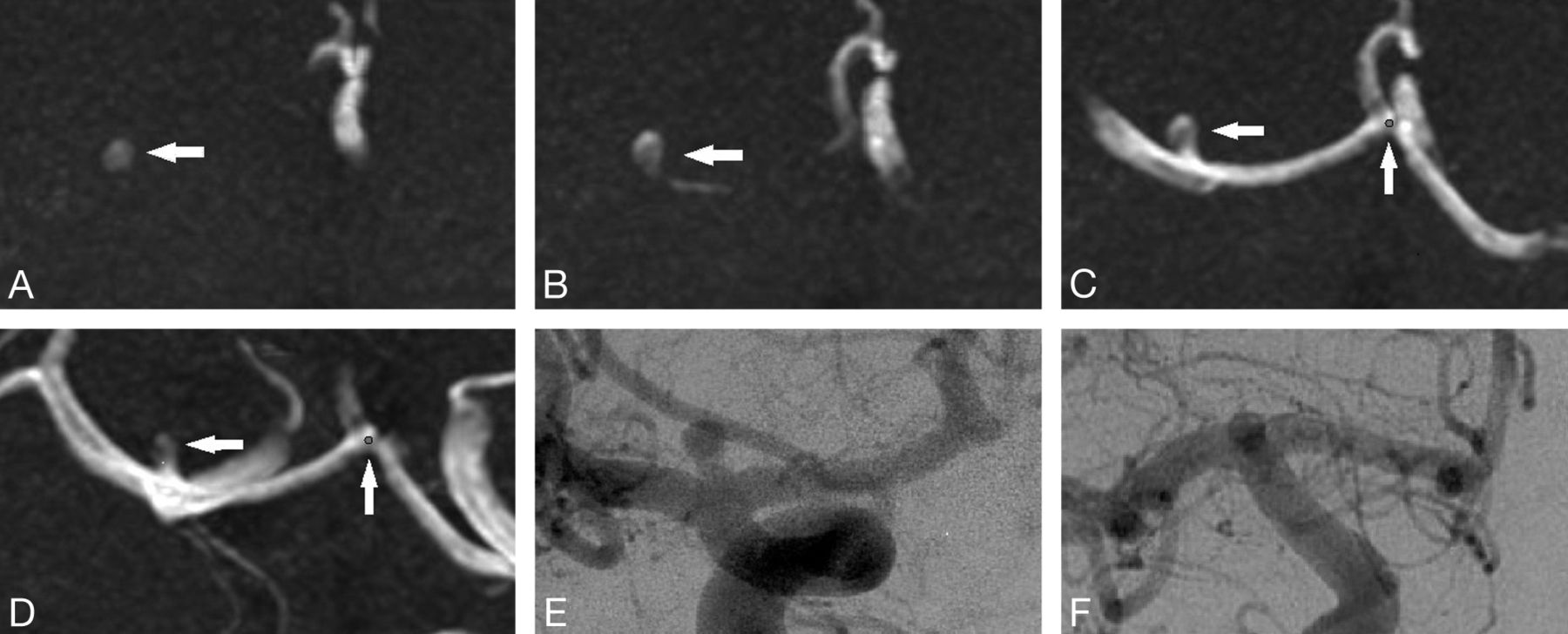
  • Fig 3.
    • Download figure
    • Open in new tab
    • Download powerpoint
    Fig 3.

    The smallest aneurysm (1.1 mm).

  • Fig 4.
    • Download figure
    • Open in new tab
    • Download powerpoint
    Fig 4.

    This aneurysm was missed by the CAD algorithm. We believe that the error was because the aneurysm had a relatively consistent diameter up to the dome, thus not meeting the curvature criteria. This was detected by all radiologists with and without CAD.

  • Fig 5.
    • Download figure
    • Open in new tab
    • Download powerpoint
    Fig 5.

    A free-response receiver operating characteristic curves aneurysm fraction.

Tables

  • Figures
    • View popup
    Table 1:

    Location and size of 11 aneurysmsa

    No.Size (mm)Location
    13.6Left middle cerebral artery (M1)
    22.5Right posterior communicating artery
    34.2Right internal carotid artery terminus
    42.1Right distal anterior cerebral artery (A1)
    52.1Right supraclinoid internal carotid
    62.2Left pericallosal artery aneurysm (A2)
    71.1Left middle cerebral artery (M1)
    86.0Right proximal cavernous internal carotid artery
    94.0Left superior cerebellar artery
    104.0Left internal carotid artery proximal to ophthalmic
    112.5Left middle cerebral artery (M1)
    • ↵a The sizes shown are the greatest dimension based on the DSA images.

    • View popup
    Table 2:

    Estimated figure of merit by reader and methoda

    Estimated FOM
    CAD−CAD+Difference95% CIP Value
    Reader 10.6670.7260.060(−0.184–0.304).62
    Reader 20.6600.8420.182(0.021–0.344).028
    Reader 30.7890.8920.103(−0.071–0.276).24
    Reader 4b0.5940.7460.152(−0.042–0.347).12
    Reader 50.5140.7170.203(−0.158–0.564).26
    Reader 60.7070.630-0.077(−0.371–0.217).60
    Overall0.6550.7590.104(0.025–0.183).011
    • ↵a For all except reader 6, there was a definite increase in the FOM when CAD results were available, though the difference was statistically significant for only 1 reader (reader 2).

    • ↵b General radiologist; all other readers are neuroradiologists.

    • View popup
    Table 3:

    Individual reader performance results

    Sensitivity (95% CI)Specificity (95% CI)Accuracy (95% CI)
    Reader 1
        CAD−55.6% (5/9) (26.7%–81.1%)76.9% (30/39) (61.7%–87.4%)72.9% (35/48) (59.0%–83.4%)
        CAD+77.8% (7/9) (45.3%–93.7%)66.7% (26/39) (51.0%–79.4%)68.8% (33/48) (54.7%–80.1%)
    Reader 2
        CAD−66.7% (6/9) (35.4%–87.9%)74.4% (29/39) (58.9%–85.4%)72.9% (35/48) (59.0%–83.4%)
        CAD+88.9% (8/9) (56.5%–98.0%)79.5% (31/39) (64.5%–89.2%)81.3% (39/48) (68.1%–89.8%)
    Reader 3
        CAD−77.8% (7/9) (45.3%–93.7%)84.6% (33/39) (70.3%–92.8%)83.3% (40/48) (70.4%–91.3%)
        CAD+100% (9/9) (70.1%–100.0%)79.5% (31/39) (64.5%–89.2%)83.3% (40/48) (70.4%–91.3%)
    Reader 4a
        CAD−66.7% (6/9) (35.4%–87.9%)82.1% (32/39) (67.3%–91.0%)79.2% (38/48) (65.7%–88.3%)
        CAD+77.8% (7/9) (45.3%–93.7%)76.9% (30/39) (61.7%–87.4%)77.1% (37/48) (63.5%–86.7%)
    Reader 5
        CAD−88.9% (8/9) (56.5%–98.0%)79.5% (31/39) (64.5%–89.2%)81.3% (39/48) (68.1%–89.8%)
        CAD+77.8% (7/9) (45.3%–93.7%)74.4% (29/39) (58.9%–85.4%)75% (36/48) (61.2%–85.1%)
    Reader 6
        CAD−66.7% (6/9) (35.4%–87.9%)79.5% (31/39) (64.5%–89.2%)77.1% (37/48) (63.5%–86.7%)
        CAD+77.8% (7/9) (45.3%–93.7%)76.9% (30/39) (61.7%–87.4%)77.1% (37/48) (63.5%–86.7%)
    • ↵a General radiologist, all other readers are neuroradiologists.

PreviousNext
Back to top

In this issue

American Journal of Neuroradiology: 35 (10)
American Journal of Neuroradiology
Vol. 35, Issue 10
1 Oct 2014
  • Table of Contents
  • Index by author
  • Complete Issue (PDF)
Advertisement
Print
Download PDF
Email Article

Thank you for your interest in spreading the word on American Journal of Neuroradiology.

NOTE: We only request your email address so that the person you are recommending the page to knows that you wanted them to see it, and that it is not junk mail. We do not capture any email address.

Enter multiple addresses on separate lines or separate them with commas.
Computer-Aided Diagnosis Improves Detection of Small Intracranial Aneurysms on MRA in a Clinical Setting
(Your Name) has sent you a message from American Journal of Neuroradiology
(Your Name) thought you would like to see the American Journal of Neuroradiology web site.
CAPTCHA
This question is for testing whether or not you are a human visitor and to prevent automated spam submissions.
Cite this article
I.L. Štep̌án-Buksakowska, J.M. Accurso, F.E. Diehn, J. Huston, T.J. Kaufmann, P.H. Luetmer, C.P. Wood, X. Yang, D.J. Blezek, R. Carter, C. Hagen, D. Hořínek, A. Hejčl, M. Roček, B.J. Erickson
Computer-Aided Diagnosis Improves Detection of Small Intracranial Aneurysms on MRA in a Clinical Setting
American Journal of Neuroradiology Oct 2014, 35 (10) 1897-1902; DOI: 10.3174/ajnr.A3996

Citation Manager Formats

  • BibTeX
  • Bookends
  • EasyBib
  • EndNote (tagged)
  • EndNote 8 (xml)
  • Medlars
  • Mendeley
  • Papers
  • RefWorks Tagged
  • Ref Manager
  • RIS
  • Zotero
0 Responses
Respond to this article
Share
Bookmark this article
Computer-Aided Diagnosis Improves Detection of Small Intracranial Aneurysms on MRA in a Clinical Setting
I.L. Štep̌án-Buksakowska, J.M. Accurso, F.E. Diehn, J. Huston, T.J. Kaufmann, P.H. Luetmer, C.P. Wood, X. Yang, D.J. Blezek, R. Carter, C. Hagen, D. Hořínek, A. Hejčl, M. Roček, B.J. Erickson
American Journal of Neuroradiology Oct 2014, 35 (10) 1897-1902; DOI: 10.3174/ajnr.A3996
del.icio.us logo Twitter logo Facebook logo Mendeley logo
  • Tweet Widget
  • Facebook Like
  • Google Plus One
Purchase

Jump to section

  • Article
    • Abstract
    • ABBREVIATIONS:
    • Materials and Methods
    • Results
    • Discussion
    • Conclusions
    • Footnotes
    • REFERENCES
  • Figures & Data
  • Info & Metrics
  • Responses
  • References
  • PDF

Related Articles

  • No related articles found.
  • PubMed
  • Google Scholar

Cited By...

  • Automated Detection of Cerebral Aneurysms on TOF-MRA Using a Deep Learning Approach: An External Validation Study
  • Detection of clustered anomalies in single-voxel morphometry as a rapid automated method for identifying intracranial aneurysms
  • Deep Learning-Based Detection of Intracranial Aneurysms in 3D TOF-MRA
  • Computer-Assisted Detection of Cerebral Aneurysms in MR Angiography in a Routine Image-Reading Environment: Effects on Diagnosis by Radiologists
  • Crossref
  • Google Scholar

This article has not yet been cited by articles in journals that are participating in Crossref Cited-by Linking.

More in this TOC Section

  • Evaluating the Effects of White Matter Multiple Sclerosis Lesions on the Volume Estimation of 6 Brain Tissue Segmentation Methods
  • Quiet PROPELLER MRI Techniques Match the Quality of Conventional PROPELLER Brain Imaging Techniques
  • Predictors of Reperfusion in Patients with Acute Ischemic Stroke
Show more Brain

Similar Articles

Advertisement

Indexed Content

  • Current Issue
  • Accepted Manuscripts
  • Article Preview
  • Past Issues
  • Editorials
  • Editor's Choice
  • Fellows' Journal Club
  • Letters to the Editor
  • Video Articles

Cases

  • Case Collection
  • Archive - Case of the Week
  • Archive - Case of the Month
  • Archive - Classic Case

More from AJNR

  • Trainee Corner
  • Imaging Protocols
  • MRI Safety Corner
  • Book Reviews

Multimedia

  • AJNR Podcasts
  • AJNR Scantastics

Resources

  • Turnaround Time
  • Submit a Manuscript
  • Submit a Video Article
  • Submit an eLetter to the Editor/Response
  • Manuscript Submission Guidelines
  • Statistical Tips
  • Fast Publishing of Accepted Manuscripts
  • Graphical Abstract Preparation
  • Imaging Protocol Submission
  • Evidence-Based Medicine Level Guide
  • Publishing Checklists
  • Author Policies
  • Become a Reviewer/Academy of Reviewers
  • News and Updates

About Us

  • About AJNR
  • Editorial Board
  • Editorial Board Alumni
  • Alerts
  • Permissions
  • Not an AJNR Subscriber? Join Now
  • Advertise with Us
  • Librarian Resources
  • Feedback
  • Terms and Conditions
  • AJNR Editorial Board Alumni

American Society of Neuroradiology

  • Not an ASNR Member? Join Now

© 2025 by the American Society of Neuroradiology All rights, including for text and data mining, AI training, and similar technologies, are reserved.
Print ISSN: 0195-6108 Online ISSN: 1936-959X

Powered by HighWire