
Article Figures & Data
Figures
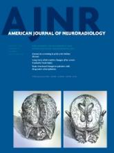
- Fig 1.
Representative images of the healthy relatives of patients with MS fulfilling Okuda criteria13 for radiologically isolated syndrome (RIS) (A) fulfilling modified Okuda criteria13 for RIS in which the dissemination in space on MR imaging was evaluated by use of Swanton criteria (RIS-Swanton),17 (B) or presenting with dirty-appearing white matter (C).

Tables
Okuda RIS criteria13 MRI criteria
Ovoid, well-circumscribed, and homogeneous foci with or without involvement of corpus callosum.
T2 hyperintensities measuring >3 mm in size fulfilling at least 3 of 4 Barkhof criteria14 for DIS, which are: 1) ≥9 lesions or ≥1 gadolinium-enhancing lesion, 2) ≥3 periventricular lesions, 3) ≥1 juxtacortical lesion, and 4) ≥1 infratentorial lesion.
CNS white matter anomalies are not consistent with a vascular pattern.
No historical accounts of remitting clinical symptoms consistent with neurologic dysfunction.
The MRI anomalies do not account for clinically apparent impairments in social, occupational, or generalized areas of functioning.
The MRI anomalies are not due to the direct physiologic effects of substances (eg, drug abuse, toxic exposure) or a medical condition.
Exclusion of individuals with MRI phenotypes suggestive of leukoaraiosis or extensive white matter pathology lacking involvement of the corpus callosum.
The MRI anomalies are not better accounted for by another disease process.
Swanton RIS criteria17 MRI criteria
DIS defined as presence of ≥1 T2 lesion in ≥2 of the following brain regions (periventricular, juxtacortical, and infratentorial) or in the spinal cord.
No historical accounts of remitting clinical symptoms consistent with neurologic dysfunction.
The MRI anomalies do not account for clinically apparent impairments in social, occupational, or generalized areas of functioning.
The MRI anomalies are not due to the direct physiologic effects of substances (eg, drug abuse, toxic exposure) or a medical condition.
Exclusion of individuals with MRI phenotypes suggestive of leukoaraiosis or extensive white matter pathology lacking involvement of the corpus callosum.
The MRI anomalies are not better accounted for by another disease process.
- Table 2:
Demographic and clinical characteristics of non-familial healthy control subjects and the healthy relatives of patients with multiple sclerosis
Non-Familial HCs (n = 82) Healthy Relatives of Patients with MS (n = 68) P Value Age, y, mean (SD) median 39.8 (14.3) 43 39.5 (16.6) 43 .917 Sex, female n (%) 57 (69.5) 45 (66.2) .663 Race/ethnicity, n (%) .180 White 64 (78.1) 62 (91.2) Black 12 (14.6) 4 (5.9) Other 6 (7.3) 2 (2.9) Vascular risk factors, n (%) Heart disease 8 (9.8) 13 (19.1) .085 Smoking 22 (27.5) 19 (27.9) .979 Hypertension 17 (20.7) 15 (22.1) .442 Obesity n (%)a .582 BMI <18.5 2 (2.4) 2 (2.9) BMI 18.5–24.9 34 (41.5) 25 (36.8) BMI 25–29.9 26 (31.7) 17 (25.0) BMI >30 13 (15.8) 18 (26.5) Autoimmune diseases, n (%) Systemic lupus erythematosus 0 0 NA Rheumatoid disorder 1 (1.2) 2 (2.9) .474 Psoriasis 0 1 (1.5) .279 Diabetes mellitus type 1 1 (1.2) 0 .352 Migraine, n (%) 10 (12.2) 13 (19.1) .282 Note:—BMI indicates body mass index.
BMI <18.5 represents underweight, BMI 18.5–24.9 represents normal weight, BMI 25–29.9 represents overweight, and BMI >30 represents obesity. The differences between the groups were compared by means of the Student t test or the χ2 test.
↵a Data missing for 13 subjects.
- Table 3:
MRI white matter signal abnormality (≥3 mm in size) characteristics of non-familial healthy control subjects and the healthy relatives of patients with multiple sclerosis
Non-Familial HCs (n = 82) Healthy Relatives of Patients With MS (n = 68) P Value Subjects with WM SAs, n (%) 21 (25.6) 20 (29.4) .603 Subjects with WM JC SAs, n (%) 4 (4.9) 7 (10.3) .205 Subjects with WM PVL SAs, n (%) 7 (8.5) 13 (19.1) .058 Subjects with WM IT SAs, n (%) 0 (0) 1 (1.5) .271 Subjects with DWM SAs, n (%) 20 (24.4) 17 (25.0) .931 Subjects with ≥9 WM SAs, n (%) 4 (4.9) 6 (8.8) .335 WM-SAN, mean (SD), median 1.5 (4.7) 0 2.1 (5.1) 0 .527 WM JC SAN, mean (SD), median 0.06 (0.3) 0 0.19 (0.7) 0 .198 WM PVL SAN, mean (SD), median 0.32 (1.8) 0 0.40 (1.2) 0 .061 WM IT SAN mean (SD) median 0.0 (0) 0 0.01 (0.1) 0 .272 WM DWM SAN, mean (SD), median 1.1 (3.0) 0 1.5 (3.9) 0 .838 WM-SAV, mean (SD), median 166.1 (892.0) 230.4 (736.8) .480 DAWM-SAV,a mean (SD), median 675.3 (554.7) 866.2 (669.3) .054 WM-SAV + DAWM-SAV, mean (SD), median 842.3 (1079.7) 1096.5 (1026.6) .024 Note:—SAs indicates signal abnormalities; SAN, signal abnormality number; JC, juxtacortical, PVL, periventricular; IT, infratentorial; DWM, deep white matter; SAV, signal abnormality volume.
Differences between the groups were compared by means of the χ2 test or Mann-Whitney U test.
The SAV is expressed in millimeters cubed (mm3).
↵a DAWM calculation was not related to the size of the hyperintensities.
- Table 4:
MRI white matter signal abnormality (all sizes) characteristics of non-familial healthy control subjects and the healthy relatives of patients with multiple sclerosis
Non-Familial HCs (n = 82) Healthy Relatives of Patients with MS (n = 68) P Value Subjects with WM SAs, n (%) 29 (35.4) 28 (41.2) .465 Subjects with WM JC SAs, n (%) 4 (4.9) 7 (10.3) .205 Subjects with WM PVL SAs, n (%) 7 (8.5) 14 (20.6) .034 Subjects with WM IT SAs, n (%) 1 (1.2) 1 (1.5) 0 .894 Subjects with DWM SAs, n (%) 28 (34.1) 26 (38.2) .603 Subjects with ≥9 WM SAs, n (%) 5 (6.1) 8 (11.8) .219 WM-SAN, mean (SD) median 2.5 (7.5) 0 3.8 (10) 0 .386 WM JC SAN, mean (SD) median 0.07 (0.3) 0 0.19 (0.7) 0 .207 WM PVL SAN, mean (SD) median 0.41 (2.1) 0 0.57 (1.9) 0 .043 WM IT SAN, mean (SD) median 0.01 (0.1) 0 0.01 (0.12) 0 .894 WM DWM SAN, mean (SD) median 1.9 (5.3) 0 3.1 (8.5) 0 .468 WM-SAV, mean (SD) median 198.2 (1085.2) 259.7 (784.4) .338 DAWM-SAV,a mean (SD) median 675.3 (554.7) 866.2 (669.3) .054 WM-SAV + DAWM-SAV, mean (SD) median 873.5 (1233.5) 1125.9 (1076.8) .025 Note:—SAs indicates signal abnormalities; SAN, signal abnormality number; JC, juxtacortical; PVL, periventricular; IT, infratentorial; DWM, deep white matter; SAV, signal abnormality volume.
Differences between the groups were compared by means of the χ2 test or Mann-Whitney U test.
The SAV is expressed in millimeters cubed (mm3).
↵a DAWM calculation was not related to the size of the hyperintensities.
- Table 5:
Non-familial healthy control subjects and the healthy relatives of patients with multiple sclerosis fulfilling Okuda criteria for RIS or fulfilling modified Okuda criteria for RIS in which the dissemination in space on MRI was evaluated by use of Swanton criteria (RIS-Swanton)
Non-Familial HCs (n = 82) Healthy Relatives of Patients with MS (n = 68) P Value RIS-Okuda criteria13 Non-RIS 80 66 .849 RIS 2 2 Subject 1 >9 WM SA: 5 JC, 3 PVL, 13 DWM >9 WM SA: 5 JC, 3 PVL, 13 DWM Subject 2 >9 WM SA: 1 JC, 8 PVL, 11 DWM >9 WM SA: 1 JC, 8 PVL, 11 DWM Total 82 68 Modified Okuda criteria (RIS-Swanton13,17) Non-RIS 79 61 .105 RIS 3 7 Subject 1 >9 WM SA: 2 JC, 5 PVL, 9 DWM >9 WM SA: 5 JC, 3 PVL, 13 DWM Subject 2 >9 WM SA: 1 JC, 15 PVL, 18 DWM >9 WM SA: 1 JC, 8 PVL, 11 DWM Subject 3 >9 WM SA: 1 JC, 1 PVL, 11 DWM >9 WM SA: 2 JC, 1 PVL, 7 DWM Subject 4 >9 WM SA: 1 JC, 1 PVL, 24 DWM Subject 5 >9 WM SA: 1 JC, 1 PVL, 7 DWM Subject 6 >9 WM SA: 1 JC, 1 PVL, 8 DWM Subject 7 2 JC, 2 PVL 3 DWM Total 82 68 Note:—SA indicates signal abnormality; JC, juxtacortical; PVL, periventricular; DWM, deep white matter.
Differences between the groups were compared by means of the χ2 test. The regional localization data for subjects fulfilling RIS criteria are presented on individual subject level.


Jump to section
Related Articles
Cited By...
- Immune signatures of prodromal multiple sclerosis in monozygotic twins
- Radiologically Isolated Syndrome: A Review for Neuroradiologists
- X linked Charcot-Marie-Tooth disease and multiple sclerosis: emerging evidence for an association
- Incidence of Radiologically Isolated Syndrome: A Population-Based Study