
Article Figures & Data
Figures
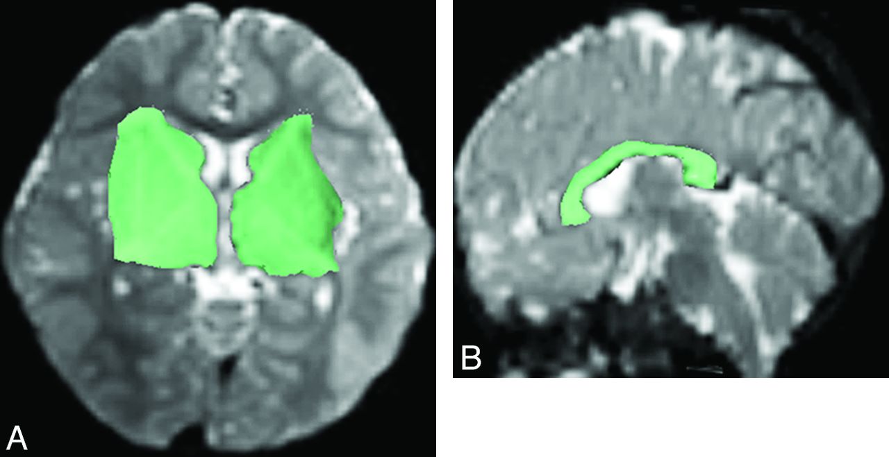
- Fig 1.
Regions of interest (green shaded areas) were manually drawn on axial B0 image (A) to include the ipsilateral caudate head, internal capsule, lentiform nucleus, external capsule, and thalamus for reconstructing the PF on one side, and on sagittal B0 image (B) to include the corpus callosum for reconstructing the CF.
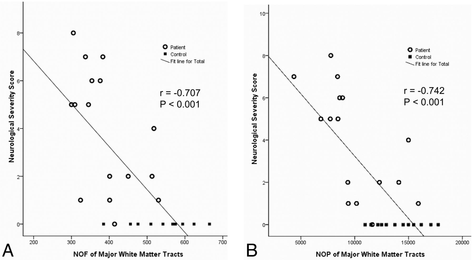
- Fig 2.
Scatterplots show moderate negative correlation between the neurologic severity score and NOF (A) and NOP (B) in the patients with tuberous sclerosis complex and control subjects.
- Fig 3.
Results of tract-based spatial statistics analysis revealed significant differences between the tuberous sclerosis complex and control groups in FA (A) and axial diffusivity (B) maps, with overlaid mean value skeleton. Regions of the skeleton in green represent areas of no significant differences in values between the tuberous sclerosis complex group and the control subjects. Regions in blue are areas in which the value was significantly lower in the tuberous sclerosis complex group.


Tables
Patient No. Seizure Developmental Disability Autism Neuropsychiatric Disorders Neurologic Severity Score 1 1 3 0 1 5 2 0 0 0 0 0 3 1 3 2 0 6 4 2 3 2 1 8 5 1 3 0 1 5 6 1 3 0 1 5 7 1 0 0 0 1 8 1 0 0 1 2 9 1 3 2 1 7 10 1 0 0 0 1 11 1 0 0 1 2 12 2 3 0 1 6 13 1 0 0 0 1 14 1 3 2 1 7 15 1 3 0 0 4 16 1 0 0 1 2 Note:—TSC indicates tuberous sclerosis complex.
- Table 2:
Mean (± SD) NOF, NOP, and FA of the commissural fiber, projection fibers, and major white matter tracts of patients with TSC and control subjects
NOF NOP FA TSC Control P TSC Control P TSC Control P CF 77.1 ± 27.4 100 ± 13.7 .01 2440 ± 1190 3280 ± 494 .02 .504 ± .026 .522 ± .016 .05 Left PF 169 ± 32.2 247 ± 61.3 .01 4200 ± 1270 6360 ± 1430 .01 .458 ± .011 .460 ± .022 .05 Right PF 146 ± 25.7 198 ± 32.7 .01 3290 ± 976 4510 ± 961 .01 .458 ± .016 .453 ± .019 .05 Bilateral PF 315 ± 53.3 445 ± 73.0 .01 7490 ± 2130 10870 ± 1870 .01 .459 ± .012 .458 ± .020 .05 MWT 391 ± 76.7 545 ± .770 .01 9900 ± 3110 14150 ± 2210 .01 .470 ± .014 .473 ± .017 .05 Note:—TSC indicates tuberous sclerosis complex.
- Table 3:
Pearson correlation coefficients between the neurologic severity score versus NOF and NOP in the commissural fiber, projection fibers, and major white matter tracts
NOF NOP CF r = −.70; P < .001 r = −.75; P < .001 Left PF r = −.55; P < .001 r = −.60; P < .001 Right PF r = −.66; P < .001 r = −.67; P < .001 Bilateral PF r = −.66; P < .001 r = −.68; P < .001 MWT r = −.71; P < .001 r = −.74; P < .001


