
Article Figures & Data
Figures
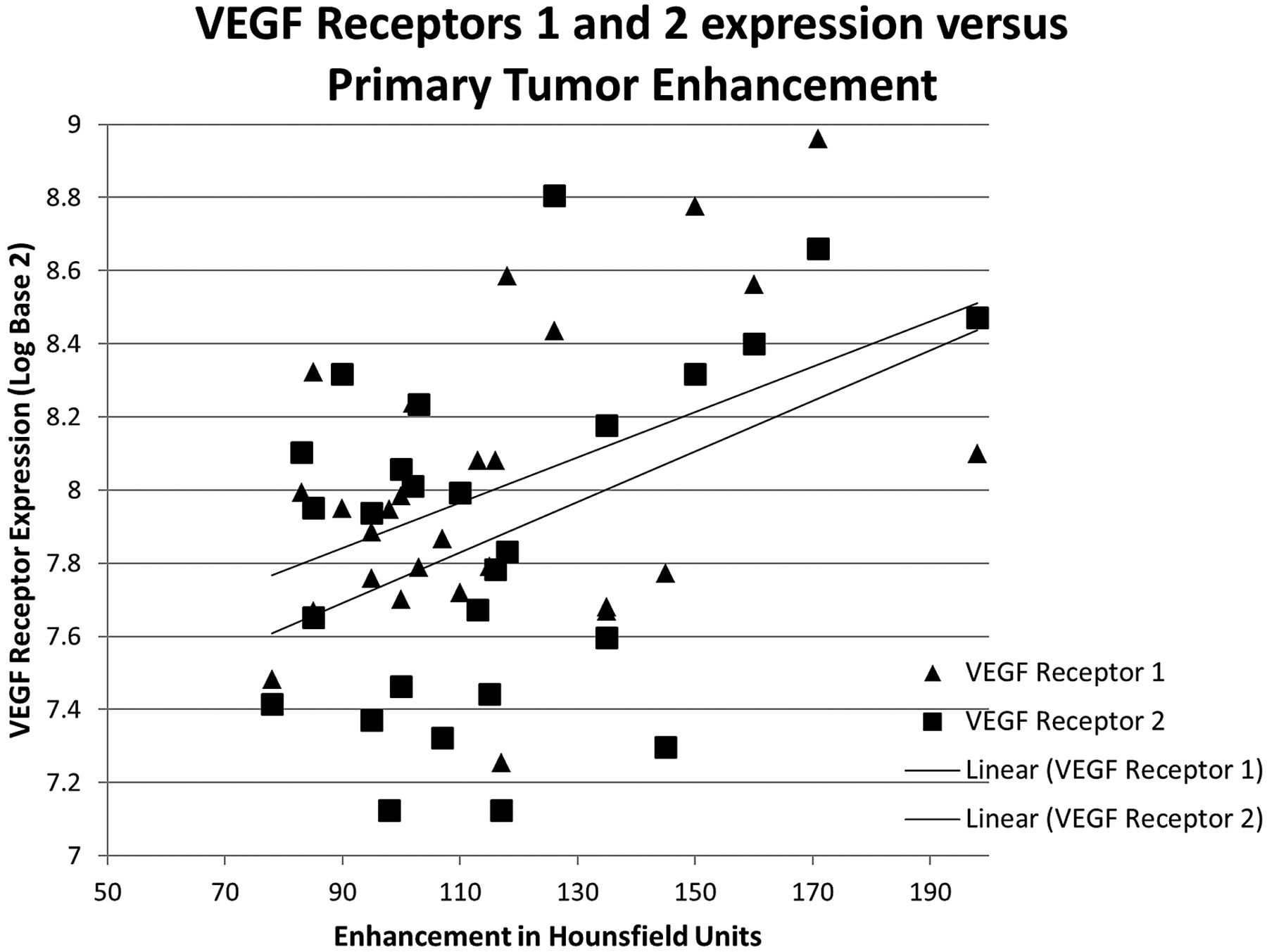
- Fig 1.
Scatterplot showing relationship of increased enhancement and VEGF receptors 1 and 2 expression. The linear fitting demonstrates that as the absolute enhancement in HU increases, the receptor expression (in log base 2) also increases.
- Fig 2.
Bar graph reflecting positive correlation between the grading of mass effect and the expression of EGFR (in log base 2) with standard deviation bars.
- Fig 3.
Scatterplot showing relationship of increasing primary tumor ellipsoid estimated cross-sectional area with increasing VEGF ligand A and decreased receptor 2 expression (in log base 2) with linear fitting.
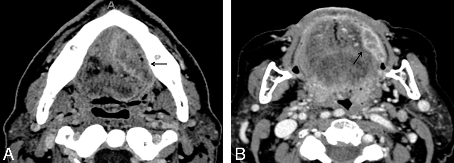
- Fig 4.
Example cases of highest expression for specific genes by use of contrast-enhanced axial CT images in different patients. A, Large OSCC located in the left tongue and at the floor of the mouth OSCC (arrow) with intrinsic tongue and perineural invasion (on histologic examination) and mass effect narrowing the airway. This tumor has the highest EGFR expression. B, Small left buccal tumor with avid enhancement with the highest VEGF receptor 1 and 2 expression.


Tables
Patient No. Age (y) Sex Stage Subsite 1 62 M T2 N0 OT 2 66 M T3 N2 OT 3 62 M T3 N0 OT 4 75 F T4 N2c OT/FOM 5 23 M T3 N2b OT 6 40 M T3 N1 OT 7 50 M T4 N2b OT 8 69 M T4 N2c OT/FOM 9 61 M T2 N1 OT 10 62 F T3 N0 OT 11 60 F T2 N2b OT/BOT 12 40 M T4 N2c OT 13 60 M T4 N0 OT/FOM 14 50 M T4a N2c OT 15 85 F T2 N0 OT 16 56 M T4 N2c FOM 17 50 F T2 N0 OT 18 74 M T4 N2b OT 19 53 M T3 N0 OT 20 61 F T2 N2b OT 21 76 F T2 N0 OT 22 56 M T3 N2b Bucca 23 48 M T2 N1 FOM 24 74 M T4 N0 Gingiva 25 64 M T2 N1 FOM 26 62 M T1 N0 Bucca 27 67 M T4 N2c FOM Note:—BOT indicates base of tongue; F, female; FOM, floor of mouth; M, male; N, nodal stage; OT, oral tongue; T, tumor stage.
Imaging Finding Gene Expressed Coefficient Value Significance (P Value) Mass effect EGFR OR: +1.2 .032a Perineural invasion EGFR OR: +3.1 .047 Axial cross-sectional area (cm2) VEGF ligand A PC: +0.49 .014 Axial orthogonal distance (cm) VEGF ligand A PC: +0.47 .010 Mass effect VEGF ligand B OR: +2.8 .026 Enhancement of primary tumor (HU) VEGF receptor 1 PC: +0.45 .018a Axial maximal diameter (cm) VEGF receptor 2 PC: −0.44 .023 Axial cross-sectional area (cm2) VEGF receptor 2 PC: −0.42 .029 Enhancement of primary tumor (HU) VEGF receptor 2 PC: +0.43 .025a Enhancement of submandibular gland (HU) VEGF receptor 2 PC: +0.43 .026 Note:—cm indicates centimeter; OR, ordinal regression; PC, Pearson correlation.
↵a Denotes confirmation of a priori hypothesis.


