
Article Figures & Data
Figures
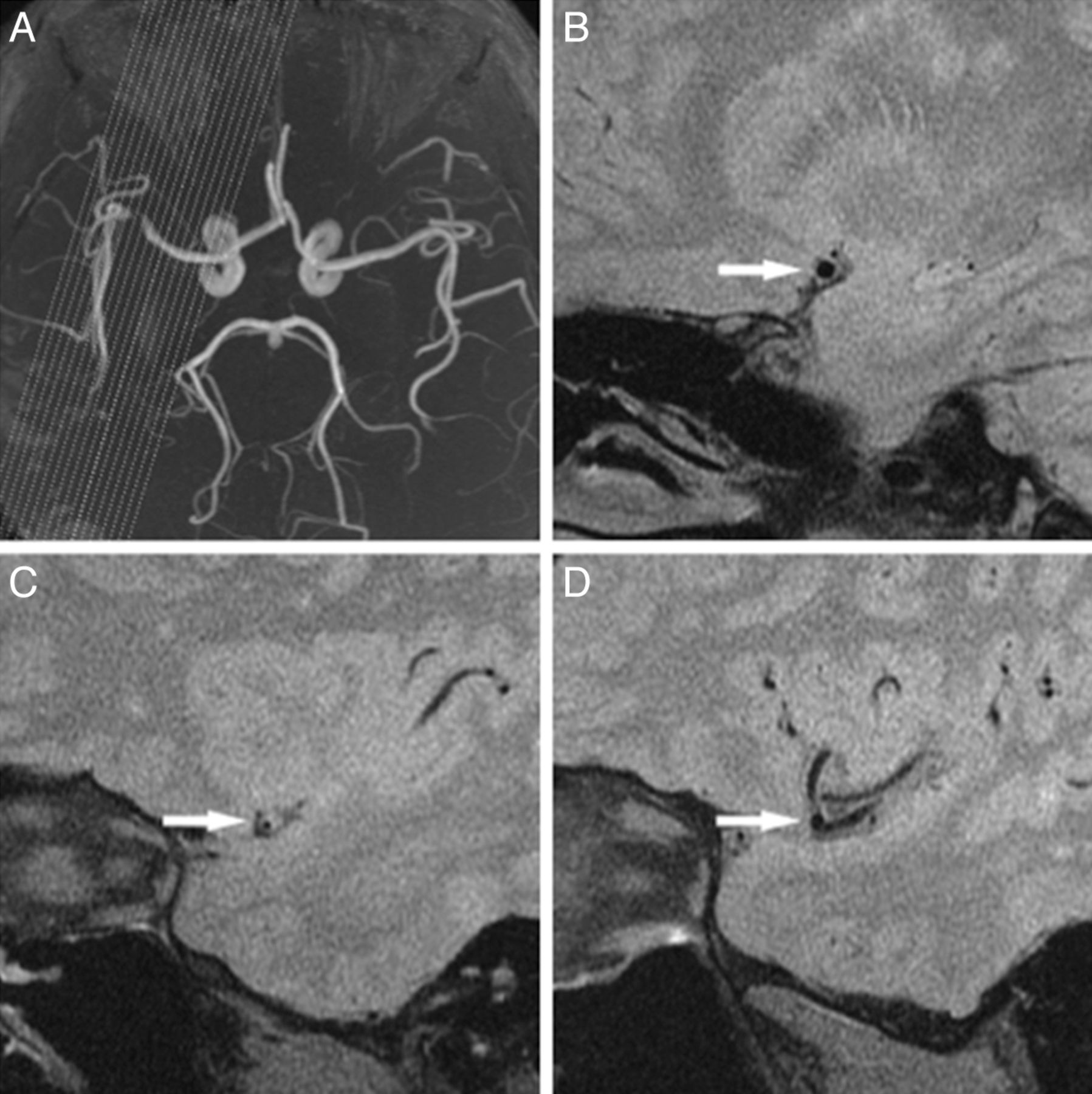
- Fig 1.
An example of an excluded case. MRA (A) shows the obvious angled stenosis located at the distal M1 segment near M2 of the MCA. Suitable cross-sectional images can be obtained at the proximal (B, arrow) and MLN (C, arrow) but not the distal (D, arrow) site.
- Fig 2.
A 37-year-old man with hypertension, hypercholesterolemia, and cigarette smoking history who presented with aphasia, a cognitive disorder, and right-side numbness for 60 days. MRA (A) shows stenosis located at the straight M1 segment of the MCA. The proximal (B, arrow), MLN (C, arrow), and distal (D, arrow) site of the left MCA are shown. The vessel area or luminal area is measured (demonstrated by the magnified view in the inset). The reference vessel area is 12.3 mm2, and the remodeling index at the MLN site is 0.8 < 1.00 (NR). The eccentricity index is 0.6. B, The plaque location is mainly on the ventral wall (arrow). The wall area is 7.6 mm2 at the proximal, 9.1 mm2 at the MLN, and 6.9 mm2 at the distal site. The reference wall area is 7.2 mm2. Plaque size is 1.9 mm2, and percentage of plaque burden is 19.1%.
- Fig 3.
A 74-year-old man with hypertension and hypercholesterolemia who presented with aphasia for 27 days. MRA (A) shows stenosis located at the nearly straight M1 segment of the MCA. The proximal (B, arrow), MLN (C, arrow), and distal (D, arrow) site of left MCA are surrounded by more CSF due to age-related brain involution compared with the case in Fig 2. The vessel area or lumen area is measured (demonstrated by the magnified view in the inset). The reference vessel area is 21.5 mm2, and the remodeling index at the MLN is 1.1 > 1.00 (PR). The eccentricity index is 0.7. B, The plaque location is mainly on the ventral and superior walls (arrow). The wall area is 12.3 mm2 at the proximal, 22.3 mm2 at the MLN, and 12.3 mm2 at the distal site. The reference wall area is 12.3 mm2. Plaque size is 10.0 mm2, and the percentage of plaque burden is 42.7%. Compared with the case in Fig 2, this case has a larger vessel area, wall area, plaque size, percentage of plaque burden, and eccentricity index.


Tables
Characteristics Included Patients (n = 44) Excluded Patients (n = 43) P Value Age (yr) (mean) (SD) 47.8 (9.9) 47.7 (11.4) .966 Men (No.) (%) 39 (88.6) 37 (86.1) .716 Hypertension (No.) (%) 29 (65.9) 26 (60.5) .599 Hypercholesterolemia (No.) (%) 42 (95.5) 42 (97.7) 1.000 Diabetes mellitus (No.) (%) 8 (18.2) 10 (23.3) .559 Smoking (No.) (%) 30 (68.2) 31 (72.1) .690 Obesity (No.) (%) 7 (15.9) 3 (7.0) .332 Three or more risk factors (No.) (%) 23 (52.3) 22 (51.2) .918 Stroke as qualifying event (No.) (%) 27 (61.4) 26 (60.5) .932 NIHSS scores at admission (median) (interquartile range) 0.0 (0.0–1.0) 0.0 (0.0–1.0) .916 Time from qualifying event to high-resolution MRI, days (mean) (SD) 33.8 (19.0) 33.3 (21.2) .905 Intraclass Correlation Coefficient (95% CI) Intraobserver Interobserver Vessel area 0.917 (0.666–0.979) 0.906 (0.620–0.977) Luminal area 0.833 (0.329–0.959) 0.898 (0.589–0.975) Maximal wall thickness 0.962 (0.847–0.991) 0.916 (0.663–0.979) Minimal wall thickness 0.876 (0.501–0.969) 0.851 (0.399–0.963) Characteristics NR Group (n = 19) PR Group (n = 25) P Value Age (yr) (mean) (SD) 48.0 (8.4) 47.8 (11.0) .951 Men (No.) (%) 15 (79.0) 24 (96.0) .198 Hypertension (No.) (%) 13 (68.4) 16 (64.0) .759 Hypercholesterolemia (No.) (%) 19 (100) 23 (92.0) .498 Diabetes mellitus (No.) (%) 5 (26.3) 3 (12.0) .409 Smoking (No.) (%) 11 (57.9) 19 (76.0) .202 Obesity (No.) (%) 5 (26.3) 2 (8.0) .219 Three or more risk factors (No.) (%) 12 (63.2) 11 (44.0) .208 Stroke as qualifying event (No.) (%) 13 (68.4) 14 (56.0) .402 NIHSS scores at admission, median (interquartile range) 0.0 (0.0–2.0) 0.0 (0.0–0.5) .419 Time from qualifying event to high-resolution MRI (days) (mean) (SD) 33.8 (17.4) 33.8 (20.5) 1.000 Variablesa NR Group (n = 19) PR Group (n = 25) P Value At reference site Vessel area (mm2) 15.5 (3.3) 14.3 (3.2) .228 Luminal area (mm2) 5.5 (1.6) 5.2 (1.9) .516 Wall area (mm2) 10.0 (1.8) 9.1 (1.6) .103 Maximal wall thickness (mm) 1.0 (0.1) 0.9 (0.1) .058 At MLN site Vessel area (mm2) 12.4 (3.3) 17.8 (4.5) <.0001 Luminal area (mm2) 0.8 (0.6) 0.8 (0.5) 0.804 Wall area (mm2) 11.6 (3.3) 16.9 (4.3) <.0001 Plaque size (mm2) 1.6 (2.3) 7.8 (3.8) <.0001 Percentage of plaque burden (%) 9.9 (16.8) 42.0 (11.1) <.0001 Degree of stenosis (%) 84.5 (13.8) 81.5 (14.6) .497 Maximal wall thickness (mm) 2.1 (0.5) 2.8 (0.5) <.0001 Minimal wall thickness, (mm) 0.9 (0.3) 1.0 (0.2) .381 Eccentricity index 0.5 (0.2) 0.6 (0.1) .023 Remodeling index 0.8 (0.1) 1.3 (0.3) <.0001 ↵a Variables are expressed as mean and SD.


Jump to section
Related Articles
Cited By...
- Development of a high resolution MRI intracranial atherosclerosis imaging phantom
- Intracranial Vessel Wall MRI: Principles and Expert Consensus Recommendations of the American Society of Neuroradiology
- Comparison of High-Resolution MR Imaging and Digital Subtraction Angiography for the Characterization and Diagnosis of Intracranial Artery Disease
- Nonatheroscleotic Isolated Middle Cerebral Artery Disease May Be Early Manifestation of Moyamoya Disease
- Magnetic Resonance Imaging of Plaque Morphology, Burden, and Distribution in Patients With Symptomatic Middle Cerebral Artery Stenosis
- Non-moyamoya vessel network formation along steno-occlusive middle cerebral artery
- Patterns and Implications of Intracranial Arterial Remodeling in Stroke Patients
- Early experience in high-resolution MRI for large vessel occlusions
- Imaging Intracranial Vessel Wall Pathology With Magnetic Resonance Imaging: Current Prospects and Future Directions