Advertisement

Main menu

  • Home
  • Content
    • Current Issue
    • Accepted Manuscripts
    • Article Preview
    • Past Issue Archive
    • Video Articles
    • AJNR Case Collection
    • Case of the Week Archive
    • Case of the Month Archive
    • Classic Case Archive
  • Special Collections
    • AJNR Awards
    • Low-Field MRI
    • Alzheimer Disease
    • ASNR Foundation Special Collection
    • Photon-Counting CT
    • View All
  • Multimedia
    • AJNR Podcasts
    • AJNR SCANtastic
    • Trainee Corner
    • MRI Safety Corner
    • Imaging Protocols
  • For Authors
    • Submit a Manuscript
    • Submit a Video Article
    • Submit an eLetter to the Editor/Response
    • Manuscript Submission Guidelines
    • Statistical Tips
    • Fast Publishing of Accepted Manuscripts
    • Graphical Abstract Preparation
    • Imaging Protocol Submission
    • Author Policies
  • About Us
    • About AJNR
    • Editorial Board
    • Editorial Board Alumni
  • More
    • Become a Reviewer/Academy of Reviewers
    • Subscribers
    • Permissions
    • Alerts
    • Feedback
    • Advertisers
    • ASNR Home

User menu

  • Alerts
  • Log in

Search

  • Advanced search
American Journal of Neuroradiology

American Journal of Neuroradiology

ASHNR American Society of Functional Neuroradiology ASHNR American Society of Pediatric Neuroradiology ASSR
  • Alerts
  • Log in

Advanced Search

  • Home
  • Content
    • Current Issue
    • Accepted Manuscripts
    • Article Preview
    • Past Issue Archive
    • Video Articles
    • AJNR Case Collection
    • Case of the Week Archive
    • Case of the Month Archive
    • Classic Case Archive
  • Special Collections
    • AJNR Awards
    • Low-Field MRI
    • Alzheimer Disease
    • ASNR Foundation Special Collection
    • Photon-Counting CT
    • View All
  • Multimedia
    • AJNR Podcasts
    • AJNR SCANtastic
    • Trainee Corner
    • MRI Safety Corner
    • Imaging Protocols
  • For Authors
    • Submit a Manuscript
    • Submit a Video Article
    • Submit an eLetter to the Editor/Response
    • Manuscript Submission Guidelines
    • Statistical Tips
    • Fast Publishing of Accepted Manuscripts
    • Graphical Abstract Preparation
    • Imaging Protocol Submission
    • Author Policies
  • About Us
    • About AJNR
    • Editorial Board
    • Editorial Board Alumni
  • More
    • Become a Reviewer/Academy of Reviewers
    • Subscribers
    • Permissions
    • Alerts
    • Feedback
    • Advertisers
    • ASNR Home
  • Follow AJNR on Twitter
  • Visit AJNR on Facebook
  • Follow AJNR on Instagram
  • Join AJNR on LinkedIn
  • RSS Feeds

AJNR Awards, New Junior Editors, and more. Read the latest AJNR updates

Research ArticleTechnical Note

A Swine Model to Analyze Arterial Structural Changes Induced by Mechanical Thrombectomy

I. Yuki, I. Kan, A. Golshan, J. Sohn, Y. Murayama, H.V. Vinters and F. Viñuela
American Journal of Neuroradiology August 2013, 34 (8) E87-E90; DOI: https://doi.org/10.3174/ajnr.A3221
I. Yuki
aFrom the Division of Interventional Neuroradiology (I.Y., I.K., A.G., F.V.), Department of Radiological Sciences
  • Find this author on Google Scholar
  • Find this author on PubMed
  • Search for this author on this site
I. Kan
aFrom the Division of Interventional Neuroradiology (I.Y., I.K., A.G., F.V.), Department of Radiological Sciences
  • Find this author on Google Scholar
  • Find this author on PubMed
  • Search for this author on this site
A. Golshan
aFrom the Division of Interventional Neuroradiology (I.Y., I.K., A.G., F.V.), Department of Radiological Sciences
  • Find this author on Google Scholar
  • Find this author on PubMed
  • Search for this author on this site
J. Sohn
bDivision of Laboratory Animal Medicine (J.S.)
  • Find this author on Google Scholar
  • Find this author on PubMed
  • Search for this author on this site
Y. Murayama
dDepartment of the Neurosurgery (Y.M.), Jikei University of Medicine, Tokyo, Japan.
  • Find this author on Google Scholar
  • Find this author on PubMed
  • Search for this author on this site
H.V. Vinters
cSection of Neuropathology and Departments of Pathology and Laboratory Medicine and Neurology (H.V.V.), David Geffen School of Medicine, University of California, Los Angeles, Los Angeles, California
  • Find this author on Google Scholar
  • Find this author on PubMed
  • Search for this author on this site
F. Viñuela
aFrom the Division of Interventional Neuroradiology (I.Y., I.K., A.G., F.V.), Department of Radiological Sciences
  • Find this author on Google Scholar
  • Find this author on PubMed
  • Search for this author on this site
  • Article
  • Figures & Data
  • Info & Metrics
  • Responses
  • References
  • PDF
Loading

Article Figures & Data

Figures

  • Fig 1.
    • Download figure
    • Open in new tab
    • Download powerpoint
    Fig 1.

    Aortogram of a Yucatan miniature pig (anteroposterior view). Bilateral superficial cervical arteries (arrows) and the main branches of the thyrocervical trunk are seen projecting superolaterally, branching into smaller vessels. Arrowheads indicate bilateral common carotid arteries.

  • Fig 2.
    • Download figure
    • Open in new tab
    • Download powerpoint
    Fig 2.

    A, Photograph of a surgically exposed left superficial cervical artery (left) and its selective angiogram (right). Note that multiple branches of the superficial cervical artery are seen in both the surgical view and the angiogram. B, Intraprocedural photograph (left) and an angiogram (right) of the left superficial cervical artery (postocclusion). The left superficial cervical artery shown in A was occluded with an injected experimental thrombus (left). Pulsation of the artery diminishes significantly after the occlusion, and the artery becomes pale. An angiogram was performed. C, Intraprocedural photograph (left) and an angiogram (right) of the left superficial cervical artery (Merci device deployment). A Merci clot retriever, deployed at the postbifurcation segment of the left superficial cervical artery (open arrow) and a microcatheter (arrow) are seen through the wall of the vessel (left). A fluoroscopic view of the same artery shows the device being deployed and reformed into its original coil shape (right). Behavior of the deployed device can be monitored during the procedure under direct vision.
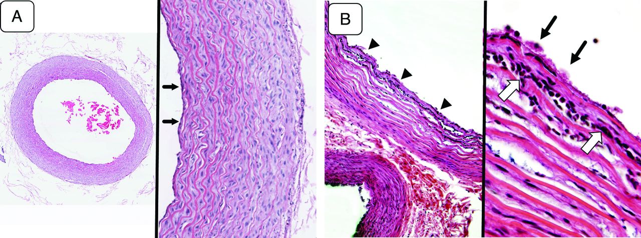
  • Fig 3.
    • Download figure
    • Open in new tab
    • Download powerpoint
    Fig 3.

    A, Microscopic view of the arterial sample (control group) (hematoxylin-eosin [H&E] staining). A low-magnification view of the harvested arterial sample (left, original magnification ×40) demonstrates well-maintained tissue integrity throughout all layers of the vessel. A magnified view of the same sample (right, original magnification ×100) shows the general histologic characteristics of a small artery. The tunica media, which constitutes most of the arterial wall, has up to 10–12 layers of smooth muscle cells. There is a very thin layer of tunica intima lined by a layer of endothelial cells (arrows). The external elastic lamina is not distinct. The structure is quite similar to that seen in the human intracranial arteries except that the layer of the tunica media is slightly thicker in this model. B, Microscopic view of an artery treated with mechanical thrombectomy (H&E staining). A low-magnification view (left, original magnification ×40) shows the separation of the smooth muscle layers in the tunica media and migration of acute inflammatory cells between the layers (arrowheads). A magnified view of the same arterial sample (right, original magnification ×200) demonstrates aggregated platelets on the surface of the arterial wall (arrows) and acute inflammatory cells migrating into the innermost layers of the tunica media (open arrows). The findings indicate an acute inflammatory reaction induced by the mechanical thrombectomy.

PreviousNext
Back to top

In this issue

American Journal of Neuroradiology: 34 (8)
American Journal of Neuroradiology
Vol. 34, Issue 8
1 Aug 2013
  • Table of Contents
  • Index by author
Advertisement
Print
Download PDF
Email Article

Thank you for your interest in spreading the word on American Journal of Neuroradiology.

NOTE: We only request your email address so that the person you are recommending the page to knows that you wanted them to see it, and that it is not junk mail. We do not capture any email address.

Enter multiple addresses on separate lines or separate them with commas.
A Swine Model to Analyze Arterial Structural Changes Induced by Mechanical Thrombectomy
(Your Name) has sent you a message from American Journal of Neuroradiology
(Your Name) thought you would like to see the American Journal of Neuroradiology web site.
CAPTCHA
This question is for testing whether or not you are a human visitor and to prevent automated spam submissions.
Cite this article
I. Yuki, I. Kan, A. Golshan, J. Sohn, Y. Murayama, H.V. Vinters, F. Viñuela
A Swine Model to Analyze Arterial Structural Changes Induced by Mechanical Thrombectomy
American Journal of Neuroradiology Aug 2013, 34 (8) E87-E90; DOI: 10.3174/ajnr.A3221

Citation Manager Formats

  • BibTeX
  • Bookends
  • EasyBib
  • EndNote (tagged)
  • EndNote 8 (xml)
  • Medlars
  • Mendeley
  • Papers
  • RefWorks Tagged
  • Ref Manager
  • RIS
  • Zotero
0 Responses
Respond to this article
Share
Bookmark this article
A Swine Model to Analyze Arterial Structural Changes Induced by Mechanical Thrombectomy
I. Yuki, I. Kan, A. Golshan, J. Sohn, Y. Murayama, H.V. Vinters, F. Viñuela
American Journal of Neuroradiology Aug 2013, 34 (8) E87-E90; DOI: 10.3174/ajnr.A3221
del.icio.us logo Twitter logo Facebook logo Mendeley logo
  • Tweet Widget
  • Facebook Like
  • Google Plus One
Purchase

Jump to section

  • Article
    • Abstract
    • ABBREVIATIONS:
    • Technique
    • Endovascular Embolization of the Superficial Cervical Artery with Experimental Thrombus
    • Discussion
    • Footnotes
    • REFERENCES
  • Figures & Data
  • Info & Metrics
  • Responses
  • References
  • PDF

Related Articles

  • No related articles found.
  • PubMed
  • Google Scholar

Cited By...

  • Impact of Target Artery Size on the Performance of Aspiration Thrombectomy: Insights from a Swine Model with Real-Time Visualization
  • Histological evaluation of acute ischemic stroke thrombi may indicate the occurrence of vessel wall injury during mechanical thrombectomy
  • A comparison of acute vascular damage caused by ADAPT versus a stent retriever device after thrombectomy in acute ischemic stroke: a histological and ultrastructural study in an animal model
  • Evaluation of the JRecan device for thrombus retrieval: efficacy and safety in a swine model of acute arterial occlusion
  • Histopathologic Evaluation of Arterial Wall Response to 5 Neurovascular Mechanical Thrombectomy Devices in a Swine Model
  • Crossref
  • Google Scholar

This article has not yet been cited by articles in journals that are participating in Crossref Cited-by Linking.

More in this TOC Section

  • Decubitus Myelography for Spinal Extradural Arachnoid Cyst—Better Classified as a Lateral Dural CSF Leak?
  • Mitigating brain MR imaging degradation due to ferumoxytol therapy in patients with iron deficiency anemia
  • Advanced Imaging of Type 2 Spinal CSF Leaks with Ultrahigh-Resolution Cone-Beam CT Myelography
Show more Technical Notes

Similar Articles

Advertisement

Indexed Content

  • Current Issue
  • Accepted Manuscripts
  • Article Preview
  • Past Issues
  • Editorials
  • Editor's Choice
  • Fellows' Journal Club
  • Letters to the Editor
  • Video Articles

Cases

  • Case Collection
  • Archive - Case of the Week
  • Archive - Case of the Month
  • Archive - Classic Case

More from AJNR

  • Trainee Corner
  • Imaging Protocols
  • MRI Safety Corner
  • Book Reviews

Multimedia

  • AJNR Podcasts
  • AJNR Scantastics

Resources

  • Turnaround Time
  • Submit a Manuscript
  • Submit a Video Article
  • Submit an eLetter to the Editor/Response
  • Manuscript Submission Guidelines
  • Statistical Tips
  • Fast Publishing of Accepted Manuscripts
  • Graphical Abstract Preparation
  • Imaging Protocol Submission
  • Evidence-Based Medicine Level Guide
  • Publishing Checklists
  • Author Policies
  • Become a Reviewer/Academy of Reviewers
  • News and Updates

About Us

  • About AJNR
  • Editorial Board
  • Editorial Board Alumni
  • Alerts
  • Permissions
  • Not an AJNR Subscriber? Join Now
  • Advertise with Us
  • Librarian Resources
  • Feedback
  • Terms and Conditions
  • AJNR Editorial Board Alumni

American Society of Neuroradiology

  • Not an ASNR Member? Join Now

© 2025 by the American Society of Neuroradiology All rights, including for text and data mining, AI training, and similar technologies, are reserved.
Print ISSN: 0195-6108 Online ISSN: 1936-959X

Powered by HighWire