
Article Figures & Data
Figures
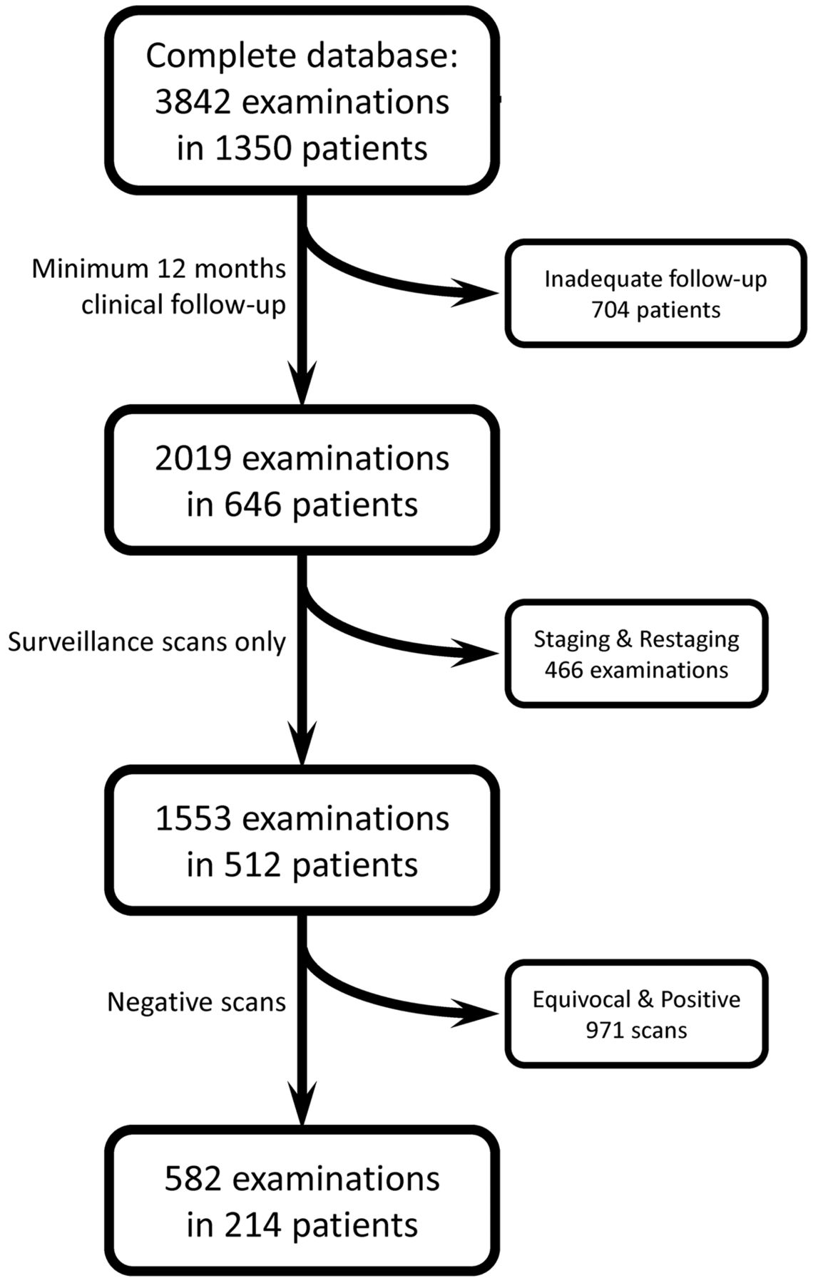
- Fig 1.
Data flow chart of patient selection.
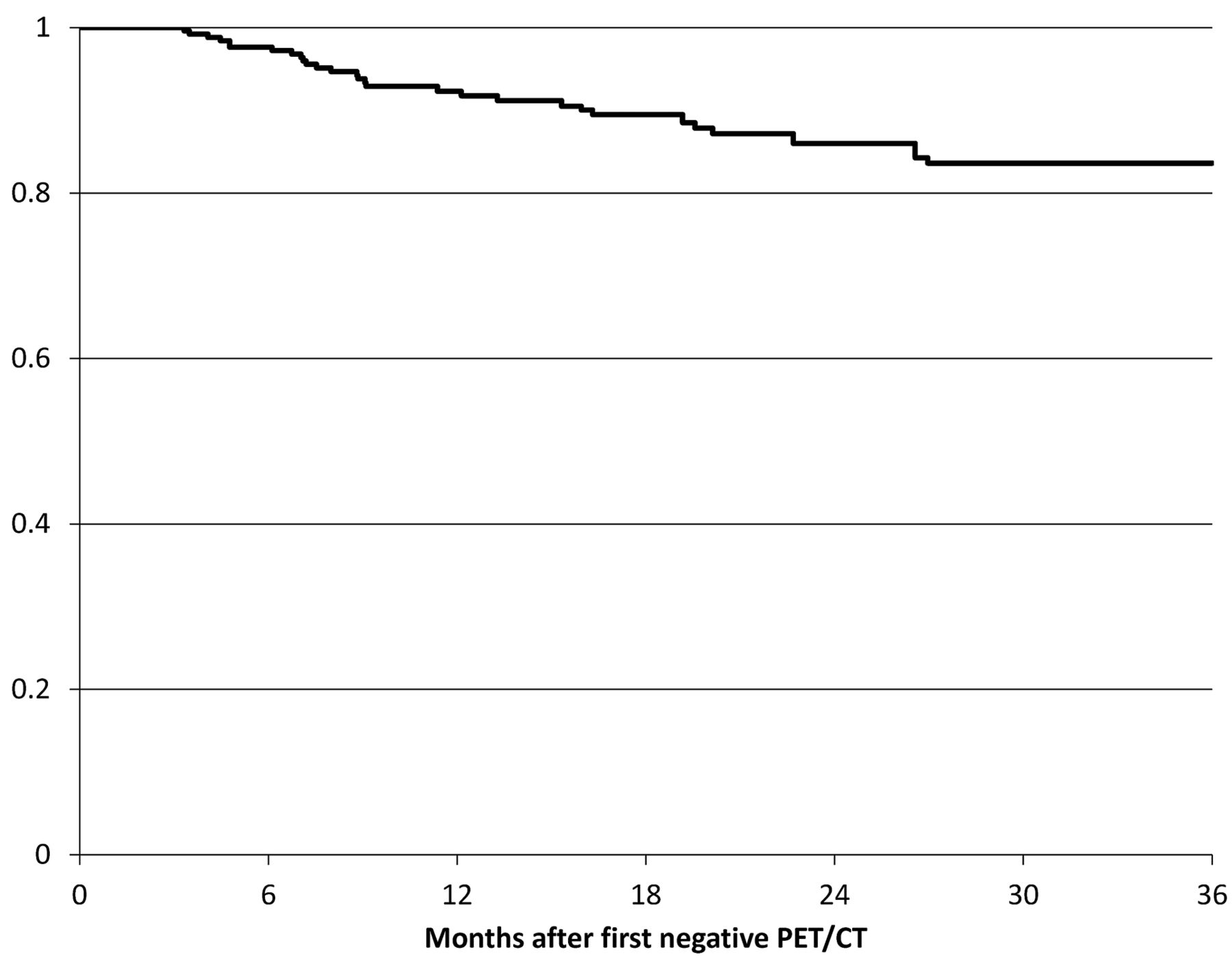
- Fig 2.
Disease-free survival curve of patients with negative findings on PET-CT surveillance examinations after treatment for HNSCC. Time zero was chosen as the date of first PET/CT examination with negative findings.


Tables
Demographics and Staging Age (yr) Median 61 Range 21–89 Sex Male 388 (76%) Female 124 (24%) Site of origin (n = 419) Oropharynx 130 (31%) Oral cavity 130 (31%) Larynx 63 (15%) Maxillofacial/sinus 37 (9%) Hypopharynx 26 (6%) Nasopharynx 12 (3%) Trans-spatial 10 (2%) Unknown primary 8 (2%) T stage (n = 428) T0 9 (2%) T1 109 (25%) T2 124 (29%) T3 85 (20%) T4 101 (24%) N stage (n = 440) N0 192 (44%) N1 69 (16%) N2 162 (37%) N3 17 (4%) M stage (n = 369) M0 365 (99%) M1 4 (1%) Therapy (n = 512) CRT alone 67 (13%) Surgery plus CRT 445 (87%) Note:—CRT indicates chemoradiation therapy; N, node; M, metastasis.
↵a A total of 512 patients were included in this study, but not all patients had initial staging data available, so fewer patients are included. The number of patients with available data is listed in the header for each section.
Site of Origin T1 T2 T3 T4 Oropharynx 25 42 21 30 Oral cavity 31 34 17 34 Larynx 10 15 21 11 Maxillofacial and sinus (parotid, skin, nasal cavity, orbit lip, sinus, maxilla) 5 5 5 10 Hypopharynx 4 4 9 5 Nasopharynx 0 1 1 6 Trans-spatial 0 2 2 6 ↵a Fewer patients are listed in Table 2 than in Table 1 because some patients had incomplete data in the registry. Three patients had unknown primaries.
- Table 3:
Primary site of origin and tumor stage for the subset of 214 patients with negative PET/CT findingsa
Site of Origin T1 T2 T3 T4 Oropharynx 25 32 7 8 Oral cavity 18 15 7 13 Larynx 3 7 9 4 Maxillofacial and sinus (parotid, skin, nasal cavity, orbit lip, sinus, maxilla) 3 3 3 3 Hypopharynx 2 4 2 3 Nasopharynx 0 1 3 4 Trans-spatial 0 1 1 1 ↵a The site of primary tumor and tumor stage were available from 182 of the 214 patients with negative PET/CT findings. No patients were recorded as having unknown primaries in the data base from this subset of patients.
- Table 4:
Clinical Information of the 19 patients with negative PET/CT findings who had a recurrence
Patient Site of Origin TNM Stage Location of Recurrence Time Interval between Negative PET/CT Findings and Recurrence (mo) 1 Oropharynx T2N2cM0 Local 7.5 2 Maxillofacial Not recorded Regional 12.7 3 Oropharynx T3N2cM0 Local 26.4 4 Oropharynx Not recorded Regional 7.5 5 Maxillofacial Not recorded Distant 11.4 6 Larynx T2N0M0 Local 9.0 7 Unknown primary T0N2bM0 Regional 27.5 8 Unknown primary T0N2bM0 Regional 23.0 9 Hypopharynx T4N2bM0 Local 6.9 10 Hypopharynx T2N2bM0 Local 3.7 11 Larynx T2N0M0 Local 3.0 12 Oropharynx T3N0M0 Local 4.6 13 Oropharynx T4N1M0 Local 9.5 14 Oral cavity Not recorded Distant 7.1 15 Oral cavity T3N1M0 Regional 37.3 16 Oral cavity T3N2cM0 Local 16.9 17 Oral cavity, larynx T2N2cM0 Regional 4.8 18 Larynx Not recorded Local 12.3 19 Oral cavity Not recorded Local 12.7 Note:—TNM indicates tumor, node, metastasis.
In this issue


Jump to section
Related Articles
Cited By...
- Posttreatment Imaging in Patients with Head and Neck Cancer without Clinical Evidence of Recurrence: Should Surveillance Imaging Extend Beyond 6 Months?
- Positive Predictive Value of Neck Imaging Reporting and Data System Categories 3 and 4 Posttreatment FDG-PET/CT in Head and Neck Squamous Cell Carcinoma
- Negative Predictive Value of NI-RADS Category 2 in the First Posttreatment FDG-PET/CT in Head and Neck Squamous Cell Carcinoma
- Surveillance Imaging in HPV-related Oropharyngeal Cancer
- Initial Performance of NI-RADS to Predict Residual or Recurrent Head and Neck Squamous Cell Carcinoma
- Evaluating the Potential Role of PET/CT in the Posttreatment Surveillance of Head and Neck Cancer
- Guideline Familiarity Predicts Variation in Self-Reported Use of Routine Surveillance PET/CT by Physicians Who Treat Head and Neck Cancer