
Article Figures & Data
Figures
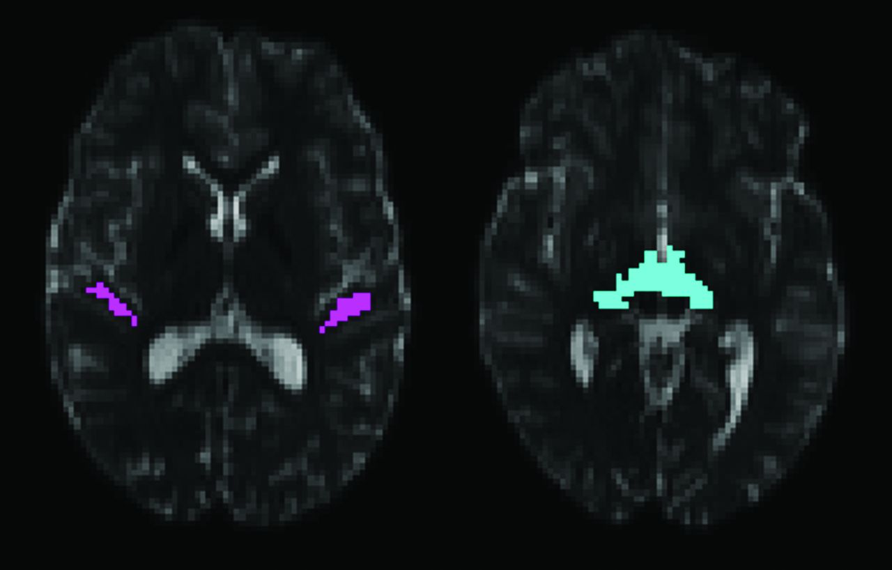
- Fig 1.
The starting region of interest for fiber tracking within the Heschl gyrus is shown on the left. The target region of interest on the inferior surface of the thalamus is shown on the right. The regions are shown overlaid on axial sections through the b=0 s/mm2 echo-planar volume from the 64-direction HARDI acquisition.
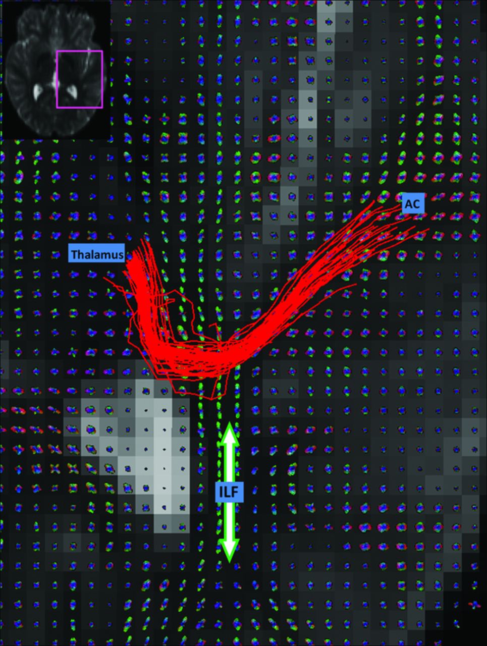
- Fig 2.
Q-ball reconstructions of the HARDI data are shown for each voxel on an axial section. HARDI fiber tracks (red streamlines) course from the AC to the thalamus. The ILF (green fiber peaks and orientation indicated with arrow) intersects the auditory radiation.
- Fig 3.
HARDI fiber tracks (red) and DTI fiber tracks (blue) are visualized in the left and right hemispheres. DTI fiber tracks are from the 64-direction dataset. The left hemisphere is on the left side of the figure. Both HARDI and DTI fiber tracks were launched from the same starting regions in the Heschl gyrus. The HARDI fiber tracks reaching the thalamic target regions are retained. For this figure, all DTI fiber tracks are retained regardless of destination. The DTI fiber tracks emerge from the Heschl gyrus and follow the ILF in either the anterior or posterior direction. No DTI fiber tracks cross the ILF to reach the thalamus in the right hemisphere.
- Fig 4.
HARDI and DTI fiber tracks connecting the AC to the thalamic target region are shown in a case where both methods were successful in each hemisphere. The number of fiber trajectories passing through each voxel is encoded with the overlay color. The yellow voxels have the highest probability of being within the auditory radiation.
- Fig 5.
The threshold for successful fiber-tracking is varied between 1 and 200 trajectories connecting the AC to the thalamus. The percentage of successful HARDI and DTI fiber-tracking trials is shown at each threshold. An asterisk indicates that the HARDI rate of success is significantly higher than the respective DTI fiber-tracking rate of success (P < .01).


Tables
Comparison of DTI and HARDI auditory fiber-tracking results. Successful connections contained 1 or more fiber trajectories
Successful Unsuccessful Left Right Total Left Right Total DTI - 30 Directions 16 5 21 9 20 29 DTI - 64 Directions 18 7 25 7 18 25 HARDI - 64 Directions 24 25 49 1 0 1
In this issue


Jump to section
Related Articles
Cited By...
- The Advanced BRain Imaging on ageing and Memory (ABRIM) data collection: Study protocol and rationale
- Resolution and b value dependent Structural Connectome in ex vivo Mouse Brain
- Auditory and Visual System White Matter Is Differentially Impacted by Normative Aging in Macaques
- A comprehensive atlas of white matter tracts in the chimpanzee
- Occipital white matter tracts in human and macaque
- Relationship between M100 Auditory Evoked Response and Auditory Radiation Microstructure in 16p11.2 Deletion and Duplication Carriers