
Article Figures & Data
Figures
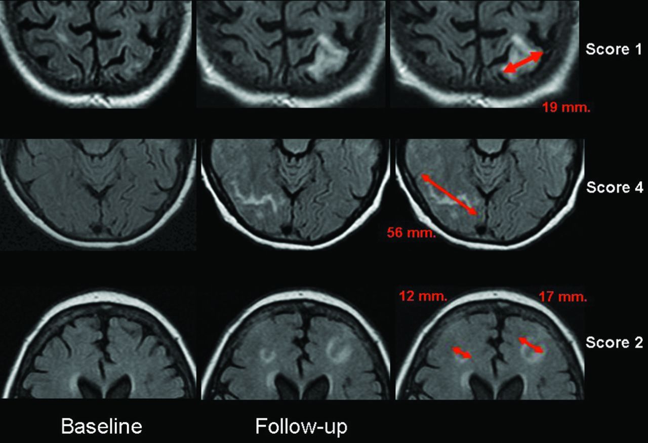
- Fig 1.
Parenchymal hyperintensity. Top row shows a left parietal lesion <2 cm in maximum diameter (score 1). Middle row shows a right occipital lesion >4 cm (score 4); lower row shows multifocal lesions, each <2 cm in diameter (score 2). All measurements were performed in-plane.
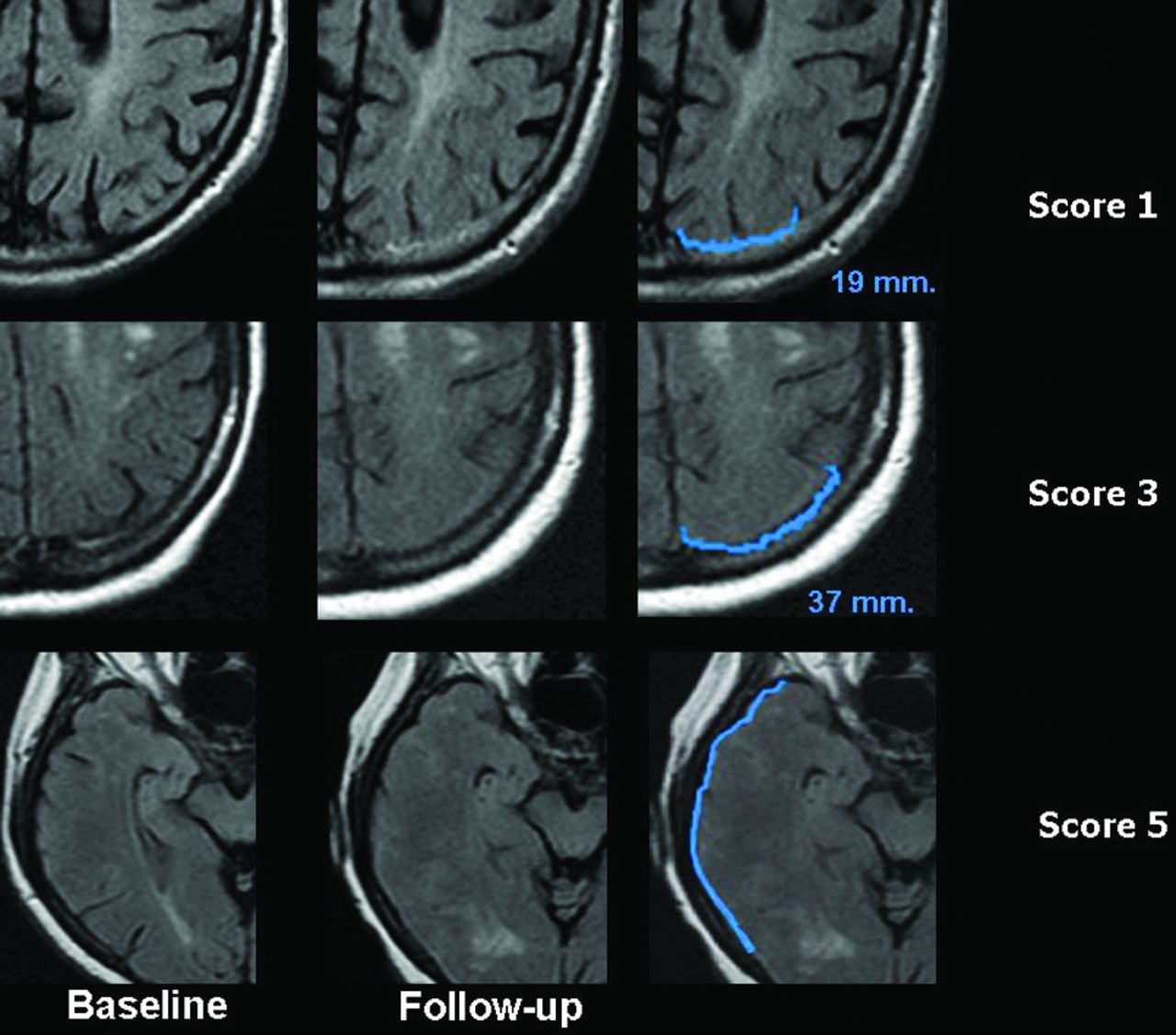
- Fig 2.
Sulcal hyperintensity. The first column shows the FLAIR images at baseline, the second column shows the FLAIR images at follow-up, and the third column shows the FLAIR images at follow-up with a very narrow contrast window/level setting accentuating the abnormalities for descriptive purposes. Upper row shows a right frontal abnormality with a diameter <2 cm (score 1). Middle row shows a left occipital abnormality with a maximum diameter between 2 and 4 cm (score 3); and lower row shows a right parietal abnormality of >4 cm (score 4).
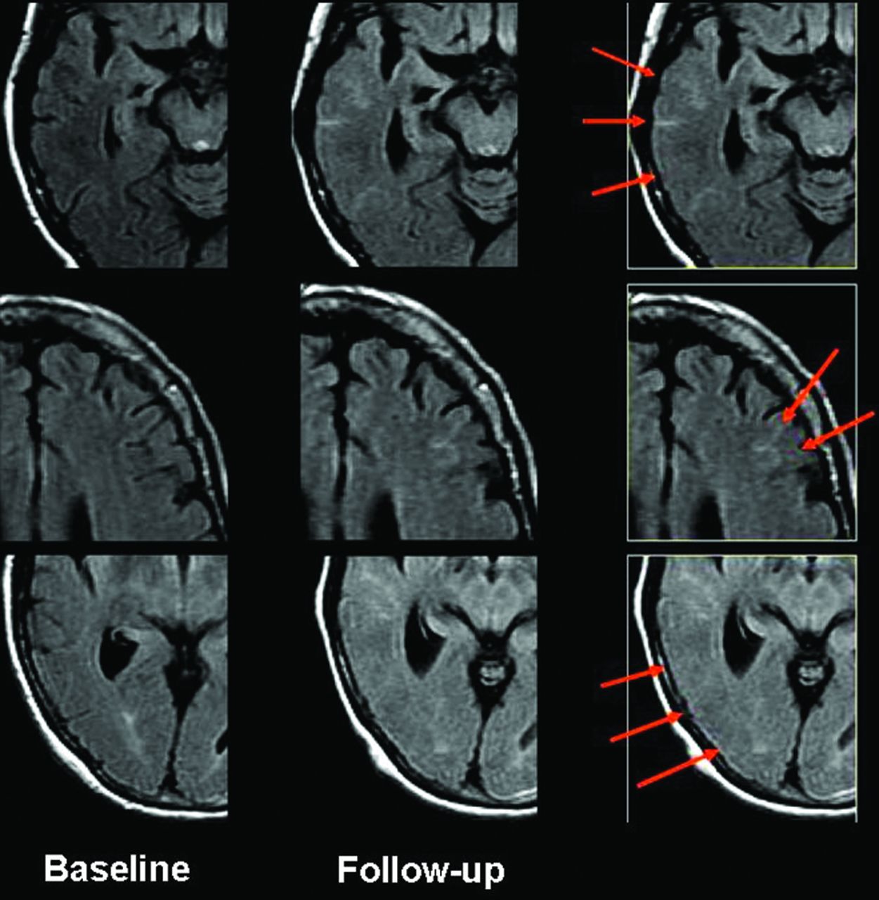
- Fig 3.
Swelling. Top row shows a left parietal area of effacement <2 cm in maximum diameter (score 1). Middle row shows a left parietal effacement of <4 cm in diameter (score 3). Lower row shows swelling of the entire right temporal lobe (score 5).
- Fig 4.
Distribution of ARIA-E scores for the 2 readers in abnormal cases. Bars represent the maximum score of each rater for either parenchymal or sulcal hyperintensity per region, summed across the 12 anatomic regions.
- Fig 5.
Discrepant readings in case 10. In this patient with widespread ARIA-E involving multiple lobes on both sides of the brain, there were discrepancies between readers regarding the extent of lesions within each affected area. Upper row shows extensive sulcal hyperintensity covering large parts of the right temporal lobe, given a score of 4 by rater 1 but only a score of 3 by rater 2. Middle row shows that a score of 2 for sulcal hyperintensity (SH) was given in the left frontal lobe by rater 1 but a score 0 for SH, by rater 2. Lower row shows that a score of 5 for sulcal hyperintensity in the right occipital lobe was given by rater 1 but only a score of 3 by rater 2.


Tables
ARIA-E Cases Non-ARIA Cases Overall No. of subjects 10 10 20 Age (mean) (±SD) 69.4 (9.5) 69.2 (8.4) 69.3 (8.7) MMSE (mean) (±SD) 20.2 (3.9) 21.2 (2.7) 20.7 (3.3) ADAS-cog11 (mean) (SD) 22.0 (1.27) 21.8 (5.6) 21.9 (9.6) DAD (mean) (±SD) 81.9 (18.8) 85.2 (11.2) 83.5 (15.2) Female (No.) (%) 7 (70.0%) 4 (40.0%) 5 (55.0%) ApoE ϵ4 status ApoE ϵ4 noncarrier (No.) (%) 3 (30.0%) 1 (10.0%) 4 (20.0%) ApoE ϵ4 (No.) (%) 3 (30.0%) 5 (50.0%) 8 (40.0%) ApoE ϵ4 homozygote (No.) (%) 4 (40.0%) 4 (40.0%) 8 (40.0%) Bapineuzumab 0.15 mg/kg (No.) (%) 1 (10.0%) 1 (10.0%) 2 (10.0%) 0.5 mg/kg (No.) (%) 1 (10.0%) 2 (20.0%) 3 (15.0%) 1.0 mg/kg (No.) (%) 2 (20.0%) 3 (30.0%) 5 (25.0%) 2.0 mg/kg (No.) (%) 6 (60.0%) 4 (40.0%) 10 (50.0%) Note:—DAD indicates Disability Assessment for Dementia; ADAS-cog11, Alzheimer's Disease Assessment Scale-cognition (11 items); MMSE, Mini-Mental State Examination.
Region Parenchymal Hyperintensity Sulcal Hyperintensity Swelling Highest Regional Score Frontal R (0–5) (0–5) (0–5) (0–5) Frontal L (0–5) (0–5) (0–5) (0–5) Parietal R (0–5) (0–5) (0–5) (0–5) Parietal L (0–5) (0–5) (0–5) (0–5) Occipital R (0–5) (0–5) (0–5) (0–5) Occipital L (0–5) (0–5) (0–5) (0–5) Temporal R (0–5) (0–5) (0–5) (0–5) Temporal L (0–5) (0–5) (0–5) (0–5) Central R (0–5) (0–5) (0–5) (0–5) Central L (0–5) (0–5) (0–5) (0–5) Infratentorial R (0–5) (0–5) (0–5) (0–5) Infratentorial L (0–5) (0–5) (0–5) (0–5) Grand total score (0–60) ↵a Data are ranges. For each region/side (L/R), enter a score depending on the largest cross-sectional diameter: 0, no abnormalities; 1, monofocal lesion ≤2 cm; 2, multifocal lesions each ≤2 cm; 3, any lesion >2 but <4 cm; 4, any lesion ≥4 cm; 5, entire lobe. All measurements were performed in-plane.
MRI Finding ICC 95% CI ARIA-E (parenchymal and sulcal hyperintensity) 0.887 0.622–0.971 Parenchymal hyperintensity 0.831 0.481–0.955 Sulcal hyperintensity 0.892 0.629–0.972 Swelling 0.542 −0.058–0.861 ARIA-E (all 3 features) 0.777 0.361–0.939
In this issue


Jump to section
Related Articles
Cited By...
- Clinical-radiological presentation and natural history of iatrogenic cerebral amyloid angiopathy
- Association of Microglial Activation With Spontaneous ARIA-E and CSF Levels of Anti-A{beta} Autoantibodies
- Amyloid-Related Imaging Abnormalities with Emerging Alzheimer Disease Therapeutics: Detection and Reporting Recommendations for Clinical Practice
- Prevalence of cortical superficial siderosis in a memory clinic population