Advertisement

Main menu

  • Home
  • Content
    • Current Issue
    • Accepted Manuscripts
    • Article Preview
    • Past Issue Archive
    • Video Articles
    • AJNR Case Collection
    • Case of the Week Archive
    • Case of the Month Archive
    • Classic Case Archive
  • Special Collections
    • AJNR Awards
    • Low-Field MRI
    • Alzheimer Disease
    • ASNR Foundation Special Collection
    • Photon-Counting CT
    • View All
  • Multimedia
    • AJNR Podcasts
    • AJNR SCANtastic
    • Trainee Corner
    • MRI Safety Corner
    • Imaging Protocols
  • For Authors
    • Submit a Manuscript
    • Submit a Video Article
    • Submit an eLetter to the Editor/Response
    • Manuscript Submission Guidelines
    • Statistical Tips
    • Fast Publishing of Accepted Manuscripts
    • Graphical Abstract Preparation
    • Imaging Protocol Submission
    • Author Policies
  • About Us
    • About AJNR
    • Editorial Board
    • Editorial Board Alumni
  • More
    • Become a Reviewer/Academy of Reviewers
    • Subscribers
    • Permissions
    • Alerts
    • Feedback
    • Advertisers
    • ASNR Home

User menu

  • Alerts
  • Log in

Search

  • Advanced search
American Journal of Neuroradiology

American Journal of Neuroradiology

ASHNR American Society of Functional Neuroradiology ASHNR American Society of Pediatric Neuroradiology ASSR
  • Alerts
  • Log in

Advanced Search

  • Home
  • Content
    • Current Issue
    • Accepted Manuscripts
    • Article Preview
    • Past Issue Archive
    • Video Articles
    • AJNR Case Collection
    • Case of the Week Archive
    • Case of the Month Archive
    • Classic Case Archive
  • Special Collections
    • AJNR Awards
    • Low-Field MRI
    • Alzheimer Disease
    • ASNR Foundation Special Collection
    • Photon-Counting CT
    • View All
  • Multimedia
    • AJNR Podcasts
    • AJNR SCANtastic
    • Trainee Corner
    • MRI Safety Corner
    • Imaging Protocols
  • For Authors
    • Submit a Manuscript
    • Submit a Video Article
    • Submit an eLetter to the Editor/Response
    • Manuscript Submission Guidelines
    • Statistical Tips
    • Fast Publishing of Accepted Manuscripts
    • Graphical Abstract Preparation
    • Imaging Protocol Submission
    • Author Policies
  • About Us
    • About AJNR
    • Editorial Board
    • Editorial Board Alumni
  • More
    • Become a Reviewer/Academy of Reviewers
    • Subscribers
    • Permissions
    • Alerts
    • Feedback
    • Advertisers
    • ASNR Home
  • Follow AJNR on Twitter
  • Visit AJNR on Facebook
  • Follow AJNR on Instagram
  • Join AJNR on LinkedIn
  • RSS Feeds

AJNR Awards, New Junior Editors, and more. Read the latest AJNR updates

Research ArticleBrain

Automated Cerebral Infarct Volume Measurement in Follow-up Noncontrast CT Scans of Patients with Acute Ischemic Stroke

A.M. Boers, H.A. Marquering, J.J. Jochem, N.J. Besselink, O.A. Berkhemer, A. van der Lugt, L.F. Beenen and C.B. Majoie on behalf of the MR CLEAN investigators
American Journal of Neuroradiology August 2013, 34 (8) 1522-1527; DOI: https://doi.org/10.3174/ajnr.A3463
A.M. Boers
aFrom the Departments of Radiology (A.M.B., H.A.M., J.J.J., N.J.B., O.A.B., L.F.B., C.B.M.)
cInstitute of Technical Medicine (A.M.B., J.J.J., N.J.B.), University of Twente, Enschede, the Netherlands
  • Find this author on Google Scholar
  • Find this author on PubMed
  • Search for this author on this site
H.A. Marquering
aFrom the Departments of Radiology (A.M.B., H.A.M., J.J.J., N.J.B., O.A.B., L.F.B., C.B.M.)
bBiomedical Engineering and Physics (H.A.M.), Academic Medical Center, Amsterdam, the Netherlands
  • Find this author on Google Scholar
  • Find this author on PubMed
  • Search for this author on this site
J.J. Jochem
aFrom the Departments of Radiology (A.M.B., H.A.M., J.J.J., N.J.B., O.A.B., L.F.B., C.B.M.)
cInstitute of Technical Medicine (A.M.B., J.J.J., N.J.B.), University of Twente, Enschede, the Netherlands
  • Find this author on Google Scholar
  • Find this author on PubMed
  • Search for this author on this site
N.J. Besselink
aFrom the Departments of Radiology (A.M.B., H.A.M., J.J.J., N.J.B., O.A.B., L.F.B., C.B.M.)
cInstitute of Technical Medicine (A.M.B., J.J.J., N.J.B.), University of Twente, Enschede, the Netherlands
  • Find this author on Google Scholar
  • Find this author on PubMed
  • Search for this author on this site
O.A. Berkhemer
aFrom the Departments of Radiology (A.M.B., H.A.M., J.J.J., N.J.B., O.A.B., L.F.B., C.B.M.)
  • Find this author on Google Scholar
  • Find this author on PubMed
  • Search for this author on this site
A. van der Lugt
dDepartment of Radiology (A.v.d.L.), Erasmus Medical Center, Rotterdam, the Netherlands.
  • Find this author on Google Scholar
  • Find this author on PubMed
  • Search for this author on this site
L.F. Beenen
aFrom the Departments of Radiology (A.M.B., H.A.M., J.J.J., N.J.B., O.A.B., L.F.B., C.B.M.)
  • Find this author on Google Scholar
  • Find this author on PubMed
  • Search for this author on this site
C.B. Majoie
aFrom the Departments of Radiology (A.M.B., H.A.M., J.J.J., N.J.B., O.A.B., L.F.B., C.B.M.)
  • Find this author on Google Scholar
  • Find this author on PubMed
  • Search for this author on this site
  • Article
  • Figures & Data
  • Supplemental
  • Info & Metrics
  • Responses
  • References
  • PDF
Loading

Article Figures & Data

Figures

  • Tables
  • Fig 1.
    • Download figure
    • Open in new tab
    • Download powerpoint
    Fig 1.

    Illustration of the automated CIV segmentation pipeline. This figure shows an infarct in the right hemisphere in a 6-day follow-up CT. A, The unprocessed NCCT. B, A seed point is positioned within the infarcted area by an observer. C, Determination of the midline. D, Segmentation of the ventricles, E, The final segmentation representing the CIV.

  • Fig 2.
    • Download figure
    • Open in new tab
    • Download powerpoint
    Fig 2.

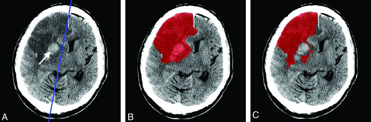
    Example of incorrect automated segmentation due to hemorrhage and erroneous midline determination. A, NCCT scan of a patient with a midline shift. The blue line indicates the midline determined by the algorithm. The hemorrhage is indicated by the white arrow. B, The infarcted area delineated by observer 1. C, Incorrect outcome of automated segmentation. The hemorrhage is unrecognized as infarct, and the infarcted tissue is shifted over the original midline. This shift results in underestimation of the CIV.

  • Fig 3.
    • Download figure
    • Open in new tab
    • Download powerpoint
    Fig 3.

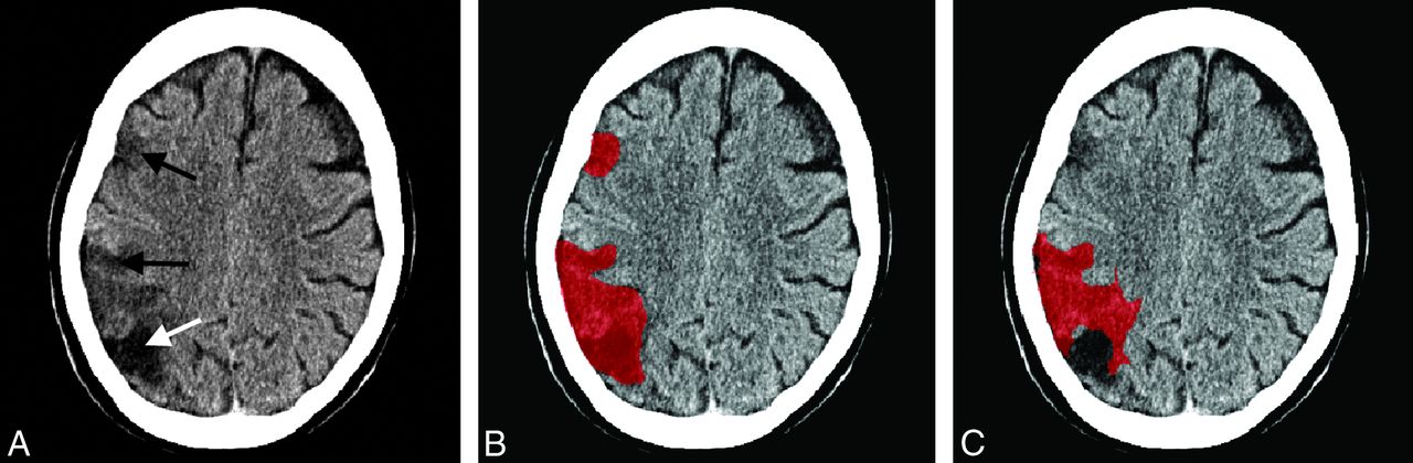
    Example of incorrect automated segmentation due to multiple infarcts. A, NCCT scan of a patient with multiple infarcts, 2 new infarcts (black arrows) and 1 old infarct (white arrow). B, The infarcted area delineated by observer 1. C, Incorrect outcome of automated segmentation. The old infarct and one of the new infarcts are unrecognized as infarcts. As a result, the automated segmentation underestimates the CIV.

Tables

  • Figures
    • View popup
    Table 1:

    List of the number of patients per hospital, CT scanner used, and reconstruction kernel

    HospitalNo. of PatientsCT ScannerKernel
    114Sensation 64aH31s
    22Brilliance 40bUB
    32iCT 256UB
    41Aquilion 64cFC23
    51Aquilion ONEFC26
    63iCT 256EC
    74Sensation 16/ Somatom Definition FlashH31s
    85LightSpeed VCTdSoft
    92Brilliance 40UB
    • ↵a Siemens, Erlangen, Germany.

    • ↵b Philips Healthcare, Best, the Netherlands.

    • ↵c Toshiba Medical Systems, Tokyo, Japan.

    • ↵d GE Healthcare, Milwaukee, Wisconsin.

    • View popup
    Table 2:

    Interobserver variability of manual CIV measurements and comparison of the manual and automated method

    Correlationa (95% CI)Dice Coefficient (Mean and Range)Bland-Altman Limits of Agreement (mL)Relative Difference (SD)No.
    Automated vs manual0.98 (0.97–0.99)0.74 (0.42–0.90)(−38.0–39.1)4% (± 20%)34
    Manual interobserver0.98 (0.96–0.99)0.84 (0.63–0.94)(−44.1–40.9)11% (± 27%)34
    • ↵a Pearson correlation; P < .01 for all coefficients.

PreviousNext
Back to top

In this issue

American Journal of Neuroradiology: 34 (8)
American Journal of Neuroradiology
Vol. 34, Issue 8
1 Aug 2013
  • Table of Contents
  • Index by author
Advertisement
Print
Download PDF
Email Article

Thank you for your interest in spreading the word on American Journal of Neuroradiology.

NOTE: We only request your email address so that the person you are recommending the page to knows that you wanted them to see it, and that it is not junk mail. We do not capture any email address.

Enter multiple addresses on separate lines or separate them with commas.
Automated Cerebral Infarct Volume Measurement in Follow-up Noncontrast CT Scans of Patients with Acute Ischemic Stroke
(Your Name) has sent you a message from American Journal of Neuroradiology
(Your Name) thought you would like to see the American Journal of Neuroradiology web site.
CAPTCHA
This question is for testing whether or not you are a human visitor and to prevent automated spam submissions.
Cite this article
A.M. Boers, H.A. Marquering, J.J. Jochem, N.J. Besselink, O.A. Berkhemer, A. van der Lugt, L.F. Beenen, C.B. Majoie
Automated Cerebral Infarct Volume Measurement in Follow-up Noncontrast CT Scans of Patients with Acute Ischemic Stroke
American Journal of Neuroradiology Aug 2013, 34 (8) 1522-1527; DOI: 10.3174/ajnr.A3463

Citation Manager Formats

  • BibTeX
  • Bookends
  • EasyBib
  • EndNote (tagged)
  • EndNote 8 (xml)
  • Medlars
  • Mendeley
  • Papers
  • RefWorks Tagged
  • Ref Manager
  • RIS
  • Zotero
0 Responses
Respond to this article
Share
Bookmark this article
Automated Cerebral Infarct Volume Measurement in Follow-up Noncontrast CT Scans of Patients with Acute Ischemic Stroke
A.M. Boers, H.A. Marquering, J.J. Jochem, N.J. Besselink, O.A. Berkhemer, A. van der Lugt, L.F. Beenen, C.B. Majoie
American Journal of Neuroradiology Aug 2013, 34 (8) 1522-1527; DOI: 10.3174/ajnr.A3463
del.icio.us logo Twitter logo Facebook logo Mendeley logo
  • Tweet Widget
  • Facebook Like
  • Google Plus One
Purchase

Jump to section

  • Article
    • Abstract
    • ABBREVIATIONS:
    • Materials and Methods
    • Results
    • Discussion
    • Conclusions
    • Acknowledgments
    • Footnotes
    • REFERENCES
  • Figures & Data
  • Supplemental
  • Info & Metrics
  • Responses
  • References
  • PDF

Related Articles

  • PubMed
  • Google Scholar

Cited By...

  • Association of White Matter Lesions and Outcome After Endovascular Stroke Treatment
  • Automatic segmentation of cerebral infarcts in follow-up computed tomography images with convolutional neural networks
  • Comparison of three commonly used CT perfusion software packages in patients with acute ischemic stroke
  • Hemorrhagic transformation is associated with poor functional outcome in patients with acute ischemic stroke due to a large vessel occlusion
  • Association of follow-up infarct volume with functional outcome in acute ischemic stroke: a pooled analysis of seven randomized trials
  • Impact of Ischemic Lesion Location on the mRS Score in Patients with Ischemic Stroke: A Voxel-Based Approach
  • Incorporation of relative cerebral blood flow into CT perfusion maps reduces false at risk' penumbra
  • Value of Quantitative Collateral Scoring on CT Angiography in Patients with Acute Ischemic Stroke
  • Opercular Index Score: a CT angiography-based predictor of capillary robustness and neurological outcomes in the endovascular management of acute ischemic stroke
  • Value of Thrombus CT Characteristics in Patients with Acute Ischemic Stroke
  • Association of Computed Tomography Ischemic Lesion Location With Functional Outcome in Acute Large Vessel Occlusion Ischemic Stroke
  • Baseline Blood Pressure Effect on the Benefit and Safety of Intra-Arterial Treatment in MR CLEAN (Multicenter Randomized Clinical Trial of Endovascular Treatment of Acute Ischemic Stroke in the Netherlands)
  • Topographic distribution of cerebral infarct probability in patients with acute ischemic stroke: mapping of intra-arterial treatment effect
  • Associations of Ischemic Lesion Volume With Functional Outcome in Patients With Acute Ischemic Stroke: 24-Hour Versus 1-Week Imaging
  • Clot Burden Score on Baseline Computerized Tomographic Angiography and Intra-Arterial Treatment Effect in Acute Ischemic Stroke
  • Influence of Device Choice on the Effect of Intra-Arterial Treatment for Acute Ischemic Stroke in MR CLEAN (Multicenter Randomized Clinical Trial of Endovascular Treatment for Acute Ischemic Stroke in the Netherlands)
  • Assessment of Collateral Status by Dynamic CT Angiography in Acute MCA Stroke: Timing of Acquisition and Relationship with Final Infarct Volume
  • Thrombus Permeability Is Associated With Improved Functional Outcome and Recanalization in Patients With Ischemic Stroke
  • Impact of Collateral Status Evaluated by Dynamic Computed Tomographic Angiography on Clinical Outcome in Patients With Ischemic Stroke
  • Crossref
  • Google Scholar

This article has not yet been cited by articles in journals that are participating in Crossref Cited-by Linking.

More in this TOC Section

  • Usefulness of Quantitative Susceptibility Mapping for the Diagnosis of Parkinson Disease
  • White Matter Alterations in the Brains of Patients with Active, Remitted, and Cured Cushing Syndrome: A DTI Study
  • Qualitative and Quantitative Analysis of MR Imaging Findings in Patients with Middle Cerebral Artery Stroke Implanted with Mesenchymal Stem Cells
Show more Brain

Similar Articles

Advertisement

Indexed Content

  • Current Issue
  • Accepted Manuscripts
  • Article Preview
  • Past Issues
  • Editorials
  • Editor's Choice
  • Fellows' Journal Club
  • Letters to the Editor
  • Video Articles

Cases

  • Case Collection
  • Archive - Case of the Week
  • Archive - Case of the Month
  • Archive - Classic Case

More from AJNR

  • Trainee Corner
  • Imaging Protocols
  • MRI Safety Corner
  • Book Reviews

Multimedia

  • AJNR Podcasts
  • AJNR Scantastics

Resources

  • Turnaround Time
  • Submit a Manuscript
  • Submit a Video Article
  • Submit an eLetter to the Editor/Response
  • Manuscript Submission Guidelines
  • Statistical Tips
  • Fast Publishing of Accepted Manuscripts
  • Graphical Abstract Preparation
  • Imaging Protocol Submission
  • Evidence-Based Medicine Level Guide
  • Publishing Checklists
  • Author Policies
  • Become a Reviewer/Academy of Reviewers
  • News and Updates

About Us

  • About AJNR
  • Editorial Board
  • Editorial Board Alumni
  • Alerts
  • Permissions
  • Not an AJNR Subscriber? Join Now
  • Advertise with Us
  • Librarian Resources
  • Feedback
  • Terms and Conditions
  • AJNR Editorial Board Alumni

American Society of Neuroradiology

  • Not an ASNR Member? Join Now

© 2025 by the American Society of Neuroradiology All rights, including for text and data mining, AI training, and similar technologies, are reserved.
Print ISSN: 0195-6108 Online ISSN: 1936-959X

Powered by HighWire