Advertisement

Main menu

  • Home
  • Content
    • Current Issue
    • Accepted Manuscripts
    • Article Preview
    • Past Issue Archive
    • Video Articles
    • AJNR Case Collection
    • Case of the Week Archive
    • Case of the Month Archive
    • Classic Case Archive
  • Special Collections
    • AJNR Awards
    • Low-Field MRI
    • Alzheimer Disease
    • ASNR Foundation Special Collection
    • Photon-Counting CT
    • View All
  • Multimedia
    • AJNR Podcasts
    • AJNR SCANtastic
    • Trainee Corner
    • MRI Safety Corner
    • Imaging Protocols
  • For Authors
    • Submit a Manuscript
    • Submit a Video Article
    • Submit an eLetter to the Editor/Response
    • Manuscript Submission Guidelines
    • Statistical Tips
    • Fast Publishing of Accepted Manuscripts
    • Graphical Abstract Preparation
    • Imaging Protocol Submission
    • Author Policies
  • About Us
    • About AJNR
    • Editorial Board
    • Editorial Board Alumni
  • More
    • Become a Reviewer/Academy of Reviewers
    • Subscribers
    • Permissions
    • Alerts
    • Feedback
    • Advertisers
    • ASNR Home

User menu

  • Alerts
  • Log in

Search

  • Advanced search
American Journal of Neuroradiology

American Journal of Neuroradiology

ASHNR American Society of Functional Neuroradiology ASHNR American Society of Pediatric Neuroradiology ASSR
  • Alerts
  • Log in

Advanced Search

  • Home
  • Content
    • Current Issue
    • Accepted Manuscripts
    • Article Preview
    • Past Issue Archive
    • Video Articles
    • AJNR Case Collection
    • Case of the Week Archive
    • Case of the Month Archive
    • Classic Case Archive
  • Special Collections
    • AJNR Awards
    • Low-Field MRI
    • Alzheimer Disease
    • ASNR Foundation Special Collection
    • Photon-Counting CT
    • View All
  • Multimedia
    • AJNR Podcasts
    • AJNR SCANtastic
    • Trainee Corner
    • MRI Safety Corner
    • Imaging Protocols
  • For Authors
    • Submit a Manuscript
    • Submit a Video Article
    • Submit an eLetter to the Editor/Response
    • Manuscript Submission Guidelines
    • Statistical Tips
    • Fast Publishing of Accepted Manuscripts
    • Graphical Abstract Preparation
    • Imaging Protocol Submission
    • Author Policies
  • About Us
    • About AJNR
    • Editorial Board
    • Editorial Board Alumni
  • More
    • Become a Reviewer/Academy of Reviewers
    • Subscribers
    • Permissions
    • Alerts
    • Feedback
    • Advertisers
    • ASNR Home
  • Follow AJNR on Twitter
  • Visit AJNR on Facebook
  • Follow AJNR on Instagram
  • Join AJNR on LinkedIn
  • RSS Feeds

AJNR Awards, New Junior Editors, and more. Read the latest AJNR updates

Getting new auth cookie, if you see this message a lot, tell someone!
Research ArticlePatient Safety
Open Access

Effects of Increased Image Noise on Image Quality and Quantitative Interpretation in Brain CT Perfusion

K. Juluru, J.C. Shih, A. Raj, J.P. Comunale, H. Delaney, E.D. Greenberg, C. Hermann, Y.B. Liu, A. Hoelscher, N. Al-Khori and P.C. Sanelli
American Journal of Neuroradiology August 2013, 34 (8) 1506-1512; DOI: https://doi.org/10.3174/ajnr.A3448
K. Juluru
aFrom the Department of Radiology, Weill Cornell Medical College, New York-Presbyterian Hospital, New York, New York.
  • Find this author on Google Scholar
  • Find this author on PubMed
  • Search for this author on this site
J.C. Shih
aFrom the Department of Radiology, Weill Cornell Medical College, New York-Presbyterian Hospital, New York, New York.
  • Find this author on Google Scholar
  • Find this author on PubMed
  • Search for this author on this site
A. Raj
aFrom the Department of Radiology, Weill Cornell Medical College, New York-Presbyterian Hospital, New York, New York.
  • Find this author on Google Scholar
  • Find this author on PubMed
  • Search for this author on this site
J.P. Comunale
aFrom the Department of Radiology, Weill Cornell Medical College, New York-Presbyterian Hospital, New York, New York.
  • Find this author on Google Scholar
  • Find this author on PubMed
  • Search for this author on this site
H. Delaney
aFrom the Department of Radiology, Weill Cornell Medical College, New York-Presbyterian Hospital, New York, New York.
  • Find this author on Google Scholar
  • Find this author on PubMed
  • Search for this author on this site
E.D. Greenberg
aFrom the Department of Radiology, Weill Cornell Medical College, New York-Presbyterian Hospital, New York, New York.
  • Find this author on Google Scholar
  • Find this author on PubMed
  • Search for this author on this site
C. Hermann
aFrom the Department of Radiology, Weill Cornell Medical College, New York-Presbyterian Hospital, New York, New York.
  • Find this author on Google Scholar
  • Find this author on PubMed
  • Search for this author on this site
Y.B. Liu
aFrom the Department of Radiology, Weill Cornell Medical College, New York-Presbyterian Hospital, New York, New York.
  • Find this author on Google Scholar
  • Find this author on PubMed
  • Search for this author on this site
A. Hoelscher
aFrom the Department of Radiology, Weill Cornell Medical College, New York-Presbyterian Hospital, New York, New York.
  • Find this author on Google Scholar
  • Find this author on PubMed
  • Search for this author on this site
N. Al-Khori
aFrom the Department of Radiology, Weill Cornell Medical College, New York-Presbyterian Hospital, New York, New York.
  • Find this author on Google Scholar
  • Find this author on PubMed
  • Search for this author on this site
P.C. Sanelli
aFrom the Department of Radiology, Weill Cornell Medical College, New York-Presbyterian Hospital, New York, New York.
  • Find this author on Google Scholar
  • Find this author on PubMed
  • Search for this author on this site
  • Article
  • Figures & Data
  • Supplemental
  • Info & Metrics
  • Responses
  • References
  • PDF
Loading

Article Figures & Data

Figures

  • Tables
  • Fig 1.
    • Download figure
    • Open in new tab
    • Download powerpoint
    Fig 1.

    Representative reference scan at 190 mAs (A) and simulated 44-mAs scan (B) in a single section of a CTP examination. Dose reduction, achieved through tube current reduction, primarily results in increased image noise, demonstrated as increased “graininess” in the simulated 44-mAs scan.
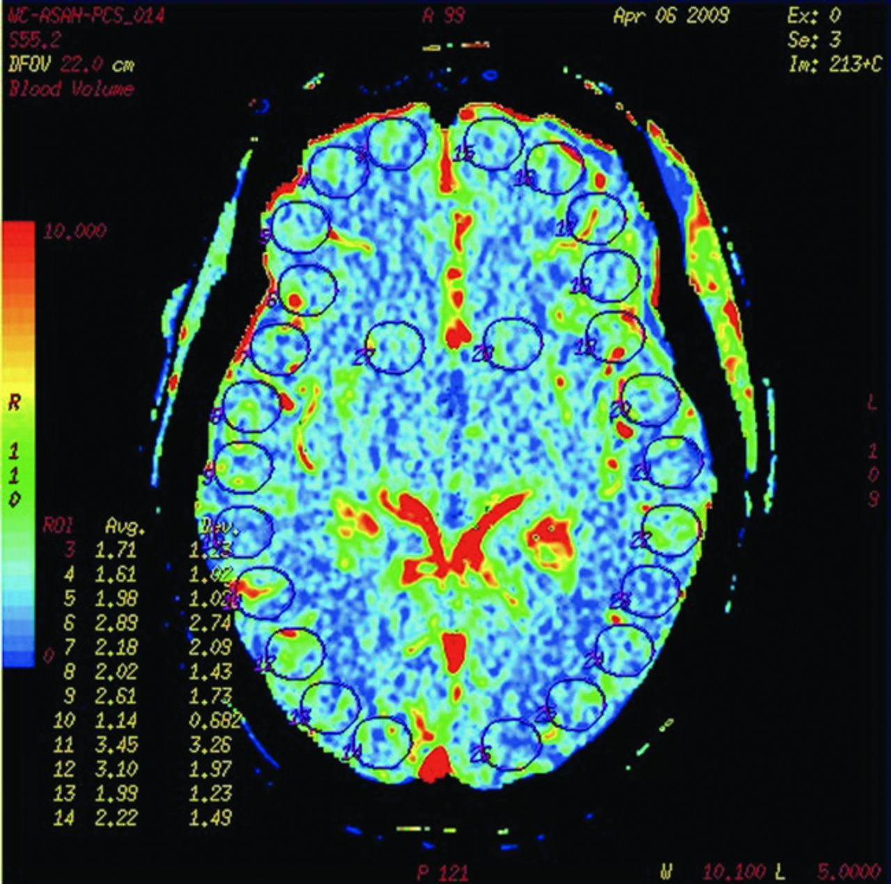
  • Fig 2.
    • Download figure
    • Open in new tab
    • Download powerpoint
    Fig 2.

    A representative blood volume map obtained in a single participant. The map shows placement of 26 uniform regions of interest: 12 in the right lobe, 12 in the left lobe, and 1 each over the right and left basal ganglia. Similar ROIs were placed over each section of a CTP dataset. The positions of ROIs from all sections in a given reference dataset were saved as a template and then were pasted onto the 5 generated noise datasets, ensuring perfect uniformity of position.
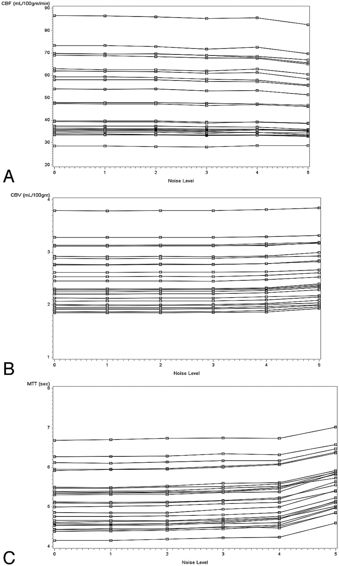
  • Fig 3.
    • Download figure
    • Open in new tab
    • Download powerpoint
    Fig 3.

    Frequency of quality scores assigned by the 6 observers to noise datasets at each noise level for (A) CBF (Spearman rank coefficient = −0.34; P < .0001), (B) CBV (Spearman rank coefficient = −0.35; P < .0001), and (C) MTT (Spearman rank coefficient = −0.44; P < .0001).

Tables

  • Figures
    • View popup
    Table 1:

    Simulated exposure for CTP examinations at each level of noise added

    Noise LevelAdded Noise Distribution, σ(add)Simulated Exposure, E2 (Range) (mAs)
    10.8188 (187–188)
    22.0177 (174–180)
    32.8167 (161–171)
    45.3127 (116–135)
    513.744 (36–51)
    • Note:—The range is determined using noise values of the reference scans at 1 SD above and below the mean, σ(E1) = 7.5 ± 0.8.

    • E2 indicates second exposure level.

    • View popup
    Table 2:

    Difference of least square means in perfusion parameters between reference dataset and noise datasets across all ROIs in all participants and across ROIs in participants with perfusion abnormalities (subanalysis)

    Perfusion ParameterSimulated Exposure (mAs)All ROIsOnly in ROIs with Perfusion Abnormalities
    Difference from Reference DatasetStandard ErrorPAdj PDifference from Reference DatasetStandard ErrorPAdj P
    CBF (mL/100 g/min)188−0.120.68.87>.99−0.350.86.69>.99
    177−0.200.68.77>.99−0.220.86.80>.99
    167−0.720.68.29.73−0.670.86.44.90
    127−0.600.68.38.85−0.010.86.99>.99
    44−1.810.68.01.040.470.86.58.97
    CBV (mL/100 g/min)1880.000.02.89>.99−0.010.02.74>.99
    1770.000.02.91>.990.000.02.99>.99
    1670.000.02.92>.990.000.02.93>.99
    1270.020.02.26.680.010.02.62.98
    440.070.02<.0001<.0010.100.02<.0001<.0001
    MTT (sec)1880.000.06.94>.990.030.10.79>.99
    1770.020.06.74>.990.050.10.66>.99
    1670.060.06.32.780.070.10.47.92
    1270.110.06.08.280.070.10.51.95
    440.460.06<.0001<.0010.330.10.00.01
    • Note:—ROIs with perfusion abnormalities were identified by a trained neuroradiologist (P.S.) with 12 years of experience.

    • Adj P indicates adjusted P value.

    • View popup
    Table 3:

    Coefficient of variance of perfusion parameters in noise datasets generated in 6 trials at each noise level

    Simulated Exposure (mAs)Coefficient of Variance (%)
    CBFCBVMTT
    1881.30.31.2
    1772.10.71.9
    1673.20.92.9
    1274.11.94.6
    447.14.56.7
    • Note:—Coefficient of variance is defined as SD/mean × 100.

    • View popup
    Table 4:

    Mean calculated interuser agreeability PABAK score for each CTP map

    Simulated Exposure (mAs)Mean PABAK Score of CBF Maps (range)Mean PABAK Score of CBV Maps (range)Mean PABAK Score of MTT Maps (range)
    1880.95 (0.88-1)1.00 (1-1)0.83 (0.70-0.98)
    1770.98 (0.93-1)1.00 (1-1)0.86 (0.70-0.98)
    1670.98 (0.93-1)1.00 (1-1)0.88 (0.73-1)
    1270.95 (0.88-1)1.00 (1-1)0.74 (0.43-1)
    440.51 (0.18-0.93)0.83 (0.58-1)0.05 (−0.87-0.68)
PreviousNext
Back to top

In this issue

American Journal of Neuroradiology: 34 (8)
American Journal of Neuroradiology
Vol. 34, Issue 8
1 Aug 2013
  • Table of Contents
  • Index by author
Advertisement
Print
Download PDF
Email Article

Thank you for your interest in spreading the word on American Journal of Neuroradiology.

NOTE: We only request your email address so that the person you are recommending the page to knows that you wanted them to see it, and that it is not junk mail. We do not capture any email address.

Enter multiple addresses on separate lines or separate them with commas.
Effects of Increased Image Noise on Image Quality and Quantitative Interpretation in Brain CT Perfusion
(Your Name) has sent you a message from American Journal of Neuroradiology
(Your Name) thought you would like to see the American Journal of Neuroradiology web site.
CAPTCHA
This question is for testing whether or not you are a human visitor and to prevent automated spam submissions.
Cite this article
K. Juluru, J.C. Shih, A. Raj, J.P. Comunale, H. Delaney, E.D. Greenberg, C. Hermann, Y.B. Liu, A. Hoelscher, N. Al-Khori, P.C. Sanelli
Effects of Increased Image Noise on Image Quality and Quantitative Interpretation in Brain CT Perfusion
American Journal of Neuroradiology Aug 2013, 34 (8) 1506-1512; DOI: 10.3174/ajnr.A3448

Citation Manager Formats

  • BibTeX
  • Bookends
  • EasyBib
  • EndNote (tagged)
  • EndNote 8 (xml)
  • Medlars
  • Mendeley
  • Papers
  • RefWorks Tagged
  • Ref Manager
  • RIS
  • Zotero
0 Responses
Respond to this article
Share
Bookmark this article
Effects of Increased Image Noise on Image Quality and Quantitative Interpretation in Brain CT Perfusion
K. Juluru, J.C. Shih, A. Raj, J.P. Comunale, H. Delaney, E.D. Greenberg, C. Hermann, Y.B. Liu, A. Hoelscher, N. Al-Khori, P.C. Sanelli
American Journal of Neuroradiology Aug 2013, 34 (8) 1506-1512; DOI: 10.3174/ajnr.A3448
del.icio.us logo Twitter logo Facebook logo Mendeley logo
  • Tweet Widget
  • Facebook Like
  • Google Plus One
Purchase

Jump to section

  • Article
    • Abstract
    • ABBREVIATIONS:
    • Materials and Methods
    • Results
    • Discussion
    • Conclusions
    • Acknowledgments
    • Footnotes
    • REFERENCES
  • Figures & Data
  • Supplemental
  • Info & Metrics
  • Responses
  • References
  • PDF

Related Articles

  • PubMed
  • Google Scholar

Cited By...

  • No citing articles found.
  • Crossref
  • Google Scholar

This article has not yet been cited by articles in journals that are participating in Crossref Cited-by Linking.

More in this TOC Section

  • Safety of Intrathecal Gadobutrol in Various Doses
  • Impact of Kidney Function on CNS Gadolinium Deposition in Patients Receiving Repeated Doses of Gadobutrol
  • Contrast-Induced Acute Kidney Injury in Radiologic Management of Acute Ischemic Stroke in the Emergency Setting
Show more Patient Safety

Similar Articles

Advertisement

Indexed Content

  • Current Issue
  • Accepted Manuscripts
  • Article Preview
  • Past Issues
  • Editorials
  • Editor's Choice
  • Fellows' Journal Club
  • Letters to the Editor
  • Video Articles

Cases

  • Case Collection
  • Archive - Case of the Week
  • Archive - Case of the Month
  • Archive - Classic Case

More from AJNR

  • Trainee Corner
  • Imaging Protocols
  • MRI Safety Corner
  • Book Reviews

Multimedia

  • AJNR Podcasts
  • AJNR Scantastics

Resources

  • Turnaround Time
  • Submit a Manuscript
  • Submit a Video Article
  • Submit an eLetter to the Editor/Response
  • Manuscript Submission Guidelines
  • Statistical Tips
  • Fast Publishing of Accepted Manuscripts
  • Graphical Abstract Preparation
  • Imaging Protocol Submission
  • Evidence-Based Medicine Level Guide
  • Publishing Checklists
  • Author Policies
  • Become a Reviewer/Academy of Reviewers
  • News and Updates

About Us

  • About AJNR
  • Editorial Board
  • Editorial Board Alumni
  • Alerts
  • Permissions
  • Not an AJNR Subscriber? Join Now
  • Advertise with Us
  • Librarian Resources
  • Feedback
  • Terms and Conditions
  • AJNR Editorial Board Alumni

American Society of Neuroradiology

  • Not an ASNR Member? Join Now

© 2025 by the American Society of Neuroradiology All rights, including for text and data mining, AI training, and similar technologies, are reserved.
Print ISSN: 0195-6108 Online ISSN: 1936-959X

Powered by HighWire