Advertisement

Main menu

  • Home
  • Content
    • Current Issue
    • Accepted Manuscripts
    • Article Preview
    • Past Issue Archive
    • Video Articles
    • AJNR Case Collection
    • Case of the Week Archive
    • Case of the Month Archive
    • Classic Case Archive
  • Special Collections
    • AJNR Awards
    • Low-Field MRI
    • Alzheimer Disease
    • ASNR Foundation Special Collection
    • Photon-Counting CT
    • View All
  • Multimedia
    • AJNR Podcasts
    • AJNR SCANtastic
    • Trainee Corner
    • MRI Safety Corner
    • Imaging Protocols
  • For Authors
    • Submit a Manuscript
    • Submit a Video Article
    • Submit an eLetter to the Editor/Response
    • Manuscript Submission Guidelines
    • Statistical Tips
    • Fast Publishing of Accepted Manuscripts
    • Graphical Abstract Preparation
    • Imaging Protocol Submission
    • Author Policies
  • About Us
    • About AJNR
    • Editorial Board
    • Editorial Board Alumni
  • More
    • Become a Reviewer/Academy of Reviewers
    • Subscribers
    • Permissions
    • Alerts
    • Feedback
    • Advertisers
    • ASNR Home

User menu

  • Alerts
  • Log in

Search

  • Advanced search
American Journal of Neuroradiology

American Journal of Neuroradiology

ASHNR American Society of Functional Neuroradiology ASHNR American Society of Pediatric Neuroradiology ASSR
  • Alerts
  • Log in

Advanced Search

  • Home
  • Content
    • Current Issue
    • Accepted Manuscripts
    • Article Preview
    • Past Issue Archive
    • Video Articles
    • AJNR Case Collection
    • Case of the Week Archive
    • Case of the Month Archive
    • Classic Case Archive
  • Special Collections
    • AJNR Awards
    • Low-Field MRI
    • Alzheimer Disease
    • ASNR Foundation Special Collection
    • Photon-Counting CT
    • View All
  • Multimedia
    • AJNR Podcasts
    • AJNR SCANtastic
    • Trainee Corner
    • MRI Safety Corner
    • Imaging Protocols
  • For Authors
    • Submit a Manuscript
    • Submit a Video Article
    • Submit an eLetter to the Editor/Response
    • Manuscript Submission Guidelines
    • Statistical Tips
    • Fast Publishing of Accepted Manuscripts
    • Graphical Abstract Preparation
    • Imaging Protocol Submission
    • Author Policies
  • About Us
    • About AJNR
    • Editorial Board
    • Editorial Board Alumni
  • More
    • Become a Reviewer/Academy of Reviewers
    • Subscribers
    • Permissions
    • Alerts
    • Feedback
    • Advertisers
    • ASNR Home
  • Follow AJNR on Twitter
  • Visit AJNR on Facebook
  • Follow AJNR on Instagram
  • Join AJNR on LinkedIn
  • RSS Feeds

AJNR Awards, New Junior Editors, and more. Read the latest AJNR updates

Review ArticleReview Articles
Open Access

Imaging Features of Midface Injectable Fillers and Associated Complications

D.T. Ginat and C.J. Schatz
American Journal of Neuroradiology August 2013, 34 (8) 1488-1495; DOI: https://doi.org/10.3174/ajnr.A3161
D.T. Ginat
aFrom the Department of Radiology (D.T.G.), Massachusetts General Hospital, Harvard Medical School, Boston, Massachusetts
  • Find this author on Google Scholar
  • Find this author on PubMed
  • Search for this author on this site
C.J. Schatz
bDepartment of Radiology (C.J.S.), Beverly Tower Wilshire Advanced Imaging, University of Southern California Keck School of Medicine, Los Angeles, California.
  • Find this author on Google Scholar
  • Find this author on PubMed
  • Search for this author on this site
  • Article
  • Figures & Data
  • Info & Metrics
  • Responses
  • References
  • PDF
Loading

Article Figures & Data

Figures

  • Fig 1.
    • Download figure
    • Open in new tab
    • Download powerpoint
    Fig 1.

    Calcium hydroxyapatite. Axial CT image (A) shows hyperattenuated material within the bilateral cheek subcutaneous tissues (arrows). Axial T1 (B) and T2 (C) MR images show that the fillers have low-to-intermediate signal on both sequences (arrows).
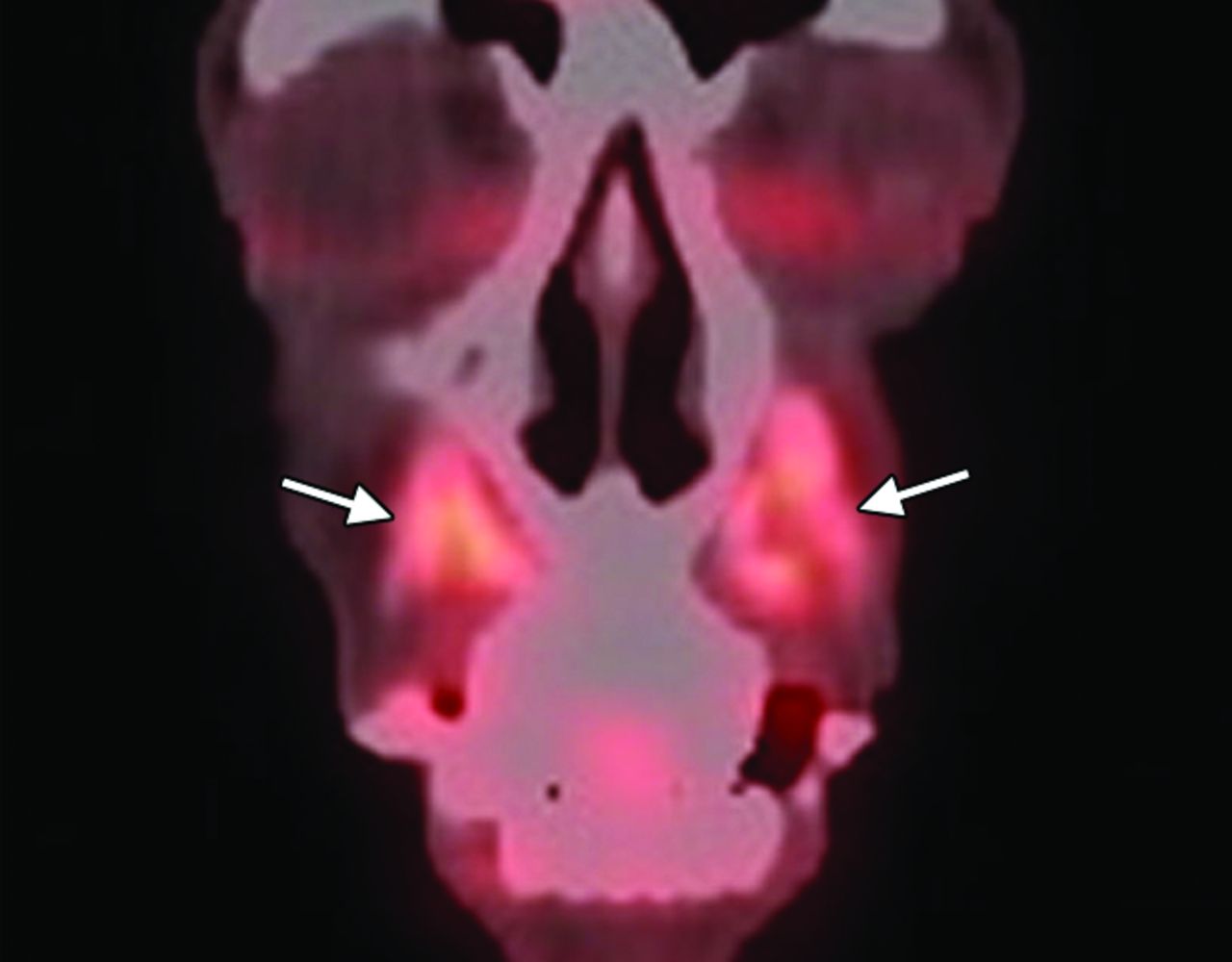
  • Fig 2.
    • Download figure
    • Open in new tab
    • Download powerpoint
    Fig 2.

    Calcium hydroxyapatite. Coronal 18FDG-PET image shows apparent hypermetabolism at the site of the fillers, which can be a false-positive finding for malignant disease (arrows).

  • Fig 3.
    • Download figure
    • Open in new tab
    • Download powerpoint
    Fig 3.

    Collagen. Axial CT image (A) shows stranding in the bilateral cheek subcutaneous tissues (arrows). Axial T1 (B), postcontrast T1 (C), and fat-saturated T2 (D) MR images show that the filler has nearly fluid signal characteristics (arrows).

  • Fig 4.
    • Download figure
    • Open in new tab
    • Download powerpoint
    Fig 4.

    Liquid silicone. Axial CT image (A) shows numerous high-attenuation foci within the bilateral cheeks (arrows). Axial T1 (B) and T2 (C) MR images show corresponding intermediate signal on both sequences.
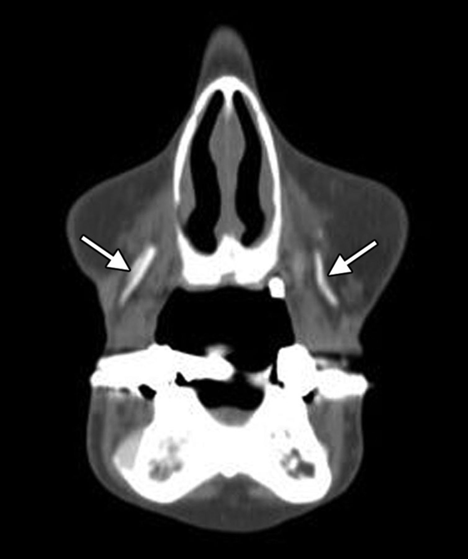
  • Fig 5.
    • Download figure
    • Open in new tab
    • Download powerpoint
    Fig 5.

    Polytetrafluoroethylene. Coronal CT image shows linear hyperattenuation in the bilateral nasolabial folds (arrows).
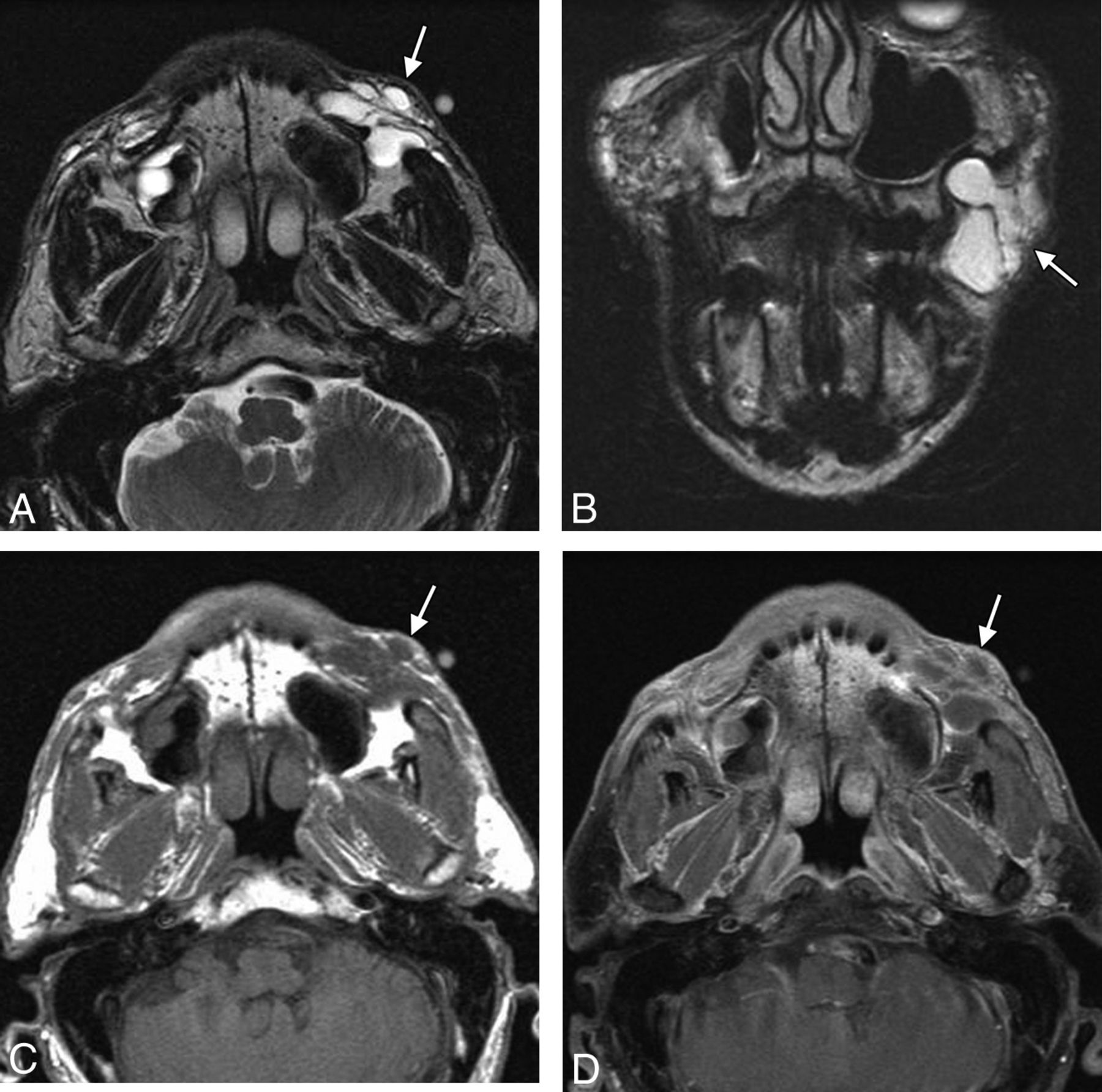
  • Fig 6.
    • Download figure
    • Open in new tab
    • Download powerpoint
    Fig 6.

    Hyaluronic acid gel. Axial T1 (A) and fat-suppressed postcontrast T1 (B) and T2 (C) MR images show bilateral fluid-intensity collections in the nasolabial folds, right greater than left (arrows).

  • Fig 7.
    • Download figure
    • Open in new tab
    • Download powerpoint
    Fig 7.

    Poly-l-lactic acid. Axial CT image shows bilateral irregular soft-tissue attenuation, with surrounding stranding in the subcutaneous tissues of the cheeks.

  • Fig 8.
    • Download figure
    • Open in new tab
    • Download powerpoint
    Fig 8.

    Polyacrylamide gel. Axial T1 (A) and T2 (B) MR images show a cluster of fluid-intensity collections surrounded by thin hypointense capsules within the left buccal space (arrows).
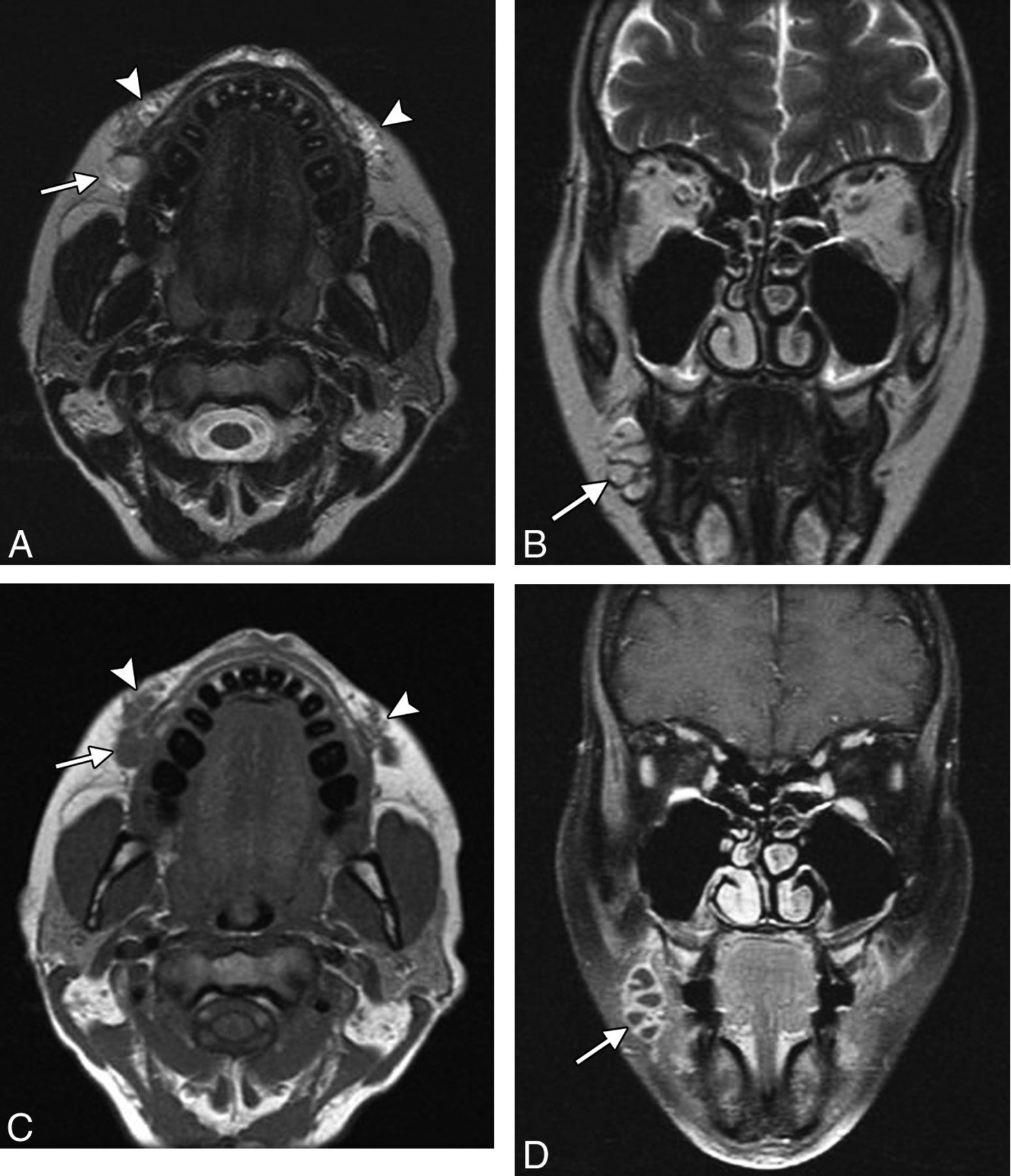
  • Fig 9.
    • Download figure
    • Open in new tab
    • Download powerpoint
    Fig 9.

    Abscess. The patient presented with painful swelling of right cheek after hyaluronic acid injection 2 months earlier. Axial (A) and coronal (B) T2, axial T1 (C), and coronal fat-saturated postcontrast T1 (D) MR images show deposits of hyaluronic acid in the bilateral oral commissures and upper lip (arrowheads). Posterior to the filler on the right, there is a multiloculated rim-enhancing fluid collection (arrows).

  • Fig 10.
    • Download figure
    • Open in new tab
    • Download powerpoint
    Fig 10.

    Overfilling. The patient presented with a facial lump and did not initially disclose a history of facial augmentation, which prompted imaging evaluation. A metallic marker was placed over the affected site. Axial CT images (A and B) show asymmetric contour deformity of the right nasolabial fold where there is a prominent deposit of filler agent. Courtesy of Gul Moonis, MD.
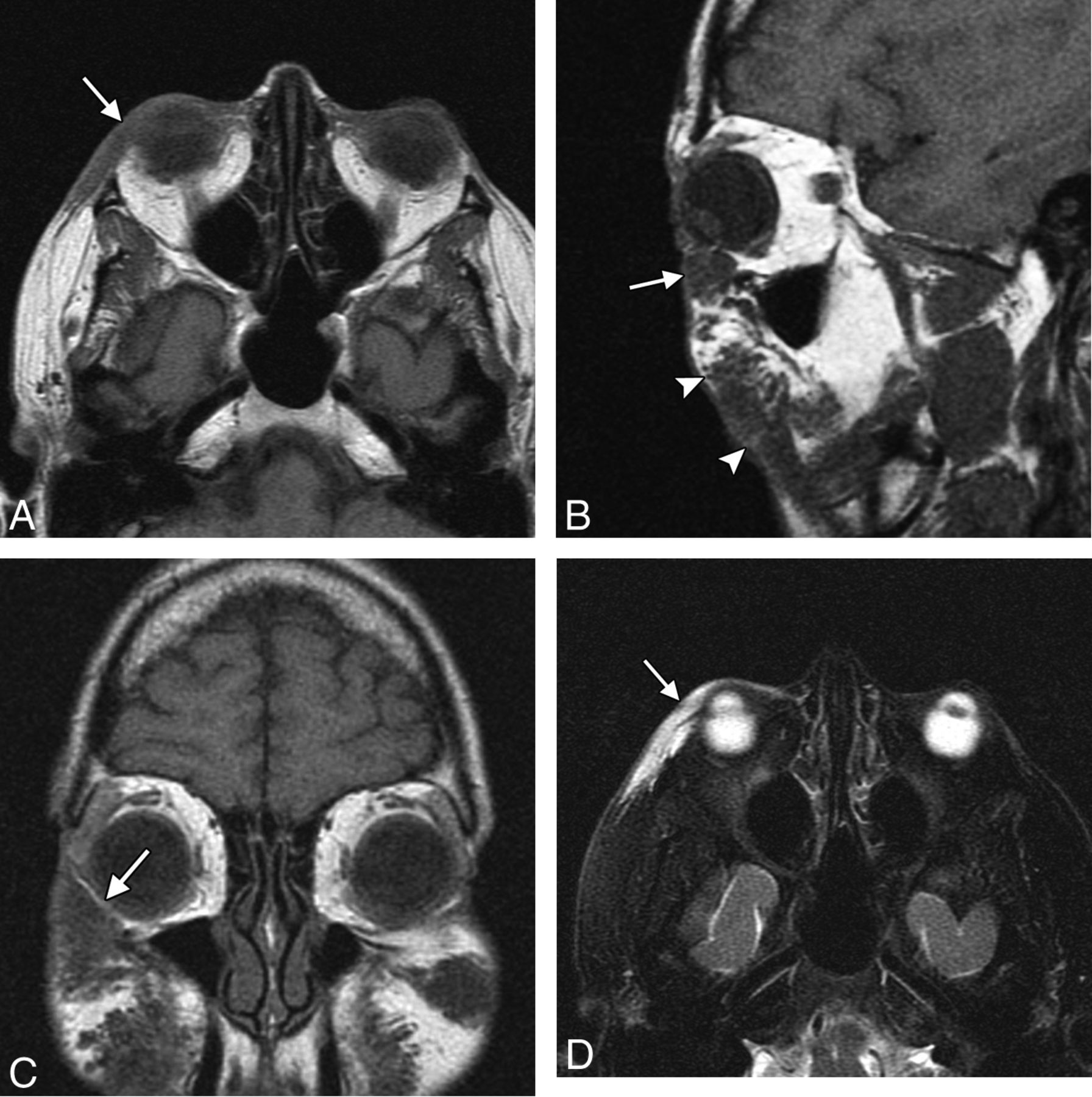
  • Fig 11.
    • Download figure
    • Open in new tab
    • Download powerpoint
    Fig 11.

    Migration. The patient presented with an eyelid “mass” after hyaluronic acid injection in the cheeks. Axial (A), sagittal (B), and coronal (C) T1 and axial fat-suppressed T2 (D) MR images show a globular collection of hyaluronic acid within the inferior eyelid (arrow), which extends superiorly from large deposits of filler in the cheeks (arrowheads).
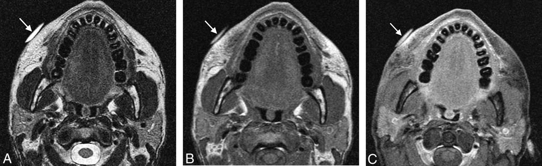
  • Fig 12.
    • Download figure
    • Open in new tab
    • Download powerpoint
    Fig 12.

    Chronic inflammation. The patient presented with right-greater-than-left facial swelling approximately 5 years after silicone injection. Axial T2 (A), T1 (B), and fat-saturated postcontrast T1 (C) MR images show an external marker overlying the lower right cheek (arrows), where there is diffuse swelling and enhancement surrounding the filler material. Milder involvement is also noted in the left side.

  • Fig 13.
    • Download figure
    • Open in new tab
    • Download powerpoint
    Fig 13.

    Scarring. The patient has a remote history of liquid silicone injection. Axial CT image shows fibrotic bands in the bilateral cheek subcutaneous tissues (arrows).
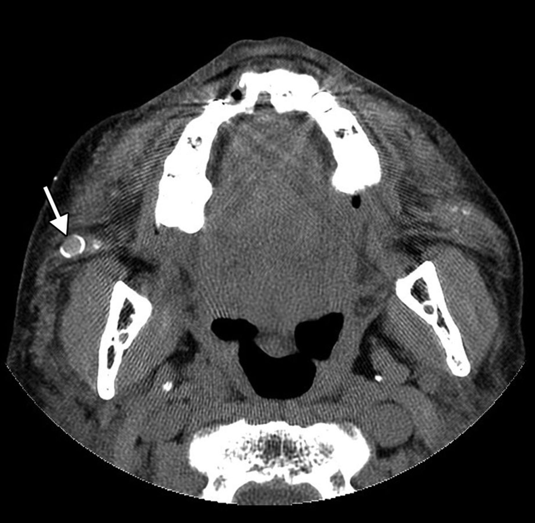
  • Fig 14.
    • Download figure
    • Open in new tab
    • Download powerpoint
    Fig 14.

    Foreign-body granuloma. The patient underwent silicone injection 50 years before. Axial CT image shows a nodule with eggshell calcification in the right cheek (arrow).

PreviousNext
Back to top

In this issue

American Journal of Neuroradiology: 34 (8)
American Journal of Neuroradiology
Vol. 34, Issue 8
1 Aug 2013
  • Table of Contents
  • Index by author
Advertisement
Print
Download PDF
Email Article

Thank you for your interest in spreading the word on American Journal of Neuroradiology.

NOTE: We only request your email address so that the person you are recommending the page to knows that you wanted them to see it, and that it is not junk mail. We do not capture any email address.

Enter multiple addresses on separate lines or separate them with commas.
Imaging Features of Midface Injectable Fillers and Associated Complications
(Your Name) has sent you a message from American Journal of Neuroradiology
(Your Name) thought you would like to see the American Journal of Neuroradiology web site.
CAPTCHA
This question is for testing whether or not you are a human visitor and to prevent automated spam submissions.
Cite this article
D.T. Ginat, C.J. Schatz
Imaging Features of Midface Injectable Fillers and Associated Complications
American Journal of Neuroradiology Aug 2013, 34 (8) 1488-1495; DOI: 10.3174/ajnr.A3161

Citation Manager Formats

  • BibTeX
  • Bookends
  • EasyBib
  • EndNote (tagged)
  • EndNote 8 (xml)
  • Medlars
  • Mendeley
  • Papers
  • RefWorks Tagged
  • Ref Manager
  • RIS
  • Zotero
0 Responses
Respond to this article
Share
Bookmark this article
Imaging Features of Midface Injectable Fillers and Associated Complications
D.T. Ginat, C.J. Schatz
American Journal of Neuroradiology Aug 2013, 34 (8) 1488-1495; DOI: 10.3174/ajnr.A3161
del.icio.us logo Twitter logo Facebook logo Mendeley logo
  • Tweet Widget
  • Facebook Like
  • Google Plus One
Purchase

Jump to section

  • Article
    • Abstract
    • Calcium Hydroxylapatite
    • Collagen and Collagen Mixed with Polymethylmethacrylate
    • Liquid Silicone
    • Polytetrafluoroethylene
    • Hyaluronic Acid
    • Poly-l-Lactic Acid
    • Polyacrylamide Gel
    • Complications
    • Footnotes
    • REFERENCES
  • Figures & Data
  • Info & Metrics
  • Responses
  • References
  • PDF

Related Articles

  • No related articles found.
  • PubMed
  • Google Scholar

Cited By...

  • Ageless beauty
  • "Finding a Voice": Imaging Features after Phonosurgical Procedures for Vocal Fold Paralysis
  • Imaging Features of Rhinoplasty
  • Crossref
  • Google Scholar

This article has not yet been cited by articles in journals that are participating in Crossref Cited-by Linking.

More in this TOC Section

  • An Atlas of Neonatal Neurovascular Imaging Anatomy as Depicted with Microvascular Imaging: The Intracranial Arteries
  • An Atlas of Neonatal Neurovascular Imaging Anatomy as Depicted with Microvascular Imaging: The Intracranial Veins
  • Clinical Translation of Hyperpolarized 13C Metabolic Probes for Glioma Imaging
Show more Review Articles

Similar Articles

Advertisement

Indexed Content

  • Current Issue
  • Accepted Manuscripts
  • Article Preview
  • Past Issues
  • Editorials
  • Editor's Choice
  • Fellows' Journal Club
  • Letters to the Editor
  • Video Articles

Cases

  • Case Collection
  • Archive - Case of the Week
  • Archive - Case of the Month
  • Archive - Classic Case

More from AJNR

  • Trainee Corner
  • Imaging Protocols
  • MRI Safety Corner
  • Book Reviews

Multimedia

  • AJNR Podcasts
  • AJNR Scantastics

Resources

  • Turnaround Time
  • Submit a Manuscript
  • Submit a Video Article
  • Submit an eLetter to the Editor/Response
  • Manuscript Submission Guidelines
  • Statistical Tips
  • Fast Publishing of Accepted Manuscripts
  • Graphical Abstract Preparation
  • Imaging Protocol Submission
  • Evidence-Based Medicine Level Guide
  • Publishing Checklists
  • Author Policies
  • Become a Reviewer/Academy of Reviewers
  • News and Updates

About Us

  • About AJNR
  • Editorial Board
  • Editorial Board Alumni
  • Alerts
  • Permissions
  • Not an AJNR Subscriber? Join Now
  • Advertise with Us
  • Librarian Resources
  • Feedback
  • Terms and Conditions
  • AJNR Editorial Board Alumni

American Society of Neuroradiology

  • Not an ASNR Member? Join Now

© 2025 by the American Society of Neuroradiology All rights, including for text and data mining, AI training, and similar technologies, are reserved.
Print ISSN: 0195-6108 Online ISSN: 1936-959X

Powered by HighWire