Advertisement

Main menu

  • Home
  • Content
    • Current Issue
    • Accepted Manuscripts
    • Article Preview
    • Past Issue Archive
    • Video Articles
    • AJNR Case Collection
    • Case of the Week Archive
    • Case of the Month Archive
    • Classic Case Archive
  • Special Collections
    • AJNR Awards
    • Low-Field MRI
    • Alzheimer Disease
    • ASNR Foundation Special Collection
    • Photon-Counting CT
    • View All
  • Multimedia
    • AJNR Podcasts
    • AJNR SCANtastic
    • Trainee Corner
    • MRI Safety Corner
    • Imaging Protocols
  • For Authors
    • Submit a Manuscript
    • Submit a Video Article
    • Submit an eLetter to the Editor/Response
    • Manuscript Submission Guidelines
    • Statistical Tips
    • Fast Publishing of Accepted Manuscripts
    • Graphical Abstract Preparation
    • Imaging Protocol Submission
    • Author Policies
  • About Us
    • About AJNR
    • Editorial Board
    • Editorial Board Alumni
  • More
    • Become a Reviewer/Academy of Reviewers
    • Subscribers
    • Permissions
    • Alerts
    • Feedback
    • Advertisers
    • ASNR Home

User menu

  • Alerts
  • Log in

Search

  • Advanced search
American Journal of Neuroradiology

American Journal of Neuroradiology

ASHNR American Society of Functional Neuroradiology ASHNR American Society of Pediatric Neuroradiology ASSR
  • Alerts
  • Log in

Advanced Search

  • Home
  • Content
    • Current Issue
    • Accepted Manuscripts
    • Article Preview
    • Past Issue Archive
    • Video Articles
    • AJNR Case Collection
    • Case of the Week Archive
    • Case of the Month Archive
    • Classic Case Archive
  • Special Collections
    • AJNR Awards
    • Low-Field MRI
    • Alzheimer Disease
    • ASNR Foundation Special Collection
    • Photon-Counting CT
    • View All
  • Multimedia
    • AJNR Podcasts
    • AJNR SCANtastic
    • Trainee Corner
    • MRI Safety Corner
    • Imaging Protocols
  • For Authors
    • Submit a Manuscript
    • Submit a Video Article
    • Submit an eLetter to the Editor/Response
    • Manuscript Submission Guidelines
    • Statistical Tips
    • Fast Publishing of Accepted Manuscripts
    • Graphical Abstract Preparation
    • Imaging Protocol Submission
    • Author Policies
  • About Us
    • About AJNR
    • Editorial Board
    • Editorial Board Alumni
  • More
    • Become a Reviewer/Academy of Reviewers
    • Subscribers
    • Permissions
    • Alerts
    • Feedback
    • Advertisers
    • ASNR Home
  • Follow AJNR on Twitter
  • Visit AJNR on Facebook
  • Follow AJNR on Instagram
  • Join AJNR on LinkedIn
  • RSS Feeds

AJNR Awards, New Junior Editors, and more. Read the latest AJNR updates

Research ArticleBrainE

Multimodal MR Imaging (Diffusion, Perfusion, and Spectroscopy): Is It Possible to Distinguish Oligodendroglial Tumor Grade and 1p/19q Codeletion in the Pretherapeutic Diagnosis?

S. Fellah, D. Caudal, A.M. De Paula, P. Dory-Lautrec, D. Figarella-Branger, O. Chinot, P. Metellus, P.J. Cozzone, S. Confort-Gouny, B. Ghattas, V. Callot and N. Girard
American Journal of Neuroradiology July 2013, 34 (7) 1326-1333; DOI: https://doi.org/10.3174/ajnr.A3352
S. Fellah
aFrom the Centre de Résonance Magnétique Biologique et Médicale (CRMBM) (S.F., P.J.C., S.C.-G., V.C.)
  • Find this author on Google Scholar
  • Find this author on PubMed
  • Search for this author on this site
D. Caudal
dDepartments of Neuroradiology (D.C., P.D.-L., N.G.)
  • Find this author on Google Scholar
  • Find this author on PubMed
  • Search for this author on this site
A.M. De Paula
ePathology and Neuropathology (A.M.D.P., D.F.-B.)
  • Find this author on Google Scholar
  • Find this author on PubMed
  • Search for this author on this site
P. Dory-Lautrec
dDepartments of Neuroradiology (D.C., P.D.-L., N.G.)
  • Find this author on Google Scholar
  • Find this author on PubMed
  • Search for this author on this site
D. Figarella-Branger
bCenter for Research in Oncobiology and Oncopharmacology (CRO2) (D.F.-B.)
ePathology and Neuropathology (A.M.D.P., D.F.-B.)
  • Find this author on Google Scholar
  • Find this author on PubMed
  • Search for this author on this site
O. Chinot
fNeurooncology (O.C.)
  • Find this author on Google Scholar
  • Find this author on PubMed
  • Search for this author on this site
P. Metellus
gNeurosurgery (P.M.), APHM, Hopital de la Timone, Marseille, France.
  • Find this author on Google Scholar
  • Find this author on PubMed
  • Search for this author on this site
P.J. Cozzone
aFrom the Centre de Résonance Magnétique Biologique et Médicale (CRMBM) (S.F., P.J.C., S.C.-G., V.C.)
  • Find this author on Google Scholar
  • Find this author on PubMed
  • Search for this author on this site
S. Confort-Gouny
aFrom the Centre de Résonance Magnétique Biologique et Médicale (CRMBM) (S.F., P.J.C., S.C.-G., V.C.)
  • Find this author on Google Scholar
  • Find this author on PubMed
  • Search for this author on this site
B. Ghattas
cMathematics Department (B.G.), Aix-Marseille University, Marseille, France
  • Find this author on Google Scholar
  • Find this author on PubMed
  • Search for this author on this site
V. Callot
aFrom the Centre de Résonance Magnétique Biologique et Médicale (CRMBM) (S.F., P.J.C., S.C.-G., V.C.)
  • Find this author on Google Scholar
  • Find this author on PubMed
  • Search for this author on this site
N. Girard
dDepartments of Neuroradiology (D.C., P.D.-L., N.G.)
  • Find this author on Google Scholar
  • Find this author on PubMed
  • Search for this author on this site
  • Article
  • Figures & Data
  • Info & Metrics
  • Responses
  • References
  • PDF
Loading

Article Figures & Data

Figures

  • Tables
  • Fig 1.
    • Download figure
    • Open in new tab
    • Download powerpoint
    Fig 1.

    Confounding examples of grade II and grade III oligodendrogliomas and oligoastrocytomas by cMRI.
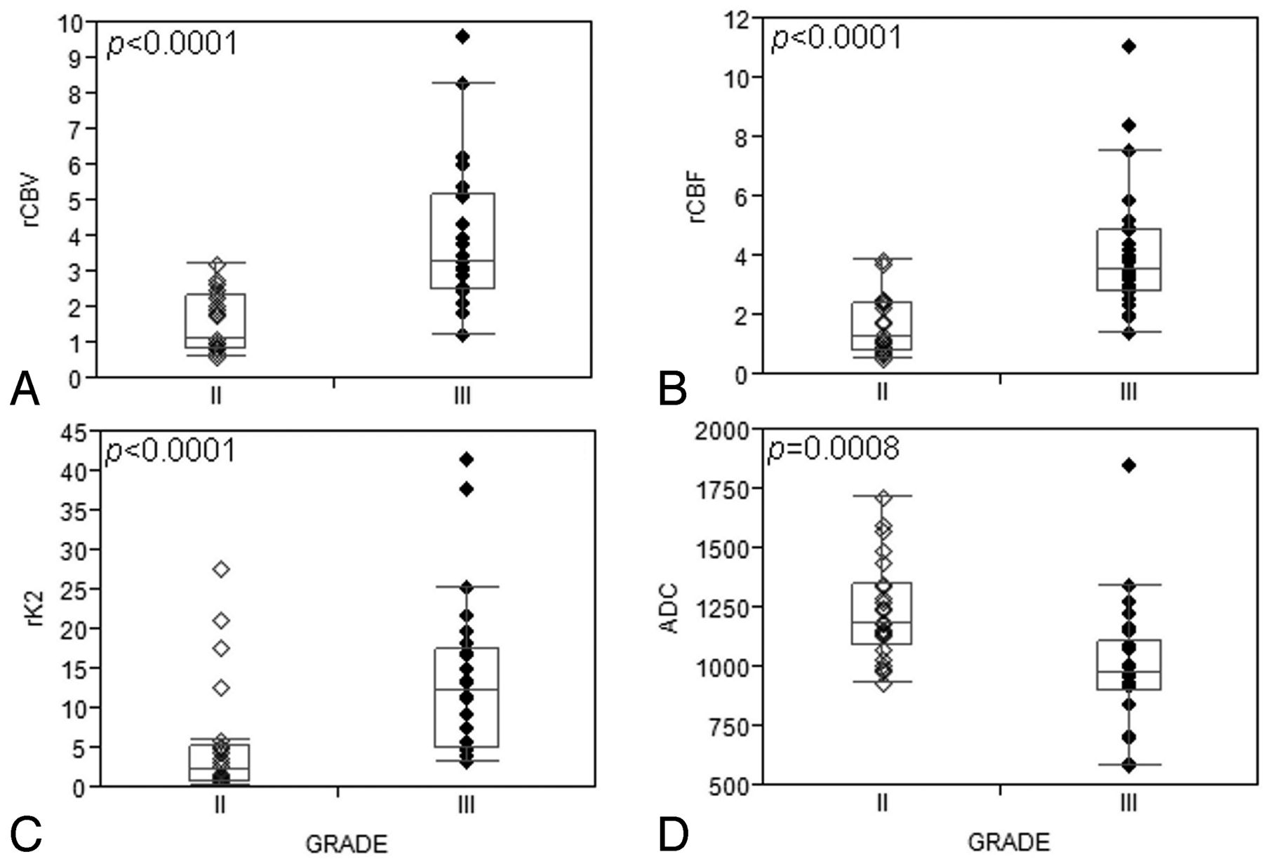
  • Fig 2.
    • Download figure
    • Open in new tab
    • Download powerpoint
    Fig 2.

    Boxplots show significant differences in rCBV, rCBF, rK2, and ADC between grade II and grade III oligodendroglial tumors: (A) rCBV, (B) rCBF, and (C) rK2 are significantly higher in grade III compared with grade II tumors, whereas (D) ADC is significantly increased in grade II tumors.

  • Fig 3.
    • Download figure
    • Open in new tab
    • Download powerpoint
    Fig 3.

    Boxplots show that grade II tumors with 1p/19q codeletion have significantly higher rCBV and rCBF than tumors with intact 1p/19q. No significant difference could be observed between genotypes in grade III oligodendroglial tumors.

Tables

  • Figures
    • View popup
    Table 1:

    Histopathologic and cMRI features of the tumors

    Grade II Oligoastrocytomas (n = 14)Grade II Oligodendrogliomas (n = 10)All Grade II (n = 24)Grade III Oligoastrocytomas (n = 14)Grade III Oligodendrogliomas (n = 12)All Grade III (n = 26)Total (n = 50)
    Contrast enhancement
        Absent8715 (62.5%)213 (11.5%)18 (36%)
        Blurry628 (33.3%)6511 (42.4%)19 (38%)
        Nodular011 (4.2%)415 (19.2%)6 (12%)
        Ringlike000 (0%)257 (26.9%)7 (14%)
    Location
        Frontal336 (25%)6612 (46.2%)18 (36%)
        Temporal123 (12.5%)336 (23.1%)9 (18%)
        Insular246 (25%)325 (19.2%)11 (22%)
        Temporoinsular718 (33.3%)112 (7.7%)10 (20%)
        Parietal101 (4.2%)000 (0%)1 (2%)
        Thalamic000 (0%)101 (3.8%)1 (2%)
    Tumor borders
        Sharp639 (37.5%)549 (34.6%)18 (36%)
        Indistinct8715 (62.5%)9817 (65.4%)32 (64%)
    Edema
        Yes9817 (70.9%)9716 (61.5%)33 (66%)
        No527 (29.1%)5510 (38.5%)17 (34%)
    Necrosis
        Yes8412 (50%)6915 (57.7%)27 (54%)
        No6612 (50%)8311 (42.3%)23 (46%)
    Hemorrhage
        Yes000 (0%)145 (19.2%)5 (10%)
        No141024 (100%)13821 (80.8%)45 (90%)
    1p/19q loss
        Yes4913 (54.2%)066 (23.1%)19 (38%)
        No10111 (45.8%)14620 (76.9%)31 (62%)
    • View popup
    Table 2:

    DWI, PWI, and MRS measurements in grade II and grade III oligodendroglial tumors and genotypes according to grade

    ParameterGradeGrade and Deletions
    II (n = 24)III (n = 26)P valueGrade II with 1p/19q Loss (n = 13)Grade II with Intact 1p/19q (n = 11)Grade III with 1p/19q Loss (n = 6)Grade III with Intact 1p/19q (n = 20)
    ADC (10–3 mm2/s)1238.50 ± 208.42995.19 ± 266.97.00081181.9 ± 201.321305.4 ± 205.381059.16 ± 127.21b976 ± 296.38a,b
    rADC1.66 ± 0.291.3 ± 0.29< .00011.57 ± 0.221.77 ± 0.331.49 ± 0.151.24 ± 0.3a,b
    rCBV1.57 ± 0.823.89 ± 1.97< .00012.09 ± 0.730.95 ± 0.33a4.43 ± 2.38a,b3.72 ± 1.86a,b
    rCBF1.65 ± 0.974.12 ± 2.15< .00012.21 ± 0.920.98 ± 0.48a4.87 ± 3.36a,b3.89 ± 1.69a,b
    rK25.18 ± 7.2613.64 ± 9.86< .00016.5 ± 8.143.57 ± 6.0512.65 ± 12.59b13.93 ± 9.27a,b
    NAA/H2Oc3.19 ± 1.522.39 ± 0.71.0233.20 ± 1.133.18 ± 1.962.53 ± 0.622.34 ± 0.75a
    Cr/H2Oc3.24 ± 1.372.84 ± 1.13.4723.03 ± 0.943.5 ± 1.772.8 ± 0.862.86 ± 1.22
    Cho/H2Oc3.87 ± 1.184.81 ± 2.65.263.74 ± 0.824.03 ± 1.534.75 ± 1.284.83 ± 2.97
    mIns/H2Oc2.27 ± 2.161.77 ± 1.19.6692.09 ± 1.62.49 ± 2.761.5 ± 0.531.84 ± 1.33
    Glx/H2Oc8.54 ± 2.967.94 ± 2.82.698.92 ± 2.742.08 ± 3.277.83 ± 1.637.97 ± 3.12
    Lipids/H2Oc14.25 ± 16.0715.88 ± 28.40.96919.13 ± 20.768.48 ± 3.0211.76 ± 5.8917.11 ± 32.32
    NAA/Crc1.02 ± 0.380.99 ± 0.61.1451.11 ± 0.400.91 ± 0.340.98 ± 0.350.99 ± 0.67a
    Cho/Crc1.33 ± 0.551.80 ± 1.00.0341.42 ± 0.691.22 ± 0.332.14 ± 1.811.7 ± 0.64b
    NAA/Choc0.86 ± 0.410.64 ± 0.40.020.91 ± 0.480.8 ± 0.340.59 ± 0.27a0.66 ± 0.43
    mIns/Crc0.67 ± 0.410.58 ± 0.31.8460.72 ± 0.50.61 ± 0.280.56 ± 0.140.59 ± 0.35
    • Note:—mIns indicates myo-inositol; rADC, relative apparent diffusion coefficient; rCBV, relative cerebral blood volume; rCBF, relative cerebral blood flow; rK2, relative permeability index.

    • ↵a Significantly different from grade II with 1p/19q loss (P < .05).

    • ↵b Significantly different from grade II with intact 1p/19q (P < .05).

    • ↵c Short TE.

    • View popup
    Table 3:

    Sensitivity, specificity, PPV, NPV, and misclassification error rate in the differentiation of tumor grades and genotypes

    Sensitivity/Specificity, PPV/NPVcMRIaMultimodal MRIb,c
    Gradeb
        II73/65%, 67/72%84/82%, 82/84%
        III65/73%, 72/67%82/84%, 84/82%
        Misclassification error rate31%17%
    Genotypec
        1p/19q Loss23/70%, 33/59%29/79%, 46/64%
        1p/19q Intact70/23%, 59/33%79/29%, 64/46%
        Misclassification error rate48%40%
    • Note:—CE indicates contrast enhancement; NPV, negative predictive value; PPV, positive predictive value.

    • The 6 most important predictors arising from the random forest analyses are:

    • ↵a CE+Location+Necrosis+Hemorrhage+Edema+Tumor borders

    • ↵b rCBF+rCBV+CE+ADC+Hemorrhage+ NAA/Cho at short TE

    • ↵c Location+ADC+rCBF+Cho/H2O+ NAA/Cr+lipids/H2O at short TE.

PreviousNext
Back to top

In this issue

American Journal of Neuroradiology: 34 (7)
American Journal of Neuroradiology
Vol. 34, Issue 7
1 Jul 2013
  • Table of Contents
  • Index by author
Advertisement
Print
Download PDF
Email Article

Thank you for your interest in spreading the word on American Journal of Neuroradiology.

NOTE: We only request your email address so that the person you are recommending the page to knows that you wanted them to see it, and that it is not junk mail. We do not capture any email address.

Enter multiple addresses on separate lines or separate them with commas.
Multimodal MR Imaging (Diffusion, Perfusion, and Spectroscopy): Is It Possible to Distinguish Oligodendroglial Tumor Grade and 1p/19q Codeletion in the Pretherapeutic Diagnosis?
(Your Name) has sent you a message from American Journal of Neuroradiology
(Your Name) thought you would like to see the American Journal of Neuroradiology web site.
CAPTCHA
This question is for testing whether or not you are a human visitor and to prevent automated spam submissions.
Cite this article
S. Fellah, D. Caudal, A.M. De Paula, P. Dory-Lautrec, D. Figarella-Branger, O. Chinot, P. Metellus, P.J. Cozzone, S. Confort-Gouny, B. Ghattas, V. Callot, N. Girard
Multimodal MR Imaging (Diffusion, Perfusion, and Spectroscopy): Is It Possible to Distinguish Oligodendroglial Tumor Grade and 1p/19q Codeletion in the Pretherapeutic Diagnosis?
American Journal of Neuroradiology Jul 2013, 34 (7) 1326-1333; DOI: 10.3174/ajnr.A3352

Citation Manager Formats

  • BibTeX
  • Bookends
  • EasyBib
  • EndNote (tagged)
  • EndNote 8 (xml)
  • Medlars
  • Mendeley
  • Papers
  • RefWorks Tagged
  • Ref Manager
  • RIS
  • Zotero
0 Responses
Respond to this article
Share
Bookmark this article
Multimodal MR Imaging (Diffusion, Perfusion, and Spectroscopy): Is It Possible to Distinguish Oligodendroglial Tumor Grade and 1p/19q Codeletion in the Pretherapeutic Diagnosis?
S. Fellah, D. Caudal, A.M. De Paula, P. Dory-Lautrec, D. Figarella-Branger, O. Chinot, P. Metellus, P.J. Cozzone, S. Confort-Gouny, B. Ghattas, V. Callot, N. Girard
American Journal of Neuroradiology Jul 2013, 34 (7) 1326-1333; DOI: 10.3174/ajnr.A3352
del.icio.us logo Twitter logo Facebook logo Mendeley logo
  • Tweet Widget
  • Facebook Like
  • Google Plus One
Purchase

Jump to section

  • Article
    • Abstract
    • ABBREVIATIONS:
    • Materials and Methods
    • Results
    • Discussion
    • Conclusions
    • Acknowledgments
    • Footnotes
    • REFERENCES
  • Figures & Data
  • Info & Metrics
  • Responses
  • References
  • PDF

Related Articles

  • No related articles found.
  • PubMed
  • Google Scholar

Cited By...

  • Molecular Subtype Classification in Lower-Grade Glioma with Accelerated DTI
  • Neuroimaging-Based Classification Algorithm for Predicting 1p/19q-Codeletion Status in IDH-Mutant Lower Grade Gliomas
  • Prediction of IDH1-Mutation and 1p/19q-Codeletion Status Using Preoperative MR Imaging Phenotypes in Lower Grade Gliomas
  • 3D Pseudocontinuous Arterial Spin-Labeling MR Imaging in the Preoperative Evaluation of Gliomas
  • Genetically Defined Oligodendroglioma Is Characterized by Indistinct Tumor Borders at MRI
  • Molecular imaging of 1p/19q deletion in oligodendroglial tumours with 11C-methionine positron emission tomography
  • Magnetic resonance spectroscopy of paragangliomas: new insights into in vivo metabolomics
  • Crossref
  • Google Scholar

This article has not yet been cited by articles in journals that are participating in Crossref Cited-by Linking.

More in this TOC Section

  • Predictors of Reperfusion in Patients with Acute Ischemic Stroke
  • Enhanced Axonal Metabolism during Early Natalizumab Treatment in Relapsing-Remitting Multiple Sclerosis
  • Progression of Microstructural Damage in Spinocerebellar Ataxia Type 2: A Longitudinal DTI Study
Show more Brain

Similar Articles

Advertisement

Indexed Content

  • Current Issue
  • Accepted Manuscripts
  • Article Preview
  • Past Issues
  • Editorials
  • Editor's Choice
  • Fellows' Journal Club
  • Letters to the Editor
  • Video Articles

Cases

  • Case Collection
  • Archive - Case of the Week
  • Archive - Case of the Month
  • Archive - Classic Case

More from AJNR

  • Trainee Corner
  • Imaging Protocols
  • MRI Safety Corner
  • Book Reviews

Multimedia

  • AJNR Podcasts
  • AJNR Scantastics

Resources

  • Turnaround Time
  • Submit a Manuscript
  • Submit a Video Article
  • Submit an eLetter to the Editor/Response
  • Manuscript Submission Guidelines
  • Statistical Tips
  • Fast Publishing of Accepted Manuscripts
  • Graphical Abstract Preparation
  • Imaging Protocol Submission
  • Evidence-Based Medicine Level Guide
  • Publishing Checklists
  • Author Policies
  • Become a Reviewer/Academy of Reviewers
  • News and Updates

About Us

  • About AJNR
  • Editorial Board
  • Editorial Board Alumni
  • Alerts
  • Permissions
  • Not an AJNR Subscriber? Join Now
  • Advertise with Us
  • Librarian Resources
  • Feedback
  • Terms and Conditions
  • AJNR Editorial Board Alumni

American Society of Neuroradiology

  • Not an ASNR Member? Join Now

© 2025 by the American Society of Neuroradiology All rights, including for text and data mining, AI training, and similar technologies, are reserved.
Print ISSN: 0195-6108 Online ISSN: 1936-959X

Powered by HighWire