Advertisement

Main menu

  • Home
  • Content
    • Current Issue
    • Accepted Manuscripts
    • Article Preview
    • Past Issue Archive
    • Video Articles
    • AJNR Case Collection
    • Case of the Week Archive
    • Case of the Month Archive
    • Classic Case Archive
  • Special Collections
    • AJNR Awards
    • Low-Field MRI
    • Alzheimer Disease
    • ASNR Foundation Special Collection
    • Photon-Counting CT
    • View All
  • Multimedia
    • AJNR Podcasts
    • AJNR SCANtastic
    • Trainee Corner
    • MRI Safety Corner
    • Imaging Protocols
  • For Authors
    • Submit a Manuscript
    • Submit a Video Article
    • Submit an eLetter to the Editor/Response
    • Manuscript Submission Guidelines
    • Statistical Tips
    • Fast Publishing of Accepted Manuscripts
    • Graphical Abstract Preparation
    • Imaging Protocol Submission
    • Author Policies
  • About Us
    • About AJNR
    • Editorial Board
    • Editorial Board Alumni
  • More
    • Become a Reviewer/Academy of Reviewers
    • Subscribers
    • Permissions
    • Alerts
    • Feedback
    • Advertisers
    • ASNR Home

User menu

  • Alerts
  • Log in

Search

  • Advanced search
American Journal of Neuroradiology

American Journal of Neuroradiology

ASHNR American Society of Functional Neuroradiology ASHNR American Society of Pediatric Neuroradiology ASSR
  • Alerts
  • Log in

Advanced Search

  • Home
  • Content
    • Current Issue
    • Accepted Manuscripts
    • Article Preview
    • Past Issue Archive
    • Video Articles
    • AJNR Case Collection
    • Case of the Week Archive
    • Case of the Month Archive
    • Classic Case Archive
  • Special Collections
    • AJNR Awards
    • Low-Field MRI
    • Alzheimer Disease
    • ASNR Foundation Special Collection
    • Photon-Counting CT
    • View All
  • Multimedia
    • AJNR Podcasts
    • AJNR SCANtastic
    • Trainee Corner
    • MRI Safety Corner
    • Imaging Protocols
  • For Authors
    • Submit a Manuscript
    • Submit a Video Article
    • Submit an eLetter to the Editor/Response
    • Manuscript Submission Guidelines
    • Statistical Tips
    • Fast Publishing of Accepted Manuscripts
    • Graphical Abstract Preparation
    • Imaging Protocol Submission
    • Author Policies
  • About Us
    • About AJNR
    • Editorial Board
    • Editorial Board Alumni
  • More
    • Become a Reviewer/Academy of Reviewers
    • Subscribers
    • Permissions
    • Alerts
    • Feedback
    • Advertisers
    • ASNR Home
  • Follow AJNR on Twitter
  • Visit AJNR on Facebook
  • Follow AJNR on Instagram
  • Join AJNR on LinkedIn
  • RSS Feeds

AJNR Awards, New Junior Editors, and more. Read the latest AJNR updates

Research ArticlePediatric Neuroimaging

Utility of Magnetization Transfer T1 Imaging in Children with Seizures

N. Kadom, A. Trofimova and G.L. Vezina
American Journal of Neuroradiology April 2013, 34 (4) 895-898; DOI: https://doi.org/10.3174/ajnr.A3396
N. Kadom
aFrom the Children's National Medical Center (N.K., G.L.V.), Washington, DC
  • Find this author on Google Scholar
  • Find this author on PubMed
  • Search for this author on this site
A. Trofimova
bInternational Clinic and Hospital MEDEM (A.T.), St. Petersburg, Russia.
  • Find this author on Google Scholar
  • Find this author on PubMed
  • Search for this author on this site
G.L. Vezina
aFrom the Children's National Medical Center (N.K., G.L.V.), Washington, DC
  • Find this author on Google Scholar
  • Find this author on PubMed
  • Search for this author on this site
  • Article
  • Figures & Data
  • Info & Metrics
  • Responses
  • References
  • PDF
Loading

Article Figures & Data

Figures

  • Fig. 1.
    • Download figure
    • Open in new tab
    • Download powerpoint
    Fig. 1.

    A 10-month-old boy with cardiac rhabdomyoma. MR imaging axial T1 with MT (A) and axial T2 (B). There is a bright lesion on T1 with MT in the medial posterior left frontal cortex, representing a cortical tuber (long arrows, A). This abnormality cannot be reliably detected on the axial T2 image (B) and other sequences (not shown) with a background of incomplete myelination. The findings are compatible with cortical tuber of tuberous sclerosis (diagnosis confirmed clinically).

  • Fig. 2.
    • Download figure
    • Open in new tab
    • Download powerpoint
    Fig. 2.

    A 3-year-old boy with increased seizure activity. MR imaging axial T1 with MT (A), axial FLAIR (B), and coronal T2 (C). Hyperintense lesion on T1 MT in the medial aspect of the left posterior frontal lobe (arrows, A) is seen. On axial FLAIR images, the lesion (arrows, B) is not readily identified and could be mistaken for volume-averaging. When one cross-references the lesion seen on T1 MT to the coronal T2 image (arrows, C), the lesion remains very subtle. The findings are compatible with focal cortical dysplasia (surgically confirmed).

  • Fig. 3.
    • Download figure
    • Open in new tab
    • Download powerpoint
    Fig. 3.

    A 9-year-old boy with seizures and a right anterior frontal focus on EEG. MR imaging axial T1 with MT (A) and axial T2 (B). T1 with MT shows a linear hyperintense lesion in the right frontal lobe, extending from the subcortical white matter toward the ventricular wall (arrows, A). On the axial T2 image, the lesion is less well-visualized (arrows, B). The findings are compatible with focal cortical dysplasia (diagnosis confirmed clinically).

  • Fig. 4.
    • Download figure
    • Open in new tab
    • Download powerpoint
    Fig. 4.

    A 12-year-old girl with tuberous sclerosis. MR imaging axial FLAIR (A) and axial T1 with MT (B). When cross-referencing the multiple bright lesions on the T1 with the MT image (B, not marked by arrows), only 1 shows a bright signal correlate on FLAIR (arrows, A and B). Without the T1 with MT sequence, disease burden in this patient would be significantly underestimated (diagnosis made clinically).
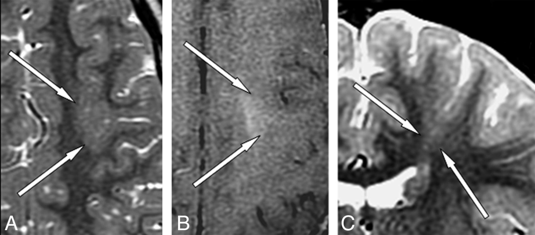
  • Fig. 5.
    • Download figure
    • Open in new tab
    • Download powerpoint
    Fig. 5.

    A 4-year-old boy with afebrile seizures. Sequential images on MR imaging axial T2 (A), axial T1 with MT (B), and coronal T2 (C). On axial T2, there is very subtle increased thickness of the left posterior frontal lobe cortex with mild blurring of the gray-white matter junction (arrows, A). MR imaging axial T1 with magnetization transfer shows a corresponding distinct area of hyperintense signal with an irregular shape, located predominantly subcortically (arrows, B). The coronal T2-weighted image shows a gray-matter isointense linear lesion extending from the thickened cortex to the wall of the left lateral ventricle (arrows, C). The findings are compatible with focal cortical dysplasia (surgically confirmed).

  • Fig. 6.
    • Download figure
    • Open in new tab
    • Download powerpoint
    Fig. 6.

    A 3-year-old boy with increasing seizure activity. MR imaging axial T1 with MT (A) and axial FLAIR (B). There is a small hyperintense lesion in the supraorbital lateral inferior right frontal lobe on T1 with MT (arrows, A). Axial FLAIR image shows local increased cortical thickness in this area and subtle hyperintense signal (arrows, B). The findings are compatible with focal cortical dysplasia (not surgically confirmed).

  • Fig. 7.
    • Download figure
    • Open in new tab
    • Download powerpoint
    Fig. 7.

    A 7-year-old boy with new seizures and an EEG abnormality in the right temporal parietal area. MR imaging axial T1 with MT (A) and axial FLAIR (B). T1 MT shows a small bright lesion in the right posterior frontal cortex (arrows, A). FLAIR image shows a focal hyperintense signal and minimal thickening of the frontal cortex without signal changes in the adjacent subcortical white matter (arrows, B), only seen on a single axial section in each sequence. The findings are compatible with focal cortical dysplasia (surgically confirmed).

PreviousNext
Back to top

In this issue

American Journal of Neuroradiology: 34 (4)
American Journal of Neuroradiology
Vol. 34, Issue 4
1 Apr 2013
  • Table of Contents
  • Index by author
Advertisement
Print
Download PDF
Email Article

Thank you for your interest in spreading the word on American Journal of Neuroradiology.

NOTE: We only request your email address so that the person you are recommending the page to knows that you wanted them to see it, and that it is not junk mail. We do not capture any email address.

Enter multiple addresses on separate lines or separate them with commas.
Utility of Magnetization Transfer T1 Imaging in Children with Seizures
(Your Name) has sent you a message from American Journal of Neuroradiology
(Your Name) thought you would like to see the American Journal of Neuroradiology web site.
CAPTCHA
This question is for testing whether or not you are a human visitor and to prevent automated spam submissions.
Cite this article
N. Kadom, A. Trofimova, G.L. Vezina
Utility of Magnetization Transfer T1 Imaging in Children with Seizures
American Journal of Neuroradiology Apr 2013, 34 (4) 895-898; DOI: 10.3174/ajnr.A3396

Citation Manager Formats

  • BibTeX
  • Bookends
  • EasyBib
  • EndNote (tagged)
  • EndNote 8 (xml)
  • Medlars
  • Mendeley
  • Papers
  • RefWorks Tagged
  • Ref Manager
  • RIS
  • Zotero
0 Responses
Respond to this article
Share
Bookmark this article
Utility of Magnetization Transfer T1 Imaging in Children with Seizures
N. Kadom, A. Trofimova, G.L. Vezina
American Journal of Neuroradiology Apr 2013, 34 (4) 895-898; DOI: 10.3174/ajnr.A3396
del.icio.us logo Twitter logo Facebook logo Mendeley logo
  • Tweet Widget
  • Facebook Like
  • Google Plus One
Purchase

Jump to section

  • Article
    • Abstract
    • ABBREVIATIONS:
    • Case Series
    • Discussion
    • Conclusions
    • References
  • Figures & Data
  • Info & Metrics
  • Responses
  • References
  • PDF

Related Articles

  • No related articles found.
  • PubMed
  • Google Scholar

Cited By...

  • MR Imaging Detection of CNS Lesions in Tuberous Sclerosis Complex: The Usefulness of T1WI with Chemical Shift Selective Images
  • Enhanced MR Conspicuity of Type IIb Focal Cortical Dysplasia by T1WI With CHESS: Two Case Reports
  • Radiologic and Pathologic Features of the Transmantle Sign in Focal Cortical Dysplasia: The T1 Signal Is Useful for Differentiating Subtypes
  • Crossref
  • Google Scholar

This article has not yet been cited by articles in journals that are participating in Crossref Cited-by Linking.

More in this TOC Section

  • Frontal Paraventricular Cysts
  • Sodium MRI in Pediatric Brain Tumors
  • FRACTURE MR in Congenital Vertebral Anomalies
Show more Pediatric Neuroimaging

Similar Articles

Advertisement

Indexed Content

  • Current Issue
  • Accepted Manuscripts
  • Article Preview
  • Past Issues
  • Editorials
  • Editor's Choice
  • Fellows' Journal Club
  • Letters to the Editor
  • Video Articles

Cases

  • Case Collection
  • Archive - Case of the Week
  • Archive - Case of the Month
  • Archive - Classic Case

More from AJNR

  • Trainee Corner
  • Imaging Protocols
  • MRI Safety Corner
  • Book Reviews

Multimedia

  • AJNR Podcasts
  • AJNR Scantastics

Resources

  • Turnaround Time
  • Submit a Manuscript
  • Submit a Video Article
  • Submit an eLetter to the Editor/Response
  • Manuscript Submission Guidelines
  • Statistical Tips
  • Fast Publishing of Accepted Manuscripts
  • Graphical Abstract Preparation
  • Imaging Protocol Submission
  • Evidence-Based Medicine Level Guide
  • Publishing Checklists
  • Author Policies
  • Become a Reviewer/Academy of Reviewers
  • News and Updates

About Us

  • About AJNR
  • Editorial Board
  • Editorial Board Alumni
  • Alerts
  • Permissions
  • Not an AJNR Subscriber? Join Now
  • Advertise with Us
  • Librarian Resources
  • Feedback
  • Terms and Conditions
  • AJNR Editorial Board Alumni

American Society of Neuroradiology

  • Not an ASNR Member? Join Now

© 2025 by the American Society of Neuroradiology All rights, including for text and data mining, AI training, and similar technologies, are reserved.
Print ISSN: 0195-6108 Online ISSN: 1936-959X

Powered by HighWire