Advertisement

Main menu

  • Home
  • Content
    • Current Issue
    • Accepted Manuscripts
    • Article Preview
    • Past Issue Archive
    • Video Articles
    • AJNR Case Collection
    • Case of the Week Archive
    • Case of the Month Archive
    • Classic Case Archive
  • Special Collections
    • AJNR Awards
    • Low-Field MRI
    • Alzheimer Disease
    • ASNR Foundation Special Collection
    • Photon-Counting CT
    • View All
  • Multimedia
    • AJNR Podcasts
    • AJNR SCANtastic
    • Trainee Corner
    • MRI Safety Corner
    • Imaging Protocols
  • For Authors
    • Submit a Manuscript
    • Submit a Video Article
    • Submit an eLetter to the Editor/Response
    • Manuscript Submission Guidelines
    • Statistical Tips
    • Fast Publishing of Accepted Manuscripts
    • Graphical Abstract Preparation
    • Imaging Protocol Submission
    • Author Policies
  • About Us
    • About AJNR
    • Editorial Board
    • Editorial Board Alumni
  • More
    • Become a Reviewer/Academy of Reviewers
    • Subscribers
    • Permissions
    • Alerts
    • Feedback
    • Advertisers
    • ASNR Home

User menu

  • Alerts
  • Log in

Search

  • Advanced search
American Journal of Neuroradiology

American Journal of Neuroradiology

ASHNR American Society of Functional Neuroradiology ASHNR American Society of Pediatric Neuroradiology ASSR
  • Alerts
  • Log in

Advanced Search

  • Home
  • Content
    • Current Issue
    • Accepted Manuscripts
    • Article Preview
    • Past Issue Archive
    • Video Articles
    • AJNR Case Collection
    • Case of the Week Archive
    • Case of the Month Archive
    • Classic Case Archive
  • Special Collections
    • AJNR Awards
    • Low-Field MRI
    • Alzheimer Disease
    • ASNR Foundation Special Collection
    • Photon-Counting CT
    • View All
  • Multimedia
    • AJNR Podcasts
    • AJNR SCANtastic
    • Trainee Corner
    • MRI Safety Corner
    • Imaging Protocols
  • For Authors
    • Submit a Manuscript
    • Submit a Video Article
    • Submit an eLetter to the Editor/Response
    • Manuscript Submission Guidelines
    • Statistical Tips
    • Fast Publishing of Accepted Manuscripts
    • Graphical Abstract Preparation
    • Imaging Protocol Submission
    • Author Policies
  • About Us
    • About AJNR
    • Editorial Board
    • Editorial Board Alumni
  • More
    • Become a Reviewer/Academy of Reviewers
    • Subscribers
    • Permissions
    • Alerts
    • Feedback
    • Advertisers
    • ASNR Home
  • Follow AJNR on Twitter
  • Visit AJNR on Facebook
  • Follow AJNR on Instagram
  • Join AJNR on LinkedIn
  • RSS Feeds

AJNR Awards, New Junior Editors, and more. Read the latest AJNR updates

Research ArticleExtracranial Vascular

Carotid Artery Plaque Characterization Using CT Multienergy Imaging

L. Saba, G.M. Argiolas, P. Siotto and M. Piga
American Journal of Neuroradiology April 2013, 34 (4) 855-859; DOI: https://doi.org/10.3174/ajnr.A3285
L. Saba
aFrom the Department of Radiology (L.S., M.P.), Azienda Ospedaliero Universitaria (A.O.U.), di Cagliari-Polo di Monserrato, Monserrato, Cagliari, Italy
  • Find this author on Google Scholar
  • Find this author on PubMed
  • Search for this author on this site
G.M. Argiolas
bDepartment of Radiology (G.M.A., P.S.), Azienda Ospedaliera Brotzu-Cagliari, Cagliari, Italy.
  • Find this author on Google Scholar
  • Find this author on PubMed
  • Search for this author on this site
P. Siotto
bDepartment of Radiology (G.M.A., P.S.), Azienda Ospedaliera Brotzu-Cagliari, Cagliari, Italy.
  • Find this author on Google Scholar
  • Find this author on PubMed
  • Search for this author on this site
M. Piga
aFrom the Department of Radiology (L.S., M.P.), Azienda Ospedaliero Universitaria (A.O.U.), di Cagliari-Polo di Monserrato, Monserrato, Cagliari, Italy
  • Find this author on Google Scholar
  • Find this author on PubMed
  • Search for this author on this site
  • Article
  • Figures & Data
  • Info & Metrics
  • Responses
  • References
  • PDF
Loading

Article Figures & Data

Figures

  • Tables
  • Fig. 1.
    • Download figure
    • Open in new tab
    • Download powerpoint
    Fig. 1.

    An 83-year-old male patient. In the 4 panels, the HU value (white arrows) is visible at the different monochromatic energy levels (gray arrowheads): 66 (A), 70 (B), 77 (C), and 86 keV (D). This plaque is in the fatty category by using all 4 energy levels.

  • Fig. 2.
    • Download figure
    • Open in new tab
    • Download powerpoint
    Fig. 2.

    A 76-year-old male patient. In the 4 panels, the HU value (white arrows) is visible at the different monochromatic energy levels (gray arrowheads): 66 (A), 70 (B), 77 (C), and 86 keV (D). This plaque is in the mixed category for 66 (A), 70 (B), and 77 keV (C) but is in the fatty category with 86 keV (D) energy.

  • Fig. 3.
    • Download figure
    • Open in new tab
    • Download powerpoint
    Fig. 3.

    Box-and-whisker plot of the attenuation values according to the different kiloelectron volt levels.

  • Fig. 4.
    • Download figure
    • Open in new tab
    • Download powerpoint
    Fig. 4.

    Graph shows the HU values of the plaque. The attenuation value of 60 HU, which is considered the threshold between mixed and fatty plaques, is visible (the HU values >60 are visible in the shaded area of the graph). The dotted rectangle indicates the plaques in which a classification shift occurred according to the energy level.
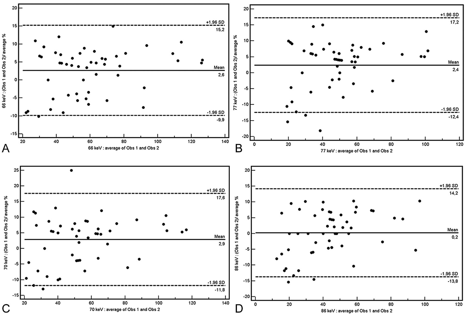
  • Fig. 5.
    • Download figure
    • Open in new tab
    • Download powerpoint
    Fig. 5.

    Bland-Altman plot analysis at 66 (A), 70 (B), 77 (C), and 86 keV (D).

Tables

  • Figures
    • View popup
    Table 1:

    Summary table of HU values at different kiloelectron volts

    Energy (keV)Mean HU95% CISDMedian95% CI2.5–97.5 PNormal Distribution
    6660,3951,598–69,18227,8545644,000–65,17121,525–129,475.028a
    7055,65947,591–63,72625,55845241,000–60,17121,050–118,425.028a
    7749,17141,969–56,37222,81554937,000–52,28518,000–105,950.036a
    8643,04936,483–49,61520,80264332,943–47,05713,575–96,750.04a
    • Note:—P indicates percentiles.

    • ↵a Statistically significant P value: normality rejected.

    • View popup
    Table 2:

    Wilcoxon analysis (P values)

    Energy (keV)66 keV70 keV77 keV86 keV
    66x.001a.001a.001a
    70.001ax.001a.001a
    77.001a.001ax.001a
    86.001a.001a.001ax
    • Note:—x indicates not calculable.

    • ↵a Statistically significant P value.

    • View popup
    Table 3:

    Plaque type according to the energy (n = 53)

    Energy (keV)Fatty PlaquesMixed Plaques
    662330
    702825
    773122
    863419
    • View popup
    Table 4:

    χ2 test to assess the variation in plaque type according to the kiloelectron volts used

    Energy (keV)66 keV70 keV77 keV86 keV
    66x0,3310,12.032a
    700,331x0,557.236
    770,120,557x.549
    860,032a0,2360,549x
    • Note:—x indicates not calculable.

    • ↵a Statistically significant P value.

PreviousNext
Back to top

In this issue

American Journal of Neuroradiology: 34 (4)
American Journal of Neuroradiology
Vol. 34, Issue 4
1 Apr 2013
  • Table of Contents
  • Index by author
Advertisement
Print
Download PDF
Email Article

Thank you for your interest in spreading the word on American Journal of Neuroradiology.

NOTE: We only request your email address so that the person you are recommending the page to knows that you wanted them to see it, and that it is not junk mail. We do not capture any email address.

Enter multiple addresses on separate lines or separate them with commas.
Carotid Artery Plaque Characterization Using CT Multienergy Imaging
(Your Name) has sent you a message from American Journal of Neuroradiology
(Your Name) thought you would like to see the American Journal of Neuroradiology web site.
CAPTCHA
This question is for testing whether or not you are a human visitor and to prevent automated spam submissions.
Cite this article
L. Saba, G.M. Argiolas, P. Siotto, M. Piga
Carotid Artery Plaque Characterization Using CT Multienergy Imaging
American Journal of Neuroradiology Apr 2013, 34 (4) 855-859; DOI: 10.3174/ajnr.A3285

Citation Manager Formats

  • BibTeX
  • Bookends
  • EasyBib
  • EndNote (tagged)
  • EndNote 8 (xml)
  • Medlars
  • Mendeley
  • Papers
  • RefWorks Tagged
  • Ref Manager
  • RIS
  • Zotero
0 Responses
Respond to this article
Share
Bookmark this article
Carotid Artery Plaque Characterization Using CT Multienergy Imaging
L. Saba, G.M. Argiolas, P. Siotto, M. Piga
American Journal of Neuroradiology Apr 2013, 34 (4) 855-859; DOI: 10.3174/ajnr.A3285
del.icio.us logo Twitter logo Facebook logo Mendeley logo
  • Tweet Widget
  • Facebook Like
  • Google Plus One
Purchase

Jump to section

  • Article
    • Abstract
    • ABBREVIATIONS:
    • Materials and Methods
    • Results
    • Discussion
    • Conclusions
    • References
  • Figures & Data
  • Info & Metrics
  • Responses
  • References
  • PDF

Related Articles

  • No related articles found.
  • PubMed
  • Google Scholar

Cited By...

  • Carotid Artery Wall Imaging: Perspective and Guidelines from the ASNR Vessel Wall Imaging Study Group and Expert Consensus Recommendations of the American Society of Neuroradiology
  • Crossref
  • Google Scholar

This article has not yet been cited by articles in journals that are participating in Crossref Cited-by Linking.

More in this TOC Section

  • Proximal Vertebral Artery Variants and Embryology
  • High-Risk Plaque Features in Carotid MRI
  • Nonstenotic Carotid Plaques and Stroke Review
Show more Extracranial Vascular

Similar Articles

Advertisement

Indexed Content

  • Current Issue
  • Accepted Manuscripts
  • Article Preview
  • Past Issues
  • Editorials
  • Editor's Choice
  • Fellows' Journal Club
  • Letters to the Editor
  • Video Articles

Cases

  • Case Collection
  • Archive - Case of the Week
  • Archive - Case of the Month
  • Archive - Classic Case

More from AJNR

  • Trainee Corner
  • Imaging Protocols
  • MRI Safety Corner
  • Book Reviews

Multimedia

  • AJNR Podcasts
  • AJNR Scantastics

Resources

  • Turnaround Time
  • Submit a Manuscript
  • Submit a Video Article
  • Submit an eLetter to the Editor/Response
  • Manuscript Submission Guidelines
  • Statistical Tips
  • Fast Publishing of Accepted Manuscripts
  • Graphical Abstract Preparation
  • Imaging Protocol Submission
  • Evidence-Based Medicine Level Guide
  • Publishing Checklists
  • Author Policies
  • Become a Reviewer/Academy of Reviewers
  • News and Updates

About Us

  • About AJNR
  • Editorial Board
  • Editorial Board Alumni
  • Alerts
  • Permissions
  • Not an AJNR Subscriber? Join Now
  • Advertise with Us
  • Librarian Resources
  • Feedback
  • Terms and Conditions
  • AJNR Editorial Board Alumni

American Society of Neuroradiology

  • Not an ASNR Member? Join Now

© 2025 by the American Society of Neuroradiology All rights, including for text and data mining, AI training, and similar technologies, are reserved.
Print ISSN: 0195-6108 Online ISSN: 1936-959X

Powered by HighWire