
Article Figures & Data
Figures
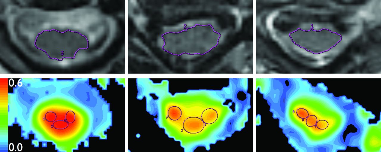
- Fig 1.
Region-of-interest placement. Top: Spinal cord area is measured on axial T2WI images (TR = 3250 ms; TE = 127 ms; noncontrast) at C2-C3 (left), stenosis (center), and C7-T1 (right). Bottom: Three isometric ROIs are placed onto FA colormaps from DTI (TR = 8100 ms, TE = 94 ms; noncontrast), same sections as Top. For each section, the 3 ROIs comprise 70% of spinal cord area.
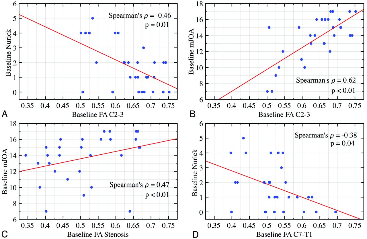
- Fig 2.
Correlation plots of baseline physical examination score and FA for all 30 subjects. A strong and significant correlation is observed between Nurick and mJOA scores and FA values at C2-C3 (upper panels), stenosis (bottom left), and C7-T1 (bottom right).
- Fig 3.
ROC curves demonstrating the accuracy of (A) mJOA, (B) FA, (C) T2 SI, and (D) DCSA for predicting surgeon's decision to operate. DCSA possesses a significantly lower area under the curve compared with the other 3 metrics.
- Fig 4.
MR imaging of subject A. Top: Axial T2WI (TR = 3250 ms; TE = 127 ms; noncontrast) at C2-C3 (left), stenosis (center), and C7-T1 (right). Bottom: FA colormaps from DTI (TR = 8100 ms, TE = 94 ms; noncontrast), same sections as Top.
- Fig 5.
MR imaging of subject B. Top: Axial T2WI (TR = 3250 ms; TE = 127 ms; noncontrast) at C2-C3 (left), stenosis (center), and C7-T1 (right). Bottom: FA colormaps from DTI (TR = 8100 ms, TE = 94 ms; noncontrast), same sections as Top.


Tables
Characteristic All Subjects (Mean ± SD) Surgery Cohort (Mean ± SD) Nonsurgery Cohort (Mean ± SD) P Value Age (years) 61.89 ± 12.37 (n = 30) 61.49 ± 9.63 (n = 15) 62.28 ± 14.96 (n = 15) .79 Sex Female 16 (53.3%) 6 (40%) 10 (66.7%) .14 Male 14 (46.7%) 9 (60%) 5 (33.3%) Follow-up (days) 180.67 ± 103.35 (n = 15) Baseline SF-36 (MCS) 6.86 ± 13.23 (n = 27) 12.64 ± 13.2 (n = 13) 1.49 ± 11.16 (n = 14) .03 Baseline SF-36 (PCS) 14.73 ± 11.17 (n = 27) 15.75 ± 10.88 (n = 13) 13.77 ± 11.76 (n = 14) .59 Baseline NDI 35.96 ± 19.19 (n = 27) 36.62 ± 17.42 (n = 13) 35.36 ± 21.28 (n = 14) .92 Baseline Nurick 1.7 ± 1.51 (n = 30) 2.2 ± 1.7 (n = 15) 1.2 ± 1.15 (n = 15) .11 Baseline mJOA 13.67 ± 2.83 (n = 30) 12.4 ± 3.04 (n = 15) 14.93 ± 1.98 (n = 15) .02 FA C2-C3 0.64 ± 0.07 (n = 30) 0.6 ± 0.07 (n = 15) 0.69 ± 0.05 (n = 15) <.01 FA stenosis level 0.51 ± 0.1 (n = 30) 0.47 ± 0.09 (n = 15) 0.55 ± 0.1 (n = 15) .05 FA C7-T1 0.52 ± 0.07 (n = 30) 0.51 ± 0.09 (n = 15) 0.53 ± 0.06 (n = 15) .4 High T2 SI 9 (30%) 7 (46.7%) 2 (13.3%) .05 DCSA (mm) 107.61 ± 22.03 (n = 30) 106.43 ± 24.82 (n = 15) 108.8 ± 19.65 (n = 15) .77 Note:—MCS indicates mental component score; PCS, physical component score.
- Table 2:
Spearman correlation coefficients relating baseline clinical scores and fractional anisotropy in various regions of the spinal cord
Anatomic Region Clinical Measure Spearman Correlation P Value n C2-C3 mJOA 0.62a <.01a 30a Nurick −0.46a .01a 30a SF-36 (MCS) −0.26 .19 27 SF-36 (PCS) −0.27 .17 27 NDI −0.19 .35 27 Level of stenosis mJOA 0.47a <.01a 30a Nurick −0.25 .18 30 SF-36 (MCS) −0.09 .64 27 SF-36 (PCS) 0.21 .29 27 NDI 0.19 .35 27 C7-T1 mJOA 0.31 .09 30 Nurick −0.38a .04a 30a SF-36 (MCS) −0.02 .92 27 SF-36 (PCS) 0.01 .96 27 NDI −0.03 .89 27 Note:—MCS indicates mental component score; PCS, physical component score.
↵a Indicates values are statistically significant.
Baseline Clinical Score High T2 SI (n = 9) Normal T2 SI (n = 21) P Value MJOA Mean ± SD 11.33 ± 3.43a 14.67 ± 1.85a .01a Median (Q1-Q3) 11 (9–15)a 15 (14–16)a SF-36 (MCS) Mean ± SD 5.95 ± 11.47 7.24 ± 14.18 .82 Median (Q1-Q3) 5 (−3.45–14.65) 9.2 (−4.1–17.8) NDI Mean ± SD 42.63 ± 22.23 33.16 ± 17.6 .25 Median (Q1-Q3) 52 (24–54) 34 (22–42) Nurick Mean ± SD 3 ± 1.32a 1.14 ± 1.24a <.01a Median (Q1-Q3) 4 (2–4)a 1 (0–2)a SF-36 (PCS) Mean ± SD 19.55 ± 12.72 12.69 ± 10.13 .15 Median (Q1-Q3) 21.35 (14.45–29.05) 9.6 (5–23.9) Change in Clinical Score (following surgery) MJOA Mean ± SD 1.86 ± 1.35 1.38 ± 1.3 .44 Median (Q1-Q3) 2 (1–3) 1 (0.5–2) Nurick Mean ± SD −0.86 ± 0.69 −0.25 ± 0.46 .07 Median (Q1-Q3) −1 (−1–0) 0 (−0.5–0) SF-36 (MCS) Mean ± SD 2.03 ± 10.24 −10.39 ± 9.9 .06 Median (Q1-Q3) 0 (−3.6–2.5) −8.1 (−17.4–0) SF-36 (PCS) Mean ± SD −0.3 ± 7.67 0.24 ± 5.24 .88 Median (Q1-Q3) 0 (−5.3–1.4) 0 (−2.5–0) NDI Mean ± SD −1 ± 27.36 −2 ± 11.2 .67 Median (Q1-Q3) 8 (−34–18) 0 (−10–6) Note:—MCS indicates mental component score; PCS, physical component score.
↵a Indicates values are significant.
- Table 4:
Spearman correlation coefficients relating postoperative clinical scores and baseline fractional anisotropy in various regions of the spinal cord
Anatomic Region Clinical Measure Spearman Correlation P Value n C7-T1 mJOA 0.02 .93 15 Nurick 0.17 .56 15 SF-36 (MCS) 0.1 .76 13 SF-36 (PCS) −0.07 .84 13 NDI 0.11 .73 13 Level of stenosis mJOA 0.06 .84 15 Nurick −0.22 .44 15 SF-36 (MCS) 0.28 .38 13 SF-36 (PCS) −0.21 .51 13 NDI −0.61a .04a 13a C2-C3 mJOA −0.26 .38 15 Nurick 0.17 .56 15 SF-36 (MCS) −0.4 .2 13 SF-36 (PCS) 0.1 .75 13 NDI 0.36 .25 13 Note:—MCS indicates mental component score; PCS, physical component score.
↵a Indicates values are significant.
In this issue


Jump to section
Related Articles
Cited By...
- Can Morphometric Analysis of Cervical Spondylotic Myelopathy Be a Tool for Surgical Outcome Prediction?
- The Evaluation and Prediction of Laminoplasty Surgery Outcome in Patients with Degenerative Cervical Myelopathy Using Diffusion Tensor MRI
- A Novel MRI Biomarker of Spinal Cord White Matter Injury: T2*-Weighted White Matter to Gray Matter Signal Intensity Ratio
- Spinal imaging update: an introduction to techniques for advanced mri