
Article Figures & Data
Figures
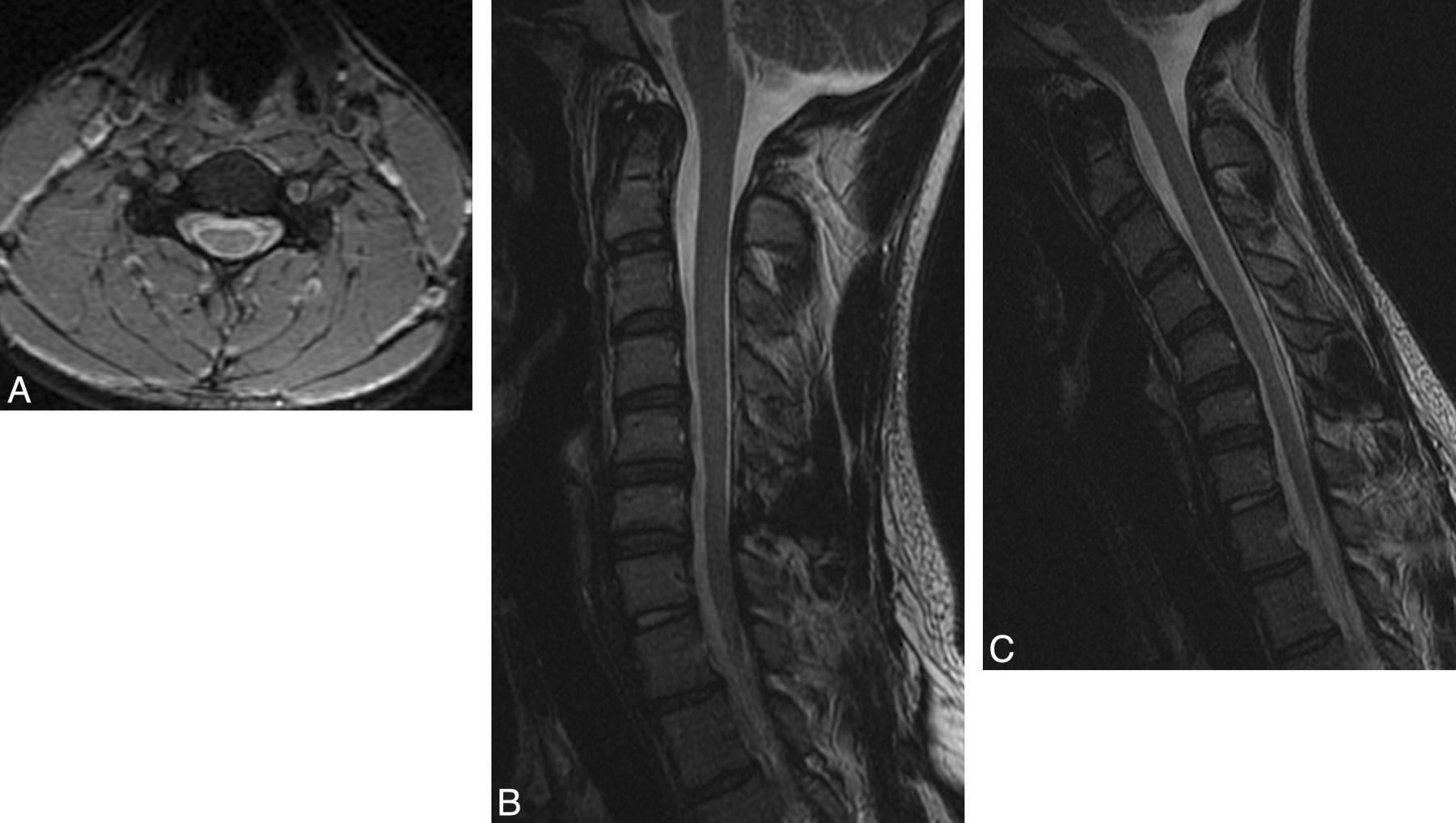
- Fig 1.
An 18-year-old man with HD. A, Axial T2-weighted image demonstrates LOA at the C5 level. B, Neutral T2-weighted image demonstrates subtle atrophy at C5-C6. C, Flexion T2-weighted image demonstrates 6 mm of anterior dural shift with near-complete obliteration of the subarachnoid space at C5-C6.
- Fig 2.
A 20-year-old man with HD. A, Neutral axial gradient-echo image at the C5 level demonstrates subtle bilateral LOA along the lateral aspects of the lamina bilaterally and spinal cord atrophy, asymmetric to the right. B, Neutral sagittal T2-weighted image also localizes this atrophy to C5-C6. C, Flexion sagittal T2-weighted image demonstrates 2 mm of anterior dural shift. The posterior subarachnoid space is not completely obliterated, and there is no direct spinal cord compression.


Tables
- Table 1:
Demographic characteristics of patients meeting clinical criteria for Hirayama disease compared with those not meeting clinical criteria
Patients with Hirayama Disease (n = 21) Patients without Hirayama Disease (n = 17) Age in yearsa (mean) (range) 24 (17–61) 41 (15–68) Male sex (No.) (%) 21 (100.0) 11 (64.7) Residence in North America, (No.) (%) 21 (100.0) 16 (94.1) White (No.) (%) 20 (95.2) 14 (82.3) Asian (No.) (%) 0 (0) 1 (5.9) Other or unknown race 1 (4.7) 2 (11.8) ↵a Age determined at time of clinical evaluation.
Sensitivity Specificity Diagnostic Odds Ratioa P Valueb Estimate 95% Confidence Interval Estimate 95% Confidence Interval Estimate 95% Confidence Interval Neutral MRIc 70% (14/20) (48%–85%) 100% (17/17) (82%–100%) 78.1 (4.0–1505.9) <.0001 Neutral and flexion MRI 71% (15/21) (50%–86%) 100% (17/17) (82%–100%) 83.5 (4.3–1605.1) <.0001 LOA on neutral imagesc 65% (13/20) (43%–82%) 94% (16/17) (73%–99%) 29.7 (3.2–273.4) .0004 Anterior dural shift with flexiond 76% (16/21) (55%–89%) 94% (16/17) (73%–99%) 51.2 (5.4–488.7) <.0001 Asymmetric cord flattening 48% (10/21) (28%–68%) 65% (11/17) (41%–83%) 1.7 (0.4–6.2) .521 Cord T2 signal 33% (7/21) (17%–55%) 94% (16/17) (73%–99%) 8.0 (0.9–73.2) .053 Abnormal curvaturec 50% (10/20) (30%–70%) 65% (11/17) (41%–83%) 1.8 (0.5–6.9) .510 C8 most affected cervical myotome at EMG with moderate-to-severe changee 50% (10/20) (30%–70%) 88% (14/16) (64%–97%) 7.0 (1.3–39.1) .0317 ↵a The odds ratio for neutral MRI and neutral and flexion MRI was computed by adding 0.5 to each cell (ie, the empiric odds ratio) to account for the zero cell that resulted from 100% specificity.
↵b P values were derived from a 2-tailed Fisher exact test.
↵c One patient did not have neutral images for assessment.
↵d Recorded as present, defined as any degree of dural shift, or absent.
↵e One patient did not have an EMG on record.


